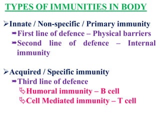
IMMUNITY
INTRODUCTION
The resistance or action of body against
invaders or harmful substances is called as
immunity.
The branch of zoology dealing with all
aspects of immunity is immunology.
 EDWARD JENER (1798) is father of
immunology.
TYPES OF IMMUNITIES IN BODY
Innate / Non-specific / Primary immunity
First line of defence – Physical barriers
Second line of defence – Internal
immunity
Acquired / Specific immunity
Third line of defence
Humoral immunity – B cell
Cell Mediated immunity – T cell
FIRST LINE OF DEFENCE
 Outermost layer of skin having dead cells prevents
entry of microbes.
 Sweat from sweat glands, oil from sebaceous
glands, acids and other metabolic wastes of
friendly bacteria on skin prevent growth of other
pathogens on it.
 Mucous of inner lining of digestive, respiratory &
urinogenital tract traps entering microbes.
 Mesh of fine hairs on nostrils filter particles from
inspired air.
 Sticky mucous secreted by cells of bronchi and
bronchioles trap microbes in inhaled air.
 Lysozyme, present in sweat, tear, saliva, nasal
discharges destroy bacterial cell wall and kill them.
SECOND LINE OF DEFENCE
Leuco
cytes
Normally human blood contains about
7000 to 8000 WBCs per cubic mm of blood.
Neutrophils (67 – 70 %), engulf foreign
bacteria. Basophils (0.5 – 1 %), engulf dust
particles and carbon. Eosinophils (3 %)
neutralise bacterial toxins and engulf
particles from antigen antibody reactions.
Lymphocytes (28 %) form antitoxins
causing wound healing. Monocytes (1.5 %)
ingest foreign living pathogens, remove
dead cells & produce antibodies.
Macro
phages
They may be fixed / wandering. Each can
engulf at least 100 bacteria.
Perfori
ns
It is a protein released from natural killer
cells. It kills the infected cell / cell into
which virus has entered. It make a hole in
plasma membrane, it allow entry of
water, cell swells and bursts.
Basic
polype
ptides
Some cells have these with antimicrobial
properties against specific microbes as
Bacillus anthracis.
Infla
mma
tion
The local changes in infected tissue
are collectively called inflammation. It
has five signs red, hot, swelling, pain
and fever in severe case. Histamine
released from broken mast cells, dilate
nearby blood vessels. It increases
more blood flow into infected tissue
causing redness and warmth. Fluid
released from damaged cells causes
swelling. Injury causes filling of pain.
Increased blood flow brings more
phagocytes that engulf microbes.
Fever Increased number of WBCs and
macrophages causes fever. Macrophages
release endogenous pyrogen or interleukin-
I that stimulate thalamus to maintain high
temperature causing fever. This is to
inhibit growth of microbes.
Comp
lemen
t-al
Protei
ns
These are secreted in liver. May rupture
cell membrane of microbes or tumour
cells. May stimulate mast cells to release
histamine causing inflammatory response.
May attract phagocytes to infected area.
May form a coat around pathogen for easy
phagocytosis.
Inter
feron
s
Almost all body cells produce δ & β
but specific lymphocytes and natural
killer cells produce γ interferons.
These are antiviral proteins released
by dead or infected cells. They enter
nearby uninfected healthy cells to
prevent viral infection in them.
Interferons produced by WBCs,
fibroblasts are not virus specific but
host specific.
THIRD LINE OF DEFENCE
Humoral Immune System – B – Cell
This system is based on B cells or B
lymphocytes or Bursa or bone marrow
derived cells.
They are formed and matured inside bone
marrow.
B cells have a life span of 5 – 7 days. With
blood, they enter lymph nodes as spleen,
thymus, tonsils etc.
They are activated by helper T cells upon
contact with the antigen. If not, die soon.
 ANTIGEN is a foreign agent that can initiate
an immune response.
Antigen may be a protein, lipoprotein or
polysaccharide molecule of the pathogen.
 Different parts of invader having such
markers are called ANTIGENIC
DETERMINATION SITES, each serving as a
separate antigen.
B cells produce ANTIBODIES in response to
antigen and react.
Antibody react with antigen by a process
called AGGLUTINATION.
It may inactivate antigen for ever or kill it or
destroy it. If antibody recognise antigen for
phagocytosis, it is called as OPSONISATION.
 COMPLEMENT MEDIATED CELL LYSIS –
immunoglobulin form pores in antigen and
breaks it.
Humoral immune system protects against
measles, small pox, cholera, cold, influenza
and tetanus.
Antibodies are proteins and collectively called
IMMUNOGLOBULINs.
IMMUNOGLOBULINS
Each immunoglobulin contains 4
polypeptide chains, 2 long / heavy / 
(kappa) & 2 short / light /  (lambda).
The chains are held together by disulphide
bonds to form a Y shaped molecule.
Top two tips of this Y bind to specific
antigen to form ANTIGEN – ANTIBODY
COMPLEX.
 Antigens may be destroyed by
agglutination / opsonisation / Complement
mediated cell.
1 IgA
Secreted in gut, respiratory & urinogenital
tract, present in milk. Prevents attachment
of pathogens to epithelial cells and mucosal
surfaces.
2 IgD
Uncertain function. Helps in B cell
activation.
3 IgE
Against allergens. Stimulate release of
histamine for allergic and inflammatory
response.
4 IgG
Gama, most abundant plasma antibody. It
only can cross placenta. Provides passive
immunity to foetus.
5 IgM Specific against bacteria and virus.
TYPES OF IMMUNOGLOBULINS
WORKING OF B – CELL
Pathogen enters into body
Macrophage brings antigen from pathogen to
lymph node
Competent B cell comes in contact of antigen
Helper T cell activates B cell in presence of
antigen
Activated B cell multiply by mitosis to form
clones
Some become plasma cells Some become
memory B cells
Plasma cell secrete specific
antibodies
Continue to
secrete
antibodies for
many years
Antibodies are transported by
blood and lymph to infected region O
They react with antigen of
pathogen to form antigen –
antibody complex
R
Response
rapidly if same
infection
reoccurs
Destroy / inactivate pathogen as
needed
THIRD LINE OF DEFENCE
Cell Mediated Immune System – T – Cell
This system is based on T cells or T lymphocytes.
They are formed inside bone marrow and
matured inside Thymus hence name T.
In thymus T cells proliferate and become
sensitive and recognise particular antigen.
 CMI is against leprosy, tuberculosis and cancer.
T cells are smaller and live for months to years
to life long.
T cells have antigen specific receptor molecule
on surface.
On contact of specific antigen, T cells proliferate
to act as needed.
FIVE TYPES OF T – CELLS
Effector T cell ( Te / TDTH ) :- These are for
Delayed Type of Hypersensitivity Reactions.
Killer / Cytotoxic T cell ( Tc / Tk ) :-
These may be induced artificially by
immunisation with allogenic tissue and naturally
by tumours and viruses.
For this, precursor must be stimulated by
antigen and class II MHC ( Major
Histocompatibility Complex ) molecules, found
on 6th chromosome.
Tk release enzyme and kill pathogen.
Helper T cell ( Th ) :- They activate B cells in
presence of particular antigen.
Memory T cells ( Tm ) :- They are induced by
first contact of an antigen and ready for rapid
action in further contact of same antigen.
Suppressor T cell ( Ts ) :- They suppress
autoimmunity & activated by antigen associated
with class II MHC molecules.
** T cells release proteins called cytokines /
lymphokines / interlukins. Interlukin-1 released
by Macrophage and T cells is needed for
activation of Tc. Interlukin-4 helps in cloning and
activation of B cells.
WORKING OF T – CELLS
Pathogen enters into body
A competent small T cell is activated by
specific antigen on pathogen
Activated T cell increase in size
Activated T cell multiply by mitosis to form
clones
Some become Tc or Th
They leave lymph nodes
and migrate to site of
infection
Tc release lytic enzymes /
Th activate B cells or
stimulates macrophages
Pathogen is killed /
destroyed
Some become
memory T
cells
Remain in
lymph nodes
Response
rapidly if
same
infection
reoccurs
CHARAC
TER
ACTIVE /
OWN
IMMUNITY
PASSIVE /
ACQUIRED
IMMUNITY
Developm
ent
After
infection in a
person
By antibodies
acquired from
others.
Example
Immunity
after
vaccination,
after measles.
Immunity of
foetus by maternal
antibodies via
placenta or
colostrum ( thick
first milk )
AUTOIMMUNITY
It is a condition when B / T cells response
to certain antigens produced by body’s
own cells.
Hasimoto’s disease – thyroid is destroyed,
rheumatic fever – heart and joint affected.
Glomerulonephritis – glomerulus
membrane affected.
Multiple sclerosis – myelin sheath affected.
VACCINATION
Vaccine contain dead or weakened
pathogens or their products.
When introduced into a person, it causes
production of antigen specific actions and
memory cells but not the illness.
It is to develop acquired immunity.
Introduction of vaccine to a person is
called as vaccination.
Introduction of antigen or pathogenic
fragment to a person is called as
immunization.
THREE TYPES OF VACCINATION
Attenuation :- Repeatedly infecting cells
with antigen until its toxic effects is
reduced. Preparing snake venom using
horse, resistance of Chandragupta to
poison.
Peptide Fragments :- Capable of
producing immune response but not
illness.
DNA Vaccine :- Introducing a gene
encoding antibody for an antigen into
body.
ALLERGY
Allergy is the hyper reactivity of immune
system to a substance. They are ALLERGENS.
Allergens are mild antigens. IgE reacts to them.
Pollen, dust and such particles may be
allergens.
When an allergen is inhaled or eaten, it causes
release of histamine & serotonin from mast
cells.
They cause dilation of blood vessels causing
inflammatory response and irritation.
Allergy may be treated with antihistamine
drugs, adrenaline or steroid.
IMMUNODEFICIENCY
It is the deficiency of immune response in the body.
It may have symptoms like recurrent and chronic
infection, unusual infecting agents and poor
response to treatment.
It is of four types :-
Phagocytic Deficiency :- Both quantitative and
qualitative deficiency of phagocytes.
B-cell Deficiency :- Deficiency of B cell or Helper
T cell. In infants of 5-9 months.
T-cell Deficiency :- Due to absence of thymus &
parathyroid glands. e.g. AIDS.
Combined B-cell & T-cell Deficiency :- e.g. SCID
( Severe Combined Deficiency Disease ).
IMMUNE SYSTEM OF BODY
The system in body that protects it from
various infections is called immune system.
It consists of lymphoid tissues, its cells and
their products – antibodies.
Origin, maturation and multiplication of
lymphocytes occur in lymphoid organs.
Lymphoid organs may be :-
Bone marrow :- It is present in cavities of
long bones. It produce lymphocytes.
Thymus :- It is present below breast bone
near heart. Here maturation of T cells
occurs.
Spleen :- It lie below stomach in U shaped
limbs of duodenumto store Lymphocytes
& phagocytes.
Lymph nodes :- These are solid organs of
lymphoid tissues. They store B cells.
Mucosal Associated Lymphoid Tissues
(MALT) :- These are lymphoid tissues
present in gut, respiratory and
urinogenital tract.
MONOCLONALANTIBODIES
These are homogeneous immunological
reagents with defined specificity.
They are made from only one type of B
cells.
Production of MA / Hybridoma
Technology :- Given by Georges J. F.
Kohler and Cesar Milstein in 1975, Nobel
prize in 1984. These are produced by
fusing B cells with tumour cells to form
hybrid cells called hybridoma. They can
produce particular antibody.B cells can’t
divide once started producing antibodies
and are of short life span. It is overcame
by this technology.
Applications of MA :- Diagnosis, screening
and treatment of diseases. Production of
vaccines and vaccination. Used as enzymes
called abzymes. Antibody may bind to
specific ligands, antibodies may be
generated by modifying such ligands and
they catalyse specific reactions as enzymes.

Immunity

  • 1.
  • 2.
    INTRODUCTION The resistance oraction of body against invaders or harmful substances is called as immunity. The branch of zoology dealing with all aspects of immunity is immunology.  EDWARD JENER (1798) is father of immunology.
  • 3.
    TYPES OF IMMUNITIESIN BODY Innate / Non-specific / Primary immunity First line of defence – Physical barriers Second line of defence – Internal immunity Acquired / Specific immunity Third line of defence Humoral immunity – B cell Cell Mediated immunity – T cell
  • 4.
    FIRST LINE OFDEFENCE  Outermost layer of skin having dead cells prevents entry of microbes.  Sweat from sweat glands, oil from sebaceous glands, acids and other metabolic wastes of friendly bacteria on skin prevent growth of other pathogens on it.  Mucous of inner lining of digestive, respiratory & urinogenital tract traps entering microbes.  Mesh of fine hairs on nostrils filter particles from inspired air.  Sticky mucous secreted by cells of bronchi and bronchioles trap microbes in inhaled air.  Lysozyme, present in sweat, tear, saliva, nasal discharges destroy bacterial cell wall and kill them.
  • 5.
    SECOND LINE OFDEFENCE Leuco cytes Normally human blood contains about 7000 to 8000 WBCs per cubic mm of blood. Neutrophils (67 – 70 %), engulf foreign bacteria. Basophils (0.5 – 1 %), engulf dust particles and carbon. Eosinophils (3 %) neutralise bacterial toxins and engulf particles from antigen antibody reactions. Lymphocytes (28 %) form antitoxins causing wound healing. Monocytes (1.5 %) ingest foreign living pathogens, remove dead cells & produce antibodies.
  • 6.
    Macro phages They may befixed / wandering. Each can engulf at least 100 bacteria. Perfori ns It is a protein released from natural killer cells. It kills the infected cell / cell into which virus has entered. It make a hole in plasma membrane, it allow entry of water, cell swells and bursts. Basic polype ptides Some cells have these with antimicrobial properties against specific microbes as Bacillus anthracis.
  • 7.
    Infla mma tion The local changesin infected tissue are collectively called inflammation. It has five signs red, hot, swelling, pain and fever in severe case. Histamine released from broken mast cells, dilate nearby blood vessels. It increases more blood flow into infected tissue causing redness and warmth. Fluid released from damaged cells causes swelling. Injury causes filling of pain. Increased blood flow brings more phagocytes that engulf microbes.
  • 8.
    Fever Increased numberof WBCs and macrophages causes fever. Macrophages release endogenous pyrogen or interleukin- I that stimulate thalamus to maintain high temperature causing fever. This is to inhibit growth of microbes. Comp lemen t-al Protei ns These are secreted in liver. May rupture cell membrane of microbes or tumour cells. May stimulate mast cells to release histamine causing inflammatory response. May attract phagocytes to infected area. May form a coat around pathogen for easy phagocytosis.
  • 9.
    Inter feron s Almost all bodycells produce δ & β but specific lymphocytes and natural killer cells produce γ interferons. These are antiviral proteins released by dead or infected cells. They enter nearby uninfected healthy cells to prevent viral infection in them. Interferons produced by WBCs, fibroblasts are not virus specific but host specific.
  • 10.
    THIRD LINE OFDEFENCE Humoral Immune System – B – Cell This system is based on B cells or B lymphocytes or Bursa or bone marrow derived cells. They are formed and matured inside bone marrow. B cells have a life span of 5 – 7 days. With blood, they enter lymph nodes as spleen, thymus, tonsils etc. They are activated by helper T cells upon contact with the antigen. If not, die soon.  ANTIGEN is a foreign agent that can initiate
  • 11.
    an immune response. Antigenmay be a protein, lipoprotein or polysaccharide molecule of the pathogen.  Different parts of invader having such markers are called ANTIGENIC DETERMINATION SITES, each serving as a separate antigen. B cells produce ANTIBODIES in response to antigen and react. Antibody react with antigen by a process called AGGLUTINATION. It may inactivate antigen for ever or kill it or destroy it. If antibody recognise antigen for
  • 12.
    phagocytosis, it iscalled as OPSONISATION.  COMPLEMENT MEDIATED CELL LYSIS – immunoglobulin form pores in antigen and breaks it. Humoral immune system protects against measles, small pox, cholera, cold, influenza and tetanus. Antibodies are proteins and collectively called IMMUNOGLOBULINs.
  • 13.
    IMMUNOGLOBULINS Each immunoglobulin contains4 polypeptide chains, 2 long / heavy /  (kappa) & 2 short / light /  (lambda). The chains are held together by disulphide bonds to form a Y shaped molecule. Top two tips of this Y bind to specific antigen to form ANTIGEN – ANTIBODY COMPLEX.  Antigens may be destroyed by agglutination / opsonisation / Complement mediated cell.
  • 14.
    1 IgA Secreted ingut, respiratory & urinogenital tract, present in milk. Prevents attachment of pathogens to epithelial cells and mucosal surfaces. 2 IgD Uncertain function. Helps in B cell activation. 3 IgE Against allergens. Stimulate release of histamine for allergic and inflammatory response. 4 IgG Gama, most abundant plasma antibody. It only can cross placenta. Provides passive immunity to foetus. 5 IgM Specific against bacteria and virus. TYPES OF IMMUNOGLOBULINS
  • 15.
    WORKING OF B– CELL Pathogen enters into body Macrophage brings antigen from pathogen to lymph node Competent B cell comes in contact of antigen Helper T cell activates B cell in presence of antigen Activated B cell multiply by mitosis to form clones
  • 16.
    Some become plasmacells Some become memory B cells Plasma cell secrete specific antibodies Continue to secrete antibodies for many years Antibodies are transported by blood and lymph to infected region O They react with antigen of pathogen to form antigen – antibody complex R Response rapidly if same infection reoccurs Destroy / inactivate pathogen as needed
  • 17.
    THIRD LINE OFDEFENCE Cell Mediated Immune System – T – Cell This system is based on T cells or T lymphocytes. They are formed inside bone marrow and matured inside Thymus hence name T. In thymus T cells proliferate and become sensitive and recognise particular antigen.  CMI is against leprosy, tuberculosis and cancer. T cells are smaller and live for months to years to life long. T cells have antigen specific receptor molecule on surface. On contact of specific antigen, T cells proliferate to act as needed.
  • 18.
    FIVE TYPES OFT – CELLS Effector T cell ( Te / TDTH ) :- These are for Delayed Type of Hypersensitivity Reactions. Killer / Cytotoxic T cell ( Tc / Tk ) :- These may be induced artificially by immunisation with allogenic tissue and naturally by tumours and viruses. For this, precursor must be stimulated by antigen and class II MHC ( Major Histocompatibility Complex ) molecules, found on 6th chromosome. Tk release enzyme and kill pathogen. Helper T cell ( Th ) :- They activate B cells in
  • 19.
    presence of particularantigen. Memory T cells ( Tm ) :- They are induced by first contact of an antigen and ready for rapid action in further contact of same antigen. Suppressor T cell ( Ts ) :- They suppress autoimmunity & activated by antigen associated with class II MHC molecules. ** T cells release proteins called cytokines / lymphokines / interlukins. Interlukin-1 released by Macrophage and T cells is needed for activation of Tc. Interlukin-4 helps in cloning and activation of B cells.
  • 20.
    WORKING OF T– CELLS Pathogen enters into body A competent small T cell is activated by specific antigen on pathogen Activated T cell increase in size Activated T cell multiply by mitosis to form clones
  • 21.
    Some become Tcor Th They leave lymph nodes and migrate to site of infection Tc release lytic enzymes / Th activate B cells or stimulates macrophages Pathogen is killed / destroyed Some become memory T cells Remain in lymph nodes Response rapidly if same infection reoccurs
  • 22.
    CHARAC TER ACTIVE / OWN IMMUNITY PASSIVE / ACQUIRED IMMUNITY Developm ent After infectionin a person By antibodies acquired from others. Example Immunity after vaccination, after measles. Immunity of foetus by maternal antibodies via placenta or colostrum ( thick first milk )
  • 23.
    AUTOIMMUNITY It is acondition when B / T cells response to certain antigens produced by body’s own cells. Hasimoto’s disease – thyroid is destroyed, rheumatic fever – heart and joint affected. Glomerulonephritis – glomerulus membrane affected. Multiple sclerosis – myelin sheath affected.
  • 24.
    VACCINATION Vaccine contain deador weakened pathogens or their products. When introduced into a person, it causes production of antigen specific actions and memory cells but not the illness. It is to develop acquired immunity. Introduction of vaccine to a person is called as vaccination. Introduction of antigen or pathogenic fragment to a person is called as immunization.
  • 25.
    THREE TYPES OFVACCINATION Attenuation :- Repeatedly infecting cells with antigen until its toxic effects is reduced. Preparing snake venom using horse, resistance of Chandragupta to poison. Peptide Fragments :- Capable of producing immune response but not illness. DNA Vaccine :- Introducing a gene encoding antibody for an antigen into body.
  • 26.
    ALLERGY Allergy is thehyper reactivity of immune system to a substance. They are ALLERGENS. Allergens are mild antigens. IgE reacts to them. Pollen, dust and such particles may be allergens. When an allergen is inhaled or eaten, it causes release of histamine & serotonin from mast cells. They cause dilation of blood vessels causing inflammatory response and irritation. Allergy may be treated with antihistamine drugs, adrenaline or steroid.
  • 27.
    IMMUNODEFICIENCY It is thedeficiency of immune response in the body. It may have symptoms like recurrent and chronic infection, unusual infecting agents and poor response to treatment. It is of four types :- Phagocytic Deficiency :- Both quantitative and qualitative deficiency of phagocytes. B-cell Deficiency :- Deficiency of B cell or Helper T cell. In infants of 5-9 months. T-cell Deficiency :- Due to absence of thymus & parathyroid glands. e.g. AIDS. Combined B-cell & T-cell Deficiency :- e.g. SCID ( Severe Combined Deficiency Disease ).
  • 28.
    IMMUNE SYSTEM OFBODY The system in body that protects it from various infections is called immune system. It consists of lymphoid tissues, its cells and their products – antibodies. Origin, maturation and multiplication of lymphocytes occur in lymphoid organs. Lymphoid organs may be :- Bone marrow :- It is present in cavities of long bones. It produce lymphocytes. Thymus :- It is present below breast bone near heart. Here maturation of T cells occurs.
  • 29.
    Spleen :- Itlie below stomach in U shaped limbs of duodenumto store Lymphocytes & phagocytes. Lymph nodes :- These are solid organs of lymphoid tissues. They store B cells. Mucosal Associated Lymphoid Tissues (MALT) :- These are lymphoid tissues present in gut, respiratory and urinogenital tract.
  • 30.
    MONOCLONALANTIBODIES These are homogeneousimmunological reagents with defined specificity. They are made from only one type of B cells. Production of MA / Hybridoma Technology :- Given by Georges J. F. Kohler and Cesar Milstein in 1975, Nobel prize in 1984. These are produced by fusing B cells with tumour cells to form hybrid cells called hybridoma. They can produce particular antibody.B cells can’t
  • 31.
    divide once startedproducing antibodies and are of short life span. It is overcame by this technology. Applications of MA :- Diagnosis, screening and treatment of diseases. Production of vaccines and vaccination. Used as enzymes called abzymes. Antibody may bind to specific ligands, antibodies may be generated by modifying such ligands and they catalyse specific reactions as enzymes.