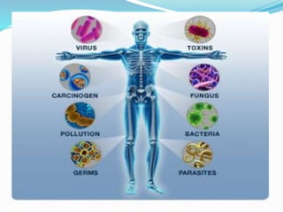
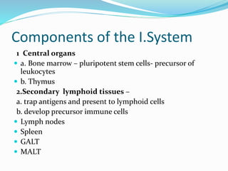
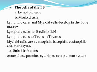
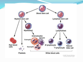
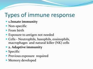
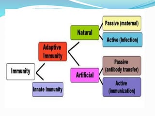
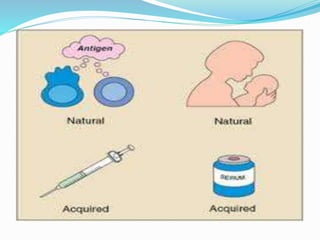
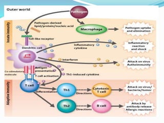
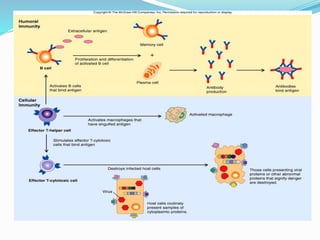
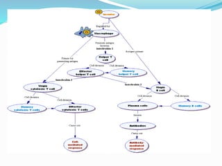
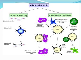
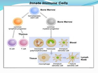
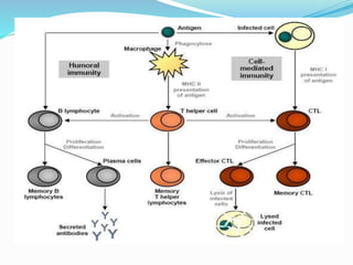
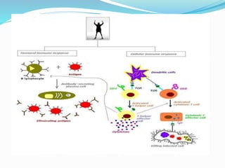
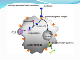
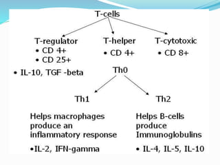
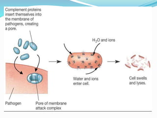
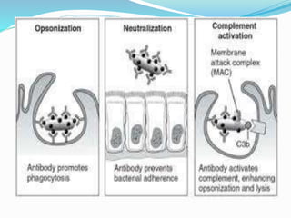
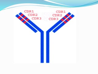
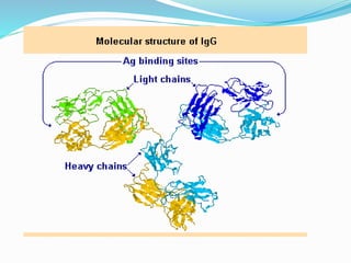
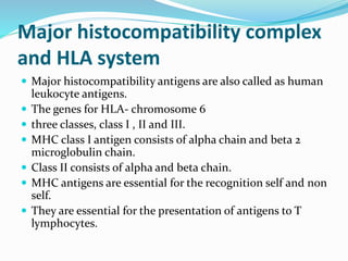
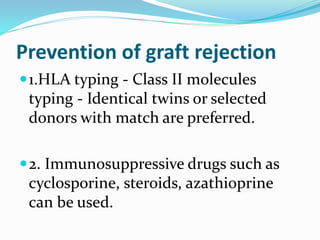
Immune System
 Dr.S.Sethupathy,
 RMMC,
 AU.
Function of immune system
To eliminate non-self
molecules
and cells such as microbes,
cancer cells, transplant tissues
and foreign substances.
Salient features
1. Recognition of self from
non- self or foreign substances.
2.Specificity of the reactions.
3.Memory of the response
Epitope
 Any substance that invokes an immunological
response is an antigen or immunogen.
 The immune response is selective against specific
spatial configurations on the antigen, the antigenic
determinant or epitopes.
 An epitope is the part of an antigen that is recognized
by specifically by antibodies, B cells, or T cells.
Components of the I.System
1 Central organs
 a. Bone marrow – pluripotent stem cells- precursor of
leukocytes
 b. Thymus
2.Secondary lymphoid tissues –
a. trap antigens and present to lymphoid cells
b. develop precursor immune cells
 Lymph nodes
 Spleen
 GALT
 MALT
3. The cells of the I.S
a. Lymphoid cells
b. Myeloid cells
Lymphoid cells and Myeloid cells develop in the Bone
marrow
Lymphoid cells to B cells in B.M
Lymphoid cells to T cells in Thymus
Myeloid cells are neutrophils, basophils, eosinophils
and monocytes.
4. Soluble factors
Acute phase proteins, cytokines, complement system
Types of immune response
 1.Innate immunity
 Non-specific
 From birth
 Exposure to antigen not needed
 Cells - Neutrophils, basophils, eosinophils,
macrophages and natural killer (NK) cells
 2. Adaptive immunity
 Specific
 Previous exposure required
 Memory developed
Components of immunity
 Humoral immunity
 Mediated by immunoglobulins produced by B- cells.
 Ig is involved in protection against microbial
infections ,
 in hyper sensitivity
 in autoimmune reactions.
Components of innate Immunity
 1.Physical barriers - skin, mucous membrane of
respiratory tract, G.I tract, flushing effects of saliva ,
tears, urine.
 They prevent entry of microbes.
 2.Chemical factors - HCl in gastric juice, lysozyme in
tears and saliva.
 They destroy microbes.
 3.Cells
 Natural killer cells (NK) kill malignant or virus
infected cells.
 Neutrophils kill bacteria by phagocytosis.
 Eosinophils are involved in allergic response.
 Basophils are involved in release of inflammatory
mediators and allergic response.
 Macrophages are involved in antigen presentation,
phagocytosis and secretion of cytokines.
NK cells
 Release perforins proteins that form holes in the
plasma membrane of the cell to be killed.
 Sodium diffuses in, then water osmoses inward,
causing the cell to swell and burst (cytolysis).
 In nucleated cells, this influx of ions may trigger
apoptosis
 Can also release granzymes that can enter the cell and
trigger apoptosis.
Dendritic cells
 Dendritic cells (DCs)- antigen-presenting cells, immune system.
 Their main function is to process antigen material and present it
on the cell surface to the T cells of the immune system.
 They act between the innate and the adaptive immune systems.
 Present in the skin ( Langerhans cell), the inner lining of the
nose, lungs, stomach and intestines.
 They are in immature state in the blood.
 Once activated, they migrate to the lymph nodes where they
interact with T cells and B cells to initiate adaptive immune
response.
 At certain development stages -w branched projections
 Dendritic cells are derived from hematopoietic bone marrow
progenitor cells.
Phagocytosis
 Phagocytes engulf the pathogenic organisms.
 Neutrophils, Eosinophils, Monocytes and
macrophages present in tissues.
 Neutrophils are the major phagocytic cells during
acute infection.
4.Soluble factors
 Acute phase proteins play a role in inflammatory
response and defense against microbes.
 Cytokines play a role in inflammatory response and
apoptosis.
 Complement proteins are involved in killing microbes,
allergic response, stimulation of inflammatory
response and attraction of phagocytes.
Immune response
 Active immunity
 It is acquired through direct interaction of immune
system with antigens by infection or vaccination.
 2.Passive immunity
 It is the immunity acquired by the administration of
antibodies or immune cells.
Cytokines
 Cytokines are the molecules involved in immune
response.
 Example : Interferons (IFN),
 Interleukins (IL-1 to 18),
 Transforming growth factor (TGF alpha and TGF
beta),
 Chemokines
 Colony stimulating factor(CSF).
 Interferons alpha, beta, gamma help the cells
overcome viral infection.
 Tumour necrosis factor(TNF) by moncytes and
macrophages.
 TNF alpha induces IL-1 production. It is cytotoxic to
tumor cells. It also induces IFN secretion.
 Transforming growth factor alpha and beta modulate
immune response.
 Colony stimulating factors -Granlocyte –monocyte
CSF (GM-CSF),granulocyte CSF(G-CSF), monocyte
CSF (M-CSF) are involved growth and differentiation
of hemopoietic and immune cells.
Interferons
 Interferons (IFNs) are released by dying, virus-
infected lymphocyte or other cell .
 Interferons "call in" other immune cells (NK cells
and macrophages) that destroy virus-infected cells
and prevent spread of viral infection
 Interferons also "interfere" with viral replication.
 Interferons are paracrine agents, regulating nearby
cells
Chemotaxis
 Chemical attractants are released from damaged tissue
cells and immune system cells to "call over" immune
cells to the site of injury.
 Neutrophils reach the site of infection as a response
to C5a, C3 complements, leukotrienes, and
chemokines.
 Neutrophils - engulf and digest microbes by the
lysozymal enzymes- oxidant independent killing.
 Using superoxide, the killing of microbes is called
oxidant dependent killing.
Chemokines
 Chemokines are the recent cytokines
 Cause chemotaxis.
 They cause chemotaxis of T- cells,
monocytes, NK cells, mast cells and
eosinophils.
 They play an important role in phagocytosis
and inflammatory response.
Less ROS
 Chronic granulomatous disease- defect in NADPH
oxidase in neutrophils – less ROS
 Leukocyte fails to adhere to endothelium due to defect
in adhesion molecule.
 Myeloperoxidase deficiency results in impaired
production of hypo chlorite ion.
 Chediak-Higashi disease is due to defective fusion of
phagosome with lysosome.
Clinical applications
 Interferon gamma is used treating for chronic
granulomatous disease , leukemia ,chronic active
hepatitis C.
 G-CSF is used for neutropenia.
 IL-2 is used in renal carcinoma.
Cell mediated immunity
 It is mediated by T- cells.

 Major defense against cancer cells and cells infected with
microbes.
 Involved in transplant rejection
 Delayed hypersensitivity reactions
 Help in proliferation and differentiation of B- lymphocytes
by presenting antigens to them.
Acute phase proteins
 Plasma proteins whose concentrations increase (positive
acute-phase proteins) or decrease (negative acute-phase
proteins) in response to inflammation.
 The acute-phase reaction or acute-phase response.
 In response to injury, local inflammatory cells (neutrophil
granulocytes and macrophages) secrete a number of
cytokines
 The interleukins IL-1, IL-6 andIL-8, and TNF-α.
 The liver produce a large number of acute-phase reactants.
 Certain proteins are reduced - "negative" acute-phase
reactants.
C-reactive protein(CRP)
 Coating of C- reactive protein to bacteria -
opsonization , complement activation and
phagocytosis.
 Serum level is lesser than 1 mg/dl .
 High sensitive CRP is a risk marker for coronary heart
disease.
 CRP is elevated in inflammatory conditions.
Alpha1 antitrypsin
 It is synthesized in liver.
 It is active against proteases such as trypsin,
chymotrypsin.
 Deficiency - chronic obstructive pulmonary disease.
 Its level is increased in bacterial infections.
 Normal serum level – 0.2-0.4 mg/dl.
Ceruloplasmin
 It is a copper containing alpha2- globulin - by liver.
 It has ferroxidase activity.
 It facilitates iron storage by ferritin and inhibits
microbe iron uptake.
 Normal serum level is 15-60 mg/dl .
 Decreased in Wilson’s disease, malnutrition,
Nephrotic syndrome, liver diseases and Menkey’s
syndrome.
 Increased in cancer, inflammatory conditions.
Haptoglobin binds
hemoglobin and inhibits iron
uptake by microbes.
Ferritin stores iron and inhibits
microbial iron uptake.
Fibrinogen
 It is a clotting factor present in blood .
 Coagulation factors trap invading microbes in blood
clots.
 Some cause chemotaxis.
 Normal plasma level is 200-450 mg/dl.
 Elevated fibrinogen level is a risk factor for CAD.
 Decreased fibrinogen level -in disseminated intra
vascular coagulation (DIC).
Complement proteins
 A group of about 20 different plasma proteins
 C1, C2, C3, and so on
 Activated in a cascade of chemical reactions triggered
by innate or adaptive mechanisms
 Can act directly, as do perforins
 Form MACs (membrane attack complexes) that poke
holes in the outer membrane
 Complement can act indirectly as a chemotactic agent
or regulating immune cells in some other way
Complement proteins
 They are involved in opsonization, lysis and
clumping of target cells and chemotaxis.
 C1 to C9 are involved in immune response.
 It is synthesized in liver and macrophages.
 Antibody opsonization is the process by which a
pathogen is marked for ingestion and destruction by a
phagocyte.
 Opsonization involves the binding of an opsonin, e.g.,
antibody, to an epitope on an antigen.
 After opsonin binds to the membrane, phagocytes are
attracted to the pathogen.
 Fab portion of the antibody binds to the antigen
 Fc portion of the antibody binds to an Fc receptor on
the phagocyte, facilitating phagocytosis.
Classical pathway
 Classical activation pathway starts from
activation of C1 followed by C4 and C2 and
the finally active C3.
 Active C3 activates C5.
 Active C5 causes activation of membrane
attack complex (MAC) composed of
C7,C8.C9.
 This MAC causes cell lysis.
Alternate pathway
In alternate activation pathway, C1,
C4, C2 is bypassed .
Activation of C3 occurs by binding
of C3 to bacterial endotoxin.
Lectin pathway
 Similar in structure to the classical complement
pathway
 After activation, it proceeds through the action of C4
and C2 further down the cascade.
 In contrast to the classical complement pathway, the
lectin pathway does not recognize antibody bound to
its target.
 The lectin pathway starts with mannose-binding
lectin or ficolin binding to certain sugars -bacteria.
Role of complements
Opsonization of microbes
Chemotaxis
Anaphylactic reactions
Cell lysis
Clearance of immune complex and
apoptotic cells
Clinical applications
 Serum complement levels are increased in
inflammatory conditions.
 Serum C3 is decreased in glomerulonephritis
 Its level is used to monitor the disease course.
 Complement deficiency is associated with serious
infections.
 Inhibitors of complement systems such as factor 1,
C1 esterase inhibitor control the level of
complements.
Humoral immunity
 The cells originated from bone marrow and processed
by the Bursa of fabricus in Avians are called B- cells.
 In humans Bursa equivalent organs are Gut associated
Peyer’s patches and lung associated lymphoid
organs.
 Immunoglobulins are secreted by plasma cells
belonging to B- lymphocytes.
 B- cells mediate the humoral immunity.
Plasma cells
 The binding of microbial antigen to appropriate
IgM type of receptor on B- lymphocytes results in
the conversion of them to plasma cells
 They secrete a selective population or clone of
immunoglobulins.
 T4 helper , T cells secrete interleukins which
mediate conversion of B- lymphocytes to plasma
cells.
 Memory B- cells are able to keep in memory of the
previous exposure to antigens.
Plasma cells
Immunoglobulins
 Antibodies
 Two identical heavy and light chains –four
polypeptides
 IgA = α heavy chain
 IgM = µ heavy chain
 IgG = γ heavy chain
 IgD = δ heavy chain
 IgE = ε heavy chain
 IgD, IgE, IgG –
monomers
 IgA – Dimer
 Pentamer- IgM
 Both heavy and light chains have a carboxyl
terminal constant region
 Amino terminal variable region.
 Heavy chains also contain a hinge region.
 Variable regions of heavy and light chains
have variable amino acid sequences
 within them hyper variable regions that act
as antigen binding site.
 It is unique to each immunoglobulin.
Hinge region allows movement of the
two antigen binding sites.
Constant regions are involved in
biological functions
CH2 of IgG and CH4 of IgM bind to
complement C1.
The carboxy terminal of
immunoglobulins (FC region) binds FC
receptors on immune cells.
Role of antibodies
 Antibodies neutralize microbial toxins and inhibit
microbes.
 Opsonization of microbes by antibody promotes
phagocytes through FC receptor of phagocytes.
 Activation of complement promotes lysis of
microbes.
 Antibody presents target cells to cytotoxic cells
through its interaction with Fc receptors on
cytotoxic cells.
IgG
 IgG- 75% of total serum immunoglobulins.
 It activates complement system
 It has antimicrobial activity.
 It can cross the placenta and protects the fetus.
 Normal serum level = 0.8-1.5 gm/dl
 Increased serum IgG - infections, cirrhosis of liver,
auto immune diseases. Monoclonal IgG is increased in
multiple myeloma.
 Decreased serum level in immune deficiency
diseases, Hodgkin’s disease, lymphoid neoplasia.
IgA
 IgA -as a monomer or dimer.
 Secretary IgA - dimer with a joining peptide called J
chain. IgA1 is in serum and IgA2 is in secretions.
 Against viral and bacterial injections by blocking them
across the mucosal cells
 Normal serum level : 0.1 to 0.3 gm/dl
 Increased serum IgA level -in liver diseases,
infections, autoimmune diseases.
 It is decreased in immune deficiency disorders.
IgM
 IgM- pentamer , J chain, the primary immune response.
 It is effective against parasitic and viral infections.
 It is mainly present in the intravascular compartment.
 It does not cross the placenta .
 Antibodies against blood group antigens - IgM.
 Normal serum level is 50 – 200 mg/dl
 Increased serum level - in autoimmune diseases,
infections, Waldenstrom’s macroglobulinemia, liver
diseases, infections, and autoimmune diseases.
 It is decreased in immune deficiency diseases.
IgD
 It is a monomer.
 The function is not known.
 Serum level is 1-4 mg/dl.
 It is increased in infections , cirrhosis of liver,
autoimmune diseases and monoclonal IgD is
increased in multiple myeloma.
IgE
 It is a monomer and involved in allergic reactions.
 It binds to Fc receptors present in mast cells and
basophils.
 It mediates immune response against parasitic
infections.
 Serum level is 0-380 IU/mL.
 Its serum level is increased in allergic conditions and
parasitic infections.
Clinical applications
Hypogammaglobulinemia, Immune
deficiency disorders
 1.Decreased synthesis of Ig
 2. Loss of Ig through urine in Nephrotic
syndrome ,through feces in protein losing
enteropathy, through the skin in burns.
Hypergammaglobulinemia
 It can be polyclonal or monoclonal.
 Polyclonal Increase in Infections, chronic liver
diseases, autoimmune diseases and sarcoidosis.
 Monoclonal increase in multiple myeloma,
Waldenstrom’s macroglobulinemia,
 cryoglobulinemia,
 benign monoclonal gammapathy,
 heavy chain disease.
Multiple myeloma
 It is a plasma cell tumor
 Increase in monoclonal immunoglobulin or Bence
Jones protein.
 In 55% of cases, IgG are overproduced. IgA in 20%
cases, light chain in 20% of cases are overproduced.
Clinical features
 Skeletal pain is seen in the back or thorax.
 Renal failure due to cast formation in renal tubules.
 Anaemia is caused by impaired erythropoiesis
 Bacterial infections are common.
Hyperviscosity syndrome
 Excess plasma globulins increase the viscosity
 fatigue, weakness, headache, mucosal bleeding and cardiac
impairment.
 Lab findings M band in EPP. Urine B- Jones positive
 Bone marrow biopsy shows clusters of plasma cells.
 Osteolytic lesions on X-ray serum calcium level high .
 Hyperuricemia .
 Increased serum beta- 2 microglobulin level - multiple
myeloma, rheumatoid arthritis ,HIV infection and renal
failure. It is small and easily filtered by the glomerulus.
 Waldenstorm’s macroglobulinemia- anemia ,
lymphadenopathy and IgM monoclonal antibodies.
 Heavy chain disease - rare and the heavy chains is
IgA alpha chain and the clinical features are diarrhea
and malabsorption.
 Cryoglobulinemia is characterized by
cryoglobulins in serum which precipitates at
temperature lower than 37oC.
 They may be polyclonal or IgM monoclonal.
 They are also observed in multiple myelin and
lymphoma.
Monoclonal antibodies
 Monoclonal antibodies -Ig produced by a single clone
 Produced by hybridoma technology.
 A specific antibody producing lymphocyte is fused
with immortal myeloma cell with the help of
polyethylene glycol.
 In HAT medium, only fused hybridoma cells will grow
 They are immortal and grown in large number
Uses of monoclonal antibodies
 Diagnostic kits
 Therapeutic drug monitoring(TDM)
 Diagnosis of infections diseases such as HIV
 Diagnosis and monitoring tumor markers.
 Diagnosis of auto immune diseases.
 Mouse monoclonal antibodies to lymphocyte surface
antigens have been used to prevent acute kidney
transplant rejection.
 Humanized monoclonal antibodies raised against
HER-2 antigen is used to treat HER-2 positive breast
cancer
T -Lymphocytes and Cell mediated
immunity
 Helper T cells (CD4+) :They respond to
antigens presented by Major
histocompatibility complex (MHC) class II
molecules containing cells.
 Cytotoxic cells (CD8+) They interact with
cells containing viral antigens or tumor cell
protein and it requires MHC I molecules on
these cells.
Major histocompatibility complex
and HLA system
 Major histocompatibility antigens are also called as human
leukocyte antigens.
 The genes for HLA- chromosome 6
 three classes, class I , II and III.
 MHC class I antigen consists of alpha chain and beta 2
microglobulin chain.
 Class II consists of alpha and beta chain.
 MHC antigens are essential for the recognition self and non
self.
 They are essential for the presentation of antigens to T
lymphocytes.
Clinical applications
 Incorrect HLA matching - rejection of transplantation.
 Some HLA antigens are associated with the disease
 Myasthenia gravis ( HLA DR3)
 Anykylosing spondilytis(HLA B27)
 Multiple sclerosis (HLA 37).
Disorders of T cells
 Deficiency of T cells:
 Primary deficiency - rare, partial or complete.
 Example. Di George syndrome, severe combined
immune deficiency, Aldrich syndrome.
 Secondary deficiency
 Diabetes mellitus, uremia, malnutrition, cancer
immunosuppressive therapy and AIDS.
HIV (AIDS)
 RNA virus. 3 major genes -5'gag-pol-env-3‘.
 Env: envelope protein gp 160. gp160 into gp120 and
gp41.
 gp 120 interacts with CD4 receptor in T cells
 gp41 mediates the fusion of the infected T cells.
 Gag codes for core protein P24, P17, P9 and P7.
 P24 is the major capsid protein,
 P9 is the nucleocapsid proteins
 P17 is a matrix protein.
 pol codes for enzymes reverse transcriptase,
integrase and a protease.
 Helper Tcells - interaction of gp120 protein
with CD4 receptor
 Fusion is mediated by gp41.
 The DNA is integrated into host DNA by
integrase.
 Once the T lymphocytes are activated by
antigens or other viruses enhanced protein
synthesis occurs along with enhanced synthesis
of viral proteins
 Replication of virus and lysis of the cell.
Complications
 Infection such as viral and fungal infections
 cancer such as Kaposi sarcoma, a neoplasm
of endothelial cell in the mucocutaneous
sites,
 Burkitt’s lymphoma,
 B- cell lymphoma etc.
Diagnosis of AIDS
 1.ELISA test for HIV antibodies .It is rapid
but false positive occurs.
 2.Western immunoblot test for HIV
antibodies is the confirmatory test.
 3.Quantitative PCR for detecting Viral RNA
is sensitive ,specific test. It is useful in
detecting the infection at very early stage
and assessing viral load.
 Absolute number of CD4+ and CD8+ T
lymphocytes
 CD4+ cells decrease as the disease progresses.
 So CD4+ count is useful to monitor the disease.
 Treatment
 Drugs used to treat HIV are reverse transcriptase
and protease inhibitors
 Example: Zidovidine is a nucleoside inhibitor and
Indinavir is a protease inhibitor.
Immunologic mechanisms of
tissue injury
1. Hypersensitivity reactions
 Harmful immune response
 four types. Type I – Anaphylactic reactions,
 Type II -antigen antibody cytotoxic
reactions,
 Type III - immune complex reactions
 Type IV- cell mediated delayed immune
reactions.
2. Autoimmunity
 It is the production of immune response to one’s own
tissues resulting in damage to them.
 Mechanism : dysregulation of immune response or
impairment in self tolerance.
 1. Cross reactivity in the microbial antigens.
 2. Polyclonal activation T lymphocytes by tumor cells or
microbes .
3. Genetic predisposition results in lack of self tolerance
4. Association of with HLA antigens.
example Rheumatoid arthritis - HLA B27 mediators of auto
immune diseases
Mediators of autoimmune
diseases
 1.Autoantibodies : Auto antibodies form antigen
antibody complexes and activate complement system
which induces inflammatory response in the affected
tissues. Eg. Systemic lupus erythematosus due to
antinuclear antibody.
 2.Activated T cells : Activated cytotoxic T cells
will target cells. Delayed hypersensitivity helper T cells
(DTH T cells) secrete lymphokines that cause
inflammatory response .
 3.Antibody dependent cytotoxic immune
response can cause cell injury.
 Examples for autoimmune diseases ( They can be
systemic or organ specific)
 Rheumatoid arthritis and systemic lupus
erythematosus are systemic.
 Grave’s disease involving thyroid, diabetes mellitus
involving pancreas, perinicious anemia involving
stomach, myasthenia gravis and Guallian Barrie
syndrome involving nervous system are organ specific
diseases.
Transplant rejection
 Transplantation is replacement of the diseased tissue or
organ by living, healthy tissues or organs from a donor. The
common transplants are kidney, liver, heart, lung and
pancreas.
 Types Auto graft refers to transfer of one’s own tissue
from one site to another site. Eg. Skin graft.
 Isograft refers to graft from one twin to another twin.
 Allograft is transplantation of tissues between the
genetically dissimilar members of the same species
 Xenograft refers to graft between members of different
species.
Transplant rejection
 Rejection of cells or tissues transplanted from are
person to another.
 It may be host versus graft disease or graft versus host
disease.
 In host versus graft disease , antigens on the
transplanted cells are recognized by the host immune
system (MHC molecules).
 Graft versus host disease Ex: Hemopoietic tissues
 Itching, diarrhea,hepatitis are features.
Prevention of graft rejection
1.HLA typing - Class II molecules
typing - Identical twins or selected
donors with match are preferred.
2. Immunosuppressive drugs such as
cyclosporine, steroids, azathioprine
can be used.
Thank
you

Immune system - concise

  • 1.
  • 3.
    Function of immunesystem To eliminate non-self molecules and cells such as microbes, cancer cells, transplant tissues and foreign substances.
  • 4.
    Salient features 1. Recognitionof self from non- self or foreign substances. 2.Specificity of the reactions. 3.Memory of the response
  • 5.
    Epitope  Any substancethat invokes an immunological response is an antigen or immunogen.  The immune response is selective against specific spatial configurations on the antigen, the antigenic determinant or epitopes.  An epitope is the part of an antigen that is recognized by specifically by antibodies, B cells, or T cells.
  • 7.
    Components of theI.System 1 Central organs  a. Bone marrow – pluripotent stem cells- precursor of leukocytes  b. Thymus 2.Secondary lymphoid tissues – a. trap antigens and present to lymphoid cells b. develop precursor immune cells  Lymph nodes  Spleen  GALT  MALT
  • 8.
    3. The cellsof the I.S a. Lymphoid cells b. Myeloid cells Lymphoid cells and Myeloid cells develop in the Bone marrow Lymphoid cells to B cells in B.M Lymphoid cells to T cells in Thymus Myeloid cells are neutrophils, basophils, eosinophils and monocytes. 4. Soluble factors Acute phase proteins, cytokines, complement system
  • 10.
    Types of immuneresponse  1.Innate immunity  Non-specific  From birth  Exposure to antigen not needed  Cells - Neutrophils, basophils, eosinophils, macrophages and natural killer (NK) cells  2. Adaptive immunity  Specific  Previous exposure required  Memory developed
  • 19.
    Components of immunity Humoral immunity  Mediated by immunoglobulins produced by B- cells.  Ig is involved in protection against microbial infections ,  in hyper sensitivity  in autoimmune reactions.
  • 21.
    Components of innateImmunity  1.Physical barriers - skin, mucous membrane of respiratory tract, G.I tract, flushing effects of saliva , tears, urine.  They prevent entry of microbes.  2.Chemical factors - HCl in gastric juice, lysozyme in tears and saliva.  They destroy microbes.
  • 22.
     3.Cells  Naturalkiller cells (NK) kill malignant or virus infected cells.  Neutrophils kill bacteria by phagocytosis.  Eosinophils are involved in allergic response.  Basophils are involved in release of inflammatory mediators and allergic response.  Macrophages are involved in antigen presentation, phagocytosis and secretion of cytokines.
  • 23.
    NK cells  Releaseperforins proteins that form holes in the plasma membrane of the cell to be killed.  Sodium diffuses in, then water osmoses inward, causing the cell to swell and burst (cytolysis).  In nucleated cells, this influx of ions may trigger apoptosis  Can also release granzymes that can enter the cell and trigger apoptosis.
  • 26.
    Dendritic cells  Dendriticcells (DCs)- antigen-presenting cells, immune system.  Their main function is to process antigen material and present it on the cell surface to the T cells of the immune system.  They act between the innate and the adaptive immune systems.  Present in the skin ( Langerhans cell), the inner lining of the nose, lungs, stomach and intestines.  They are in immature state in the blood.  Once activated, they migrate to the lymph nodes where they interact with T cells and B cells to initiate adaptive immune response.  At certain development stages -w branched projections  Dendritic cells are derived from hematopoietic bone marrow progenitor cells.
  • 29.
    Phagocytosis  Phagocytes engulfthe pathogenic organisms.  Neutrophils, Eosinophils, Monocytes and macrophages present in tissues.  Neutrophils are the major phagocytic cells during acute infection.
  • 30.
    4.Soluble factors  Acutephase proteins play a role in inflammatory response and defense against microbes.  Cytokines play a role in inflammatory response and apoptosis.  Complement proteins are involved in killing microbes, allergic response, stimulation of inflammatory response and attraction of phagocytes.
  • 31.
    Immune response  Activeimmunity  It is acquired through direct interaction of immune system with antigens by infection or vaccination.  2.Passive immunity  It is the immunity acquired by the administration of antibodies or immune cells.
  • 32.
    Cytokines  Cytokines arethe molecules involved in immune response.  Example : Interferons (IFN),  Interleukins (IL-1 to 18),  Transforming growth factor (TGF alpha and TGF beta),  Chemokines  Colony stimulating factor(CSF).
  • 33.
     Interferons alpha,beta, gamma help the cells overcome viral infection.  Tumour necrosis factor(TNF) by moncytes and macrophages.  TNF alpha induces IL-1 production. It is cytotoxic to tumor cells. It also induces IFN secretion.  Transforming growth factor alpha and beta modulate immune response.  Colony stimulating factors -Granlocyte –monocyte CSF (GM-CSF),granulocyte CSF(G-CSF), monocyte CSF (M-CSF) are involved growth and differentiation of hemopoietic and immune cells.
  • 34.
    Interferons  Interferons (IFNs)are released by dying, virus- infected lymphocyte or other cell .  Interferons "call in" other immune cells (NK cells and macrophages) that destroy virus-infected cells and prevent spread of viral infection  Interferons also "interfere" with viral replication.  Interferons are paracrine agents, regulating nearby cells
  • 35.
    Chemotaxis  Chemical attractantsare released from damaged tissue cells and immune system cells to "call over" immune cells to the site of injury.  Neutrophils reach the site of infection as a response to C5a, C3 complements, leukotrienes, and chemokines.  Neutrophils - engulf and digest microbes by the lysozymal enzymes- oxidant independent killing.  Using superoxide, the killing of microbes is called oxidant dependent killing.
  • 36.
    Chemokines  Chemokines arethe recent cytokines  Cause chemotaxis.  They cause chemotaxis of T- cells, monocytes, NK cells, mast cells and eosinophils.  They play an important role in phagocytosis and inflammatory response.
  • 37.
    Less ROS  Chronicgranulomatous disease- defect in NADPH oxidase in neutrophils – less ROS  Leukocyte fails to adhere to endothelium due to defect in adhesion molecule.  Myeloperoxidase deficiency results in impaired production of hypo chlorite ion.  Chediak-Higashi disease is due to defective fusion of phagosome with lysosome.
  • 38.
    Clinical applications  Interferongamma is used treating for chronic granulomatous disease , leukemia ,chronic active hepatitis C.  G-CSF is used for neutropenia.  IL-2 is used in renal carcinoma.
  • 39.
    Cell mediated immunity It is mediated by T- cells.   Major defense against cancer cells and cells infected with microbes.  Involved in transplant rejection  Delayed hypersensitivity reactions  Help in proliferation and differentiation of B- lymphocytes by presenting antigens to them.
  • 42.
    Acute phase proteins Plasma proteins whose concentrations increase (positive acute-phase proteins) or decrease (negative acute-phase proteins) in response to inflammation.  The acute-phase reaction or acute-phase response.  In response to injury, local inflammatory cells (neutrophil granulocytes and macrophages) secrete a number of cytokines  The interleukins IL-1, IL-6 andIL-8, and TNF-α.  The liver produce a large number of acute-phase reactants.  Certain proteins are reduced - "negative" acute-phase reactants.
  • 43.
    C-reactive protein(CRP)  Coatingof C- reactive protein to bacteria - opsonization , complement activation and phagocytosis.  Serum level is lesser than 1 mg/dl .  High sensitive CRP is a risk marker for coronary heart disease.  CRP is elevated in inflammatory conditions.
  • 44.
    Alpha1 antitrypsin  Itis synthesized in liver.  It is active against proteases such as trypsin, chymotrypsin.  Deficiency - chronic obstructive pulmonary disease.  Its level is increased in bacterial infections.  Normal serum level – 0.2-0.4 mg/dl.
  • 45.
    Ceruloplasmin  It isa copper containing alpha2- globulin - by liver.  It has ferroxidase activity.  It facilitates iron storage by ferritin and inhibits microbe iron uptake.  Normal serum level is 15-60 mg/dl .  Decreased in Wilson’s disease, malnutrition, Nephrotic syndrome, liver diseases and Menkey’s syndrome.  Increased in cancer, inflammatory conditions.
  • 46.
    Haptoglobin binds hemoglobin andinhibits iron uptake by microbes. Ferritin stores iron and inhibits microbial iron uptake.
  • 47.
    Fibrinogen  It isa clotting factor present in blood .  Coagulation factors trap invading microbes in blood clots.  Some cause chemotaxis.  Normal plasma level is 200-450 mg/dl.  Elevated fibrinogen level is a risk factor for CAD.  Decreased fibrinogen level -in disseminated intra vascular coagulation (DIC).
  • 48.
    Complement proteins  Agroup of about 20 different plasma proteins  C1, C2, C3, and so on  Activated in a cascade of chemical reactions triggered by innate or adaptive mechanisms  Can act directly, as do perforins  Form MACs (membrane attack complexes) that poke holes in the outer membrane  Complement can act indirectly as a chemotactic agent or regulating immune cells in some other way
  • 50.
    Complement proteins  Theyare involved in opsonization, lysis and clumping of target cells and chemotaxis.  C1 to C9 are involved in immune response.  It is synthesized in liver and macrophages.
  • 51.
     Antibody opsonizationis the process by which a pathogen is marked for ingestion and destruction by a phagocyte.  Opsonization involves the binding of an opsonin, e.g., antibody, to an epitope on an antigen.  After opsonin binds to the membrane, phagocytes are attracted to the pathogen.  Fab portion of the antibody binds to the antigen  Fc portion of the antibody binds to an Fc receptor on the phagocyte, facilitating phagocytosis.
  • 52.
    Classical pathway  Classicalactivation pathway starts from activation of C1 followed by C4 and C2 and the finally active C3.  Active C3 activates C5.  Active C5 causes activation of membrane attack complex (MAC) composed of C7,C8.C9.  This MAC causes cell lysis.
  • 55.
    Alternate pathway In alternateactivation pathway, C1, C4, C2 is bypassed . Activation of C3 occurs by binding of C3 to bacterial endotoxin.
  • 56.
    Lectin pathway  Similarin structure to the classical complement pathway  After activation, it proceeds through the action of C4 and C2 further down the cascade.  In contrast to the classical complement pathway, the lectin pathway does not recognize antibody bound to its target.  The lectin pathway starts with mannose-binding lectin or ficolin binding to certain sugars -bacteria.
  • 58.
    Role of complements Opsonizationof microbes Chemotaxis Anaphylactic reactions Cell lysis Clearance of immune complex and apoptotic cells
  • 59.
    Clinical applications  Serumcomplement levels are increased in inflammatory conditions.  Serum C3 is decreased in glomerulonephritis  Its level is used to monitor the disease course.  Complement deficiency is associated with serious infections.  Inhibitors of complement systems such as factor 1, C1 esterase inhibitor control the level of complements.
  • 61.
    Humoral immunity  Thecells originated from bone marrow and processed by the Bursa of fabricus in Avians are called B- cells.  In humans Bursa equivalent organs are Gut associated Peyer’s patches and lung associated lymphoid organs.  Immunoglobulins are secreted by plasma cells belonging to B- lymphocytes.  B- cells mediate the humoral immunity.
  • 62.
    Plasma cells  Thebinding of microbial antigen to appropriate IgM type of receptor on B- lymphocytes results in the conversion of them to plasma cells  They secrete a selective population or clone of immunoglobulins.  T4 helper , T cells secrete interleukins which mediate conversion of B- lymphocytes to plasma cells.  Memory B- cells are able to keep in memory of the previous exposure to antigens.
  • 63.
  • 64.
    Immunoglobulins  Antibodies  Twoidentical heavy and light chains –four polypeptides  IgA = α heavy chain  IgM = µ heavy chain  IgG = γ heavy chain  IgD = δ heavy chain  IgE = ε heavy chain
  • 70.
     IgD, IgE,IgG – monomers  IgA – Dimer  Pentamer- IgM
  • 71.
     Both heavyand light chains have a carboxyl terminal constant region  Amino terminal variable region.  Heavy chains also contain a hinge region.  Variable regions of heavy and light chains have variable amino acid sequences  within them hyper variable regions that act as antigen binding site.  It is unique to each immunoglobulin.
  • 72.
    Hinge region allowsmovement of the two antigen binding sites. Constant regions are involved in biological functions CH2 of IgG and CH4 of IgM bind to complement C1. The carboxy terminal of immunoglobulins (FC region) binds FC receptors on immune cells.
  • 73.
    Role of antibodies Antibodies neutralize microbial toxins and inhibit microbes.  Opsonization of microbes by antibody promotes phagocytes through FC receptor of phagocytes.  Activation of complement promotes lysis of microbes.  Antibody presents target cells to cytotoxic cells through its interaction with Fc receptors on cytotoxic cells.
  • 74.
    IgG  IgG- 75%of total serum immunoglobulins.  It activates complement system  It has antimicrobial activity.  It can cross the placenta and protects the fetus.  Normal serum level = 0.8-1.5 gm/dl  Increased serum IgG - infections, cirrhosis of liver, auto immune diseases. Monoclonal IgG is increased in multiple myeloma.  Decreased serum level in immune deficiency diseases, Hodgkin’s disease, lymphoid neoplasia.
  • 75.
    IgA  IgA -asa monomer or dimer.  Secretary IgA - dimer with a joining peptide called J chain. IgA1 is in serum and IgA2 is in secretions.  Against viral and bacterial injections by blocking them across the mucosal cells  Normal serum level : 0.1 to 0.3 gm/dl  Increased serum IgA level -in liver diseases, infections, autoimmune diseases.  It is decreased in immune deficiency disorders.
  • 76.
    IgM  IgM- pentamer, J chain, the primary immune response.  It is effective against parasitic and viral infections.  It is mainly present in the intravascular compartment.  It does not cross the placenta .  Antibodies against blood group antigens - IgM.  Normal serum level is 50 – 200 mg/dl  Increased serum level - in autoimmune diseases, infections, Waldenstrom’s macroglobulinemia, liver diseases, infections, and autoimmune diseases.  It is decreased in immune deficiency diseases.
  • 77.
    IgD  It isa monomer.  The function is not known.  Serum level is 1-4 mg/dl.  It is increased in infections , cirrhosis of liver, autoimmune diseases and monoclonal IgD is increased in multiple myeloma.
  • 78.
    IgE  It isa monomer and involved in allergic reactions.  It binds to Fc receptors present in mast cells and basophils.  It mediates immune response against parasitic infections.  Serum level is 0-380 IU/mL.  Its serum level is increased in allergic conditions and parasitic infections.
  • 79.
    Clinical applications Hypogammaglobulinemia, Immune deficiencydisorders  1.Decreased synthesis of Ig  2. Loss of Ig through urine in Nephrotic syndrome ,through feces in protein losing enteropathy, through the skin in burns.
  • 80.
    Hypergammaglobulinemia  It canbe polyclonal or monoclonal.  Polyclonal Increase in Infections, chronic liver diseases, autoimmune diseases and sarcoidosis.  Monoclonal increase in multiple myeloma, Waldenstrom’s macroglobulinemia,  cryoglobulinemia,  benign monoclonal gammapathy,  heavy chain disease.
  • 81.
    Multiple myeloma  Itis a plasma cell tumor  Increase in monoclonal immunoglobulin or Bence Jones protein.  In 55% of cases, IgG are overproduced. IgA in 20% cases, light chain in 20% of cases are overproduced. Clinical features  Skeletal pain is seen in the back or thorax.  Renal failure due to cast formation in renal tubules.  Anaemia is caused by impaired erythropoiesis  Bacterial infections are common.
  • 82.
    Hyperviscosity syndrome  Excessplasma globulins increase the viscosity  fatigue, weakness, headache, mucosal bleeding and cardiac impairment.  Lab findings M band in EPP. Urine B- Jones positive  Bone marrow biopsy shows clusters of plasma cells.  Osteolytic lesions on X-ray serum calcium level high .  Hyperuricemia .  Increased serum beta- 2 microglobulin level - multiple myeloma, rheumatoid arthritis ,HIV infection and renal failure. It is small and easily filtered by the glomerulus.
  • 83.
     Waldenstorm’s macroglobulinemia-anemia , lymphadenopathy and IgM monoclonal antibodies.  Heavy chain disease - rare and the heavy chains is IgA alpha chain and the clinical features are diarrhea and malabsorption.  Cryoglobulinemia is characterized by cryoglobulins in serum which precipitates at temperature lower than 37oC.  They may be polyclonal or IgM monoclonal.  They are also observed in multiple myelin and lymphoma.
  • 84.
    Monoclonal antibodies  Monoclonalantibodies -Ig produced by a single clone  Produced by hybridoma technology.  A specific antibody producing lymphocyte is fused with immortal myeloma cell with the help of polyethylene glycol.  In HAT medium, only fused hybridoma cells will grow  They are immortal and grown in large number
  • 85.
    Uses of monoclonalantibodies  Diagnostic kits  Therapeutic drug monitoring(TDM)  Diagnosis of infections diseases such as HIV  Diagnosis and monitoring tumor markers.  Diagnosis of auto immune diseases.  Mouse monoclonal antibodies to lymphocyte surface antigens have been used to prevent acute kidney transplant rejection.  Humanized monoclonal antibodies raised against HER-2 antigen is used to treat HER-2 positive breast cancer
  • 86.
    T -Lymphocytes andCell mediated immunity  Helper T cells (CD4+) :They respond to antigens presented by Major histocompatibility complex (MHC) class II molecules containing cells.  Cytotoxic cells (CD8+) They interact with cells containing viral antigens or tumor cell protein and it requires MHC I molecules on these cells.
  • 87.
    Major histocompatibility complex andHLA system  Major histocompatibility antigens are also called as human leukocyte antigens.  The genes for HLA- chromosome 6  three classes, class I , II and III.  MHC class I antigen consists of alpha chain and beta 2 microglobulin chain.  Class II consists of alpha and beta chain.  MHC antigens are essential for the recognition self and non self.  They are essential for the presentation of antigens to T lymphocytes.
  • 88.
    Clinical applications  IncorrectHLA matching - rejection of transplantation.  Some HLA antigens are associated with the disease  Myasthenia gravis ( HLA DR3)  Anykylosing spondilytis(HLA B27)  Multiple sclerosis (HLA 37).
  • 89.
    Disorders of Tcells  Deficiency of T cells:  Primary deficiency - rare, partial or complete.  Example. Di George syndrome, severe combined immune deficiency, Aldrich syndrome.  Secondary deficiency  Diabetes mellitus, uremia, malnutrition, cancer immunosuppressive therapy and AIDS.
  • 90.
    HIV (AIDS)  RNAvirus. 3 major genes -5'gag-pol-env-3‘.  Env: envelope protein gp 160. gp160 into gp120 and gp41.  gp 120 interacts with CD4 receptor in T cells  gp41 mediates the fusion of the infected T cells.  Gag codes for core protein P24, P17, P9 and P7.  P24 is the major capsid protein,  P9 is the nucleocapsid proteins  P17 is a matrix protein.
  • 95.
     pol codesfor enzymes reverse transcriptase, integrase and a protease.  Helper Tcells - interaction of gp120 protein with CD4 receptor  Fusion is mediated by gp41.  The DNA is integrated into host DNA by integrase.  Once the T lymphocytes are activated by antigens or other viruses enhanced protein synthesis occurs along with enhanced synthesis of viral proteins  Replication of virus and lysis of the cell.
  • 96.
    Complications  Infection suchas viral and fungal infections  cancer such as Kaposi sarcoma, a neoplasm of endothelial cell in the mucocutaneous sites,  Burkitt’s lymphoma,  B- cell lymphoma etc.
  • 97.
    Diagnosis of AIDS 1.ELISA test for HIV antibodies .It is rapid but false positive occurs.  2.Western immunoblot test for HIV antibodies is the confirmatory test.  3.Quantitative PCR for detecting Viral RNA is sensitive ,specific test. It is useful in detecting the infection at very early stage and assessing viral load.
  • 98.
     Absolute numberof CD4+ and CD8+ T lymphocytes  CD4+ cells decrease as the disease progresses.  So CD4+ count is useful to monitor the disease.  Treatment  Drugs used to treat HIV are reverse transcriptase and protease inhibitors  Example: Zidovidine is a nucleoside inhibitor and Indinavir is a protease inhibitor.
  • 99.
    Immunologic mechanisms of tissueinjury 1. Hypersensitivity reactions  Harmful immune response  four types. Type I – Anaphylactic reactions,  Type II -antigen antibody cytotoxic reactions,  Type III - immune complex reactions  Type IV- cell mediated delayed immune reactions.
  • 100.
    2. Autoimmunity  Itis the production of immune response to one’s own tissues resulting in damage to them.  Mechanism : dysregulation of immune response or impairment in self tolerance.  1. Cross reactivity in the microbial antigens.  2. Polyclonal activation T lymphocytes by tumor cells or microbes . 3. Genetic predisposition results in lack of self tolerance 4. Association of with HLA antigens. example Rheumatoid arthritis - HLA B27 mediators of auto immune diseases
  • 102.
    Mediators of autoimmune diseases 1.Autoantibodies : Auto antibodies form antigen antibody complexes and activate complement system which induces inflammatory response in the affected tissues. Eg. Systemic lupus erythematosus due to antinuclear antibody.  2.Activated T cells : Activated cytotoxic T cells will target cells. Delayed hypersensitivity helper T cells (DTH T cells) secrete lymphokines that cause inflammatory response .
  • 103.
     3.Antibody dependentcytotoxic immune response can cause cell injury.  Examples for autoimmune diseases ( They can be systemic or organ specific)  Rheumatoid arthritis and systemic lupus erythematosus are systemic.  Grave’s disease involving thyroid, diabetes mellitus involving pancreas, perinicious anemia involving stomach, myasthenia gravis and Guallian Barrie syndrome involving nervous system are organ specific diseases.
  • 104.
    Transplant rejection  Transplantationis replacement of the diseased tissue or organ by living, healthy tissues or organs from a donor. The common transplants are kidney, liver, heart, lung and pancreas.  Types Auto graft refers to transfer of one’s own tissue from one site to another site. Eg. Skin graft.  Isograft refers to graft from one twin to another twin.  Allograft is transplantation of tissues between the genetically dissimilar members of the same species  Xenograft refers to graft between members of different species.
  • 106.
    Transplant rejection  Rejectionof cells or tissues transplanted from are person to another.  It may be host versus graft disease or graft versus host disease.  In host versus graft disease , antigens on the transplanted cells are recognized by the host immune system (MHC molecules).  Graft versus host disease Ex: Hemopoietic tissues  Itching, diarrhea,hepatitis are features.
  • 107.
    Prevention of graftrejection 1.HLA typing - Class II molecules typing - Identical twins or selected donors with match are preferred. 2. Immunosuppressive drugs such as cyclosporine, steroids, azathioprine can be used.
  • 108.