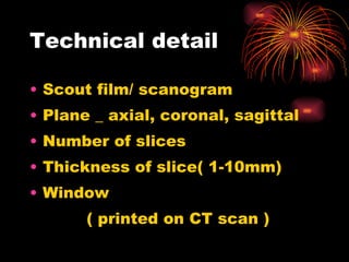
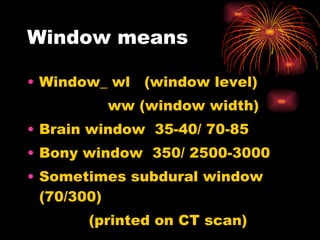
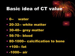
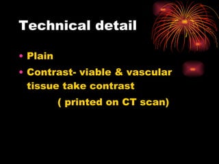
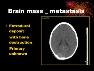
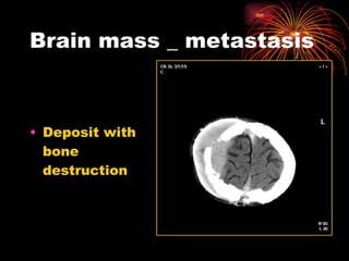
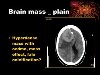
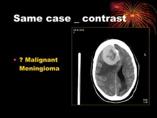
Basics for medical students By_ Dr. Rekha Khare M.D. radiology
Intracranial Lesion How to image?
Imaging modality Simple radiography -cheap preliminary screening  procedure  -accuracy & specificity is low
contd. Computerized tomography- basic choice  MRI – tissue specification is better Ultrasound scan- choice in neonate & infant
contd. Angiography- mainly for interventional radiology
contd. Radionuclide scan- - basic technique is superseded by CT & MRI -Technique for mapping regional cerebral blood flow has important clinical application as  in CVA, Dementia, Alzhiemers etc
contd. Positron emission tomography (PET) expensive & available in very advance centre ( cyclotron) PET tracer like carbon, nitrogen, oxygen - specific & showing difference in focal brain metabolism  - Major use in oncology
contd. SPECT  -  Imaging of dopamine transport system in movement disorder (like Parkinsonism) by injecting specific tracer agent working at neuromuscular junction
When to ask brain CT scan Head injury Cerebro-vascular accident Unconscious Epilepsy Pyrexia of unknown origin Suspecting intracranial mass lesion
How to read CT scan brain Identification Technical details Examine the image Localize the lesion Analyse the lesion Possible diagnosis D/D if needed
Identification Name, age, sex, of patient Date of exam. (printed on scan)
Technical detail Scout film/ scanogram Plane _ axial, coronal, sagittal Number of slices Thickness of slice( 1-10mm) Window  ( printed on CT scan )
Window means Window_ wl  (window level) ww (window width) Brain window  35-40/ 70-85 Bony window  350/ 2500-3000 Sometimes subdural window  (70/300) (printed on CT scan)
Basic idea of CT value 0--  water 20-32-- white matter 30-40-- grey matter 56-70-- blood 80-1000-- calcification to bone +100-- fat -1000-- air
Technical detail Plain Contrast- viable & vascular tissue take contrast ( printed on CT scan)
Examine the image_ a systematic approach  Midline_ shift Ventricle, cistern, sulci_ enlarged, compressed, displaced  Brain tissue_ iso/hypo/hyper/hetero  _ mass effect _ gyri enhanced _ white matter
Contd. Bones/ vault( # /mass/erosion) Scalp/ soft tissue
Localize the lesion Commonly look for_  -air sinus_ frontal/ethmoid/sphenoid -ventricle_ LV ant./ temporal/ occipital horn _ 3 rd  and aqueduct _4 th   and cisterna magna
Contd. cistern_ cerebellopontine _ suprasellar _ quadrigeminal _ interpeduncular sulci and gyri sylvian fissure
Contd. Cerebral lobes_ frontal/temporal/occipital/ parietal Mid brain Brain stem cerebellum
Contd. Basal ganglion -caudate nucleus (head indenting ant. horn) -lentiform nucleus -external capsule (in between  insula and lentiform nucleus) -internal capsule ( in between  caudate and lentiform nucleus)
Contd. thalamus_ on either side of 3 rd  ventricle pineal body choroid plexus hypothalmus basilar artery great vein of galen ( post.  to pineal)
Contd. corpus callosum -genu ( ant. to ant. horn of LV) -splenium (post. to occ. horn of LV)  septum pellucidum tentorium( dura seperating cerebellum from cerebrum)
Contd. venous sinus -sigmoid ( along petrous/ temporal bone) -transverse( between sigmoid and sup. sagittal sinus) -superior sagittal sinus( midline, falx meets inner skull table)
Contd. white matter/ corona radiata foramen magnum  -spinal cord -vertebral artery
Analyse the lesion Intra/ extra axial Supra/ infra tentorial Single/ multiple/diffuse White/ grey matter/ both Enhancement  -non enhanced_ avascular -hyper enhanced_ vascular -heterogenous enhanced_ mass
Possible pathology Mass Infarction Haemorrhage Hypoxic demyelinating/ degenerative
Possible etiology congenital/ developmental traumatic infection/inflammatory mass/ neoplasm parasitic vascular degenerative metabolic/ toxic iatrogenic/ post procedural
Onset of lesion acute sub acute chronic recurrent residual old healed
Possible diagnosis   In correlation with_ age, sex clinical signs and symptoms other imaging results any lab tests
Differential diagnosis_  common Intra axial_  m_ mass/ metastasis a_ abscess/ angioma g_ glioma/ gliosis i_ infarction/ infection/ inflamation c_ contusion/ congenital dr_ demyelination/degenerative
Contd. Extra axial_ meningioma pit. Adenoma craniopharyngioma chordoma schwanoma lipoma dermoid/ epidermoid
Sample report This is CT scan of head done on ----- for ----- age --- Axial cuts in----mm slices from foramen magnum to vertex Plain and enhanced views with brain and bone window are included Radiological findings are----- In clinical context of ----- duration these findings suggest -------- Other D/D( if needed) could be----
Few pictures Material collected while working as consultant radiologist in university teaching hospital Lusaka Zambia
Brain trauma depressed # with brain haematoma causing mass effect
Brain trauma Subdural haematoma
Mass at top of head Predominantly extracranial mass destruction of bones
Intracranial  mass-plain Well outlined hyperdense mass in frontal lobe ?Meningioma
Same case- contrast Huge hyperdense enhanced mass from falx with oedma  in surrounding brain tissue ? meningioma
Brain mass  Heterogenous enhanced cystic mass compressing 4 th  ventricle causing hydrocephalus
Brain mass  Heterogenous enhanced solid mass , compressing 4 th  ventricle causing hydrocephalus
Brain mass Strongly enhanced mid line lesion within 4 th  ventricle causing obstructive hydrocephalus  ? ependymoma
Brain mass Hypodense  nonenhanced mass in cerebellar region displacing 4 th  ventricle ? Glioma
Brain mass  Cystic mass with calcification compressing/ displacing 4 th  ventricle with hydrocephalus ? ependymoma
Brain mass Heterogenous cystic mass with mass effect
Brain mass Cystic mass lesion causing dilated LV ? colloidal cyst
Brain mass Calcified mass at level of 3 rd  ventricle
Brain mass Strongly enhanced mass lesion at pitutary level ? Adenoma
Brain mass _ metastasis Extradural deposit  with bone destruction_  Primary unknown
Brain mass _ metastasis Deposit with bone destruction
Brain mass _ plain Hyperdense mass with oedma, mass effect, falx calcification?
Same case _ contrast ? Malignant Meningioma
CVA_ acute hemiplegia  Intracerebral haematoma basal nuclei region with mass effect
CVA _ acute hemiplegia Acute infarct
Old case hemiplegia Hypodense lesion with no mass effect Overfilled lateral ventricle ? Few days old infarction
CVA_ Acute hemiplegia Serpegenious strongly enhanced lesion with mass effect ?AVM  Angiography is next modality to confirm
Unconscious  Brain oedma_ (White cerebellar sign)
Dementia Multiple hypodense lesion mainly peripheral ? Infarcts in old age
Neonatal brain  Dilated ventricle compressing  brain tissue, few calcification  HIV+ve mother ? CMV
Neonatal brain Dying/ hypoxic brain
Brain infection _ plain Acute  hydrocephalus
Same case _ contrast Acute hydrocephalus with meningeal enhancement
Brain infection _ plain Chronic SDE with mass/ pressure effect
Same case _ contrast Enhanced membrane _ subdural empyema
Infection_ plain Vague hypodensity with mass effect
Same case _ contrast Multiple ring enhanced lesion ? abscess
Fever_ plain Thin ring lesion with massive oedma & mass effect
Same case_ contrast Multiple ring enhanced lesion with midline shift & mass effect- ? abscesses
HIV+ve case_ plain Multifocal hypodense lesion with no mass effect
Same case- contrast Enhanced nodular lesion? opportunistic infection
HIV+ve case Multinodular enhanced lesion with massive oedma
HIV+ve case Multinodular hyperdense lesion with massive oedma
HIV+ve case Basal ganlion calcification
Epilepsy/ fits calcifiation in periventricular region ? Tuberous sclerosis
Epilepsy/fits Cysticercosis
Acute Headache Dilated ventricles with meningeal enhancement ? meningitis
Post procedural Shunt seen Bilateral subdural effusion Common complication Rabbit ear appearance
Unsteady gait Cerebellar atrophy

Intracranial radiology general presentation

  • 1.
    Basics for medicalstudents By_ Dr. Rekha Khare M.D. radiology
  • 2.
  • 3.
    Imaging modality Simpleradiography -cheap preliminary screening procedure -accuracy & specificity is low
  • 4.
    contd. Computerized tomography-basic choice MRI – tissue specification is better Ultrasound scan- choice in neonate & infant
  • 5.
    contd. Angiography- mainlyfor interventional radiology
  • 6.
    contd. Radionuclide scan-- basic technique is superseded by CT & MRI -Technique for mapping regional cerebral blood flow has important clinical application as in CVA, Dementia, Alzhiemers etc
  • 7.
    contd. Positron emissiontomography (PET) expensive & available in very advance centre ( cyclotron) PET tracer like carbon, nitrogen, oxygen - specific & showing difference in focal brain metabolism - Major use in oncology
  • 8.
    contd. SPECT - Imaging of dopamine transport system in movement disorder (like Parkinsonism) by injecting specific tracer agent working at neuromuscular junction
  • 9.
    When to askbrain CT scan Head injury Cerebro-vascular accident Unconscious Epilepsy Pyrexia of unknown origin Suspecting intracranial mass lesion
  • 10.
    How to readCT scan brain Identification Technical details Examine the image Localize the lesion Analyse the lesion Possible diagnosis D/D if needed
  • 11.
    Identification Name, age,sex, of patient Date of exam. (printed on scan)
  • 12.
    Technical detail Scoutfilm/ scanogram Plane _ axial, coronal, sagittal Number of slices Thickness of slice( 1-10mm) Window ( printed on CT scan )
  • 13.
    Window means Window_wl (window level) ww (window width) Brain window 35-40/ 70-85 Bony window 350/ 2500-3000 Sometimes subdural window (70/300) (printed on CT scan)
  • 14.
    Basic idea ofCT value 0-- water 20-32-- white matter 30-40-- grey matter 56-70-- blood 80-1000-- calcification to bone +100-- fat -1000-- air
  • 15.
    Technical detail PlainContrast- viable & vascular tissue take contrast ( printed on CT scan)
  • 16.
    Examine the image_a systematic approach Midline_ shift Ventricle, cistern, sulci_ enlarged, compressed, displaced Brain tissue_ iso/hypo/hyper/hetero _ mass effect _ gyri enhanced _ white matter
  • 17.
    Contd. Bones/ vault(# /mass/erosion) Scalp/ soft tissue
  • 18.
    Localize the lesionCommonly look for_ -air sinus_ frontal/ethmoid/sphenoid -ventricle_ LV ant./ temporal/ occipital horn _ 3 rd and aqueduct _4 th and cisterna magna
  • 19.
    Contd. cistern_ cerebellopontine_ suprasellar _ quadrigeminal _ interpeduncular sulci and gyri sylvian fissure
  • 20.
    Contd. Cerebral lobes_frontal/temporal/occipital/ parietal Mid brain Brain stem cerebellum
  • 21.
    Contd. Basal ganglion-caudate nucleus (head indenting ant. horn) -lentiform nucleus -external capsule (in between insula and lentiform nucleus) -internal capsule ( in between caudate and lentiform nucleus)
  • 22.
    Contd. thalamus_ oneither side of 3 rd ventricle pineal body choroid plexus hypothalmus basilar artery great vein of galen ( post. to pineal)
  • 23.
    Contd. corpus callosum-genu ( ant. to ant. horn of LV) -splenium (post. to occ. horn of LV) septum pellucidum tentorium( dura seperating cerebellum from cerebrum)
  • 24.
    Contd. venous sinus-sigmoid ( along petrous/ temporal bone) -transverse( between sigmoid and sup. sagittal sinus) -superior sagittal sinus( midline, falx meets inner skull table)
  • 25.
    Contd. white matter/corona radiata foramen magnum -spinal cord -vertebral artery
  • 26.
    Analyse the lesionIntra/ extra axial Supra/ infra tentorial Single/ multiple/diffuse White/ grey matter/ both Enhancement -non enhanced_ avascular -hyper enhanced_ vascular -heterogenous enhanced_ mass
  • 27.
    Possible pathology MassInfarction Haemorrhage Hypoxic demyelinating/ degenerative
  • 28.
    Possible etiology congenital/developmental traumatic infection/inflammatory mass/ neoplasm parasitic vascular degenerative metabolic/ toxic iatrogenic/ post procedural
  • 29.
    Onset of lesionacute sub acute chronic recurrent residual old healed
  • 30.
    Possible diagnosis In correlation with_ age, sex clinical signs and symptoms other imaging results any lab tests
  • 31.
    Differential diagnosis_ common Intra axial_ m_ mass/ metastasis a_ abscess/ angioma g_ glioma/ gliosis i_ infarction/ infection/ inflamation c_ contusion/ congenital dr_ demyelination/degenerative
  • 32.
    Contd. Extra axial_meningioma pit. Adenoma craniopharyngioma chordoma schwanoma lipoma dermoid/ epidermoid
  • 33.
    Sample report Thisis CT scan of head done on ----- for ----- age --- Axial cuts in----mm slices from foramen magnum to vertex Plain and enhanced views with brain and bone window are included Radiological findings are----- In clinical context of ----- duration these findings suggest -------- Other D/D( if needed) could be----
  • 34.
    Few pictures Materialcollected while working as consultant radiologist in university teaching hospital Lusaka Zambia
  • 35.
    Brain trauma depressed# with brain haematoma causing mass effect
  • 36.
  • 37.
    Mass at topof head Predominantly extracranial mass destruction of bones
  • 38.
    Intracranial mass-plainWell outlined hyperdense mass in frontal lobe ?Meningioma
  • 39.
    Same case- contrastHuge hyperdense enhanced mass from falx with oedma in surrounding brain tissue ? meningioma
  • 40.
    Brain mass Heterogenous enhanced cystic mass compressing 4 th ventricle causing hydrocephalus
  • 41.
    Brain mass Heterogenous enhanced solid mass , compressing 4 th ventricle causing hydrocephalus
  • 42.
    Brain mass Stronglyenhanced mid line lesion within 4 th ventricle causing obstructive hydrocephalus ? ependymoma
  • 43.
    Brain mass Hypodense nonenhanced mass in cerebellar region displacing 4 th ventricle ? Glioma
  • 44.
    Brain mass Cystic mass with calcification compressing/ displacing 4 th ventricle with hydrocephalus ? ependymoma
  • 45.
    Brain mass Heterogenouscystic mass with mass effect
  • 46.
    Brain mass Cysticmass lesion causing dilated LV ? colloidal cyst
  • 47.
    Brain mass Calcifiedmass at level of 3 rd ventricle
  • 48.
    Brain mass Stronglyenhanced mass lesion at pitutary level ? Adenoma
  • 49.
    Brain mass _metastasis Extradural deposit with bone destruction_ Primary unknown
  • 50.
    Brain mass _metastasis Deposit with bone destruction
  • 51.
    Brain mass _plain Hyperdense mass with oedma, mass effect, falx calcification?
  • 52.
    Same case _contrast ? Malignant Meningioma
  • 53.
    CVA_ acute hemiplegia Intracerebral haematoma basal nuclei region with mass effect
  • 54.
    CVA _ acutehemiplegia Acute infarct
  • 55.
    Old case hemiplegiaHypodense lesion with no mass effect Overfilled lateral ventricle ? Few days old infarction
  • 56.
    CVA_ Acute hemiplegiaSerpegenious strongly enhanced lesion with mass effect ?AVM Angiography is next modality to confirm
  • 57.
    Unconscious Brainoedma_ (White cerebellar sign)
  • 58.
    Dementia Multiple hypodenselesion mainly peripheral ? Infarcts in old age
  • 59.
    Neonatal brain Dilated ventricle compressing brain tissue, few calcification HIV+ve mother ? CMV
  • 60.
    Neonatal brain Dying/hypoxic brain
  • 61.
    Brain infection _plain Acute hydrocephalus
  • 62.
    Same case _contrast Acute hydrocephalus with meningeal enhancement
  • 63.
    Brain infection _plain Chronic SDE with mass/ pressure effect
  • 64.
    Same case _contrast Enhanced membrane _ subdural empyema
  • 65.
    Infection_ plain Vaguehypodensity with mass effect
  • 66.
    Same case _contrast Multiple ring enhanced lesion ? abscess
  • 67.
    Fever_ plain Thinring lesion with massive oedma & mass effect
  • 68.
    Same case_ contrastMultiple ring enhanced lesion with midline shift & mass effect- ? abscesses
  • 69.
    HIV+ve case_ plainMultifocal hypodense lesion with no mass effect
  • 70.
    Same case- contrastEnhanced nodular lesion? opportunistic infection
  • 71.
    HIV+ve case Multinodularenhanced lesion with massive oedma
  • 72.
    HIV+ve case Multinodularhyperdense lesion with massive oedma
  • 73.
    HIV+ve case Basalganlion calcification
  • 74.
    Epilepsy/ fits calcifiationin periventricular region ? Tuberous sclerosis
  • 75.
  • 76.
    Acute Headache Dilatedventricles with meningeal enhancement ? meningitis
  • 77.
    Post procedural Shuntseen Bilateral subdural effusion Common complication Rabbit ear appearance
  • 78.