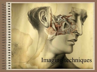
2. Important diagnostic tool for taking images of
dense parts of the body such as bones
is a transmission-based technique in which X-
rays from a source pass through the patient
and are detected either by film or an ionization
chamber on the opposite side of the body
3. Dual-Energy Imaging
◦ produces two separate images corresponding to soft tissue
and bone
◦ imaging the chest region because both soft-tissue
abnormalities and small calcifications can be visualized more
clearly
X-Ray Angiography
◦ produce images that show selectively the blood vessels in the
body
◦ is used to investigate diseases such as stenoses and clotting
of arteries and veins and irregularities in systemic and
pulmonary blood flow
X-Ray Fluoroscopy
◦ is used for placement of stents and catheters, patient
positioning for interventional surgery, and many studies of the
GI tract
4.
5. Uses
• determining the presence and severity of
fractures or cracks in the bone structure
• Dual-energy scanning is used for
diagnosing lung disease and detecting other
masses within the chest wall
• Vascular imaging is performed to study
compromised blood flow in the brain and
heart
• Diseases of the GI tract and the urinary tract
• Mammography
6. It involves sending very high frequency sound
waves through obtaining pictures or images from
inside the human body
also called ultrasound scanning or sonography
sound waves are reflected off the internal organs
and are sensed using appropriate equipments
Ultrasound images are captured in real time and
displayed on a television monitor
7.
8. For the imaging of
Cardiology
Obstetrics
Gynaecology
Gastroenterology
Musculoskeletal
Urology
Vascular
Intervention
Breast
Small Parts
Endocrinology
9. X-ray computed tomography, computed
tomography (CT scan), computed axial
tomography (CAT scan)
is a medical imaging procedure that utilizes
computer-processed X-rays to
produce tomographic images or 'slices' of specific
areas of the body
10.
11. Diagnostic uses
◦ Head
◦ Lungs
◦ Pulmonary angiogram
◦ Cardiac
◦ Abdominal and pelvic
◦ Extremities
Advantages
◦ eliminates the superimposition of images
◦ differences between tissues that differ in physical density
can be distinguished
◦ can be viewed as images in the axial, coronal, or sagittal
planes
Adverse effects
◦ a small increased risk of cancer with CT scans
◦ Causes nausea, vomiting and itching rashes
12. Magnetic resonance imaging (MRI), nuclear
magnetic resonance imaging (NMRI), or magnetic
resonance tomography (MRT)
Developed by Felix Bloch and Edward M. Purcel
Based on the phenomenon nuclear magnetic
resonance
is a medical imaging technique used in
radiology to visualize internal structures of the
body in detail
Specialized MRI scans are available like Diffusion
MRI, Magnetization transfer MRI, real time MRI
etc
13. Applications and uses
◦ To detect tiny lesions of multiple sclerosis on Brain and
Spinal cord
◦ To examine joint injuries and slipped discs in vertebral
column
◦ To detect minute cancerous tumors such as brain tumors
14.
15. Is a diagnostic examination that involves the
acquisition of physiologic images based on the
detection of radiation from the emission of
positrons
system detects pairs of gamma rays emitted
indirectly by a positron-emitting radionuclide
8F- fluorodeoxyglucose(FDG) has been used as
active molecule
16. Applications
◦ Oncology
◦ Neuroimaging
◦ Cardiology
◦ Pharmacology
◦ Small animal imaging
◦ Musculo-skeletal imaging
Limitations
◦ risk from the test radiation
◦ high costs
◦ Need careful planning with respect to patient scheduling
since the half life of the active molecule is less
17.
18. Single-photon emission computed tomography
(SPECT)
is a nuclear medicine tomographic imaging technique
using gamma rays
It is able t
information is typically presented as cross-sectional
slices through the patien to provide true 3D information
a radioisotope of gallium(III) is used as active molecule
19. tumor imaging, infection (leukocyte) imaging,
thyroid imaging or bone scintigraphy
to provide information about localised function in
internal organs, such as functional cardiac or brain
imaging
used for the diagnosis of ischemic heart disease
used in functional brain imaging