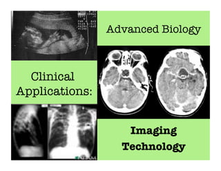
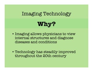
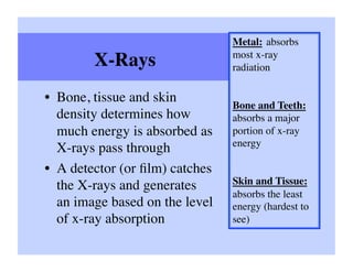
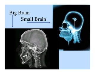
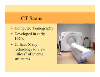
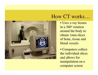
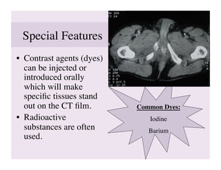
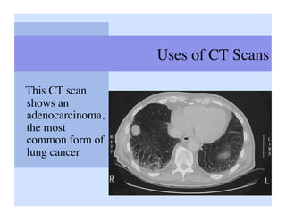
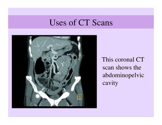
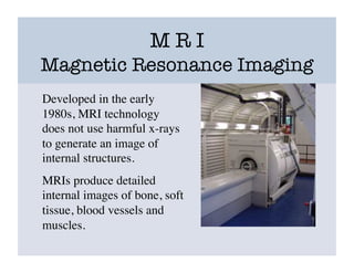
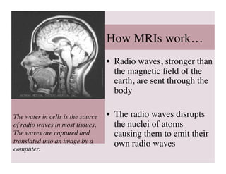
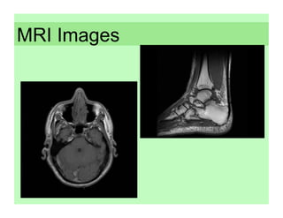
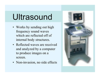
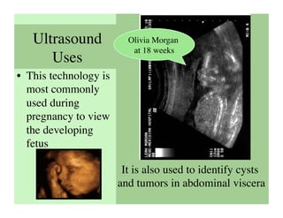
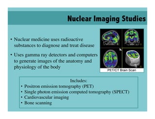
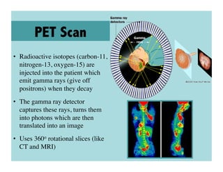
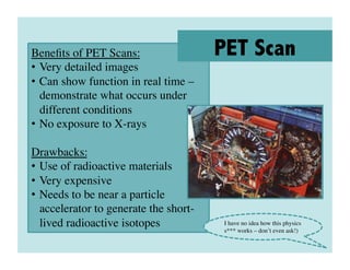
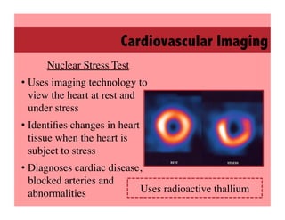
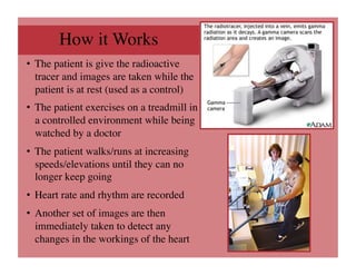
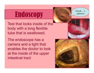
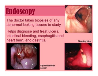
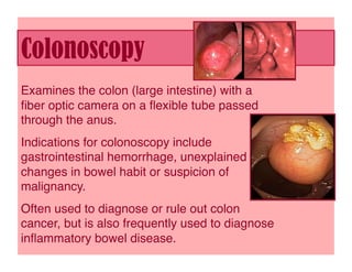
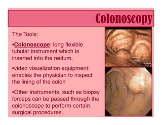
Imaging technologies allow physicians to view internal structures and diagnose diseases. X-rays were discovered in 1895 and can pass through objects. Computed tomography (CT) scans provide cross-sectional slices using X-rays while magnetic resonance imaging (MRI) uses radio waves and magnetic fields without X-rays. Ultrasound uses sound waves and nuclear imaging employs radioactive tracers to generate images. Endoscopy and colonoscopy examine the digestive tract using cameras on flexible tubes. These technologies revolutionized medicine by enabling non-invasive visualization of internal structures.
2. Imaging Technology
Why?
• Imaging allows physicians to view
internal structures and diagnose
diseases and conditions
• Technology has steadily improved
throughout the 20th century
3. Background
• X-Ray Technology
– Discovered in 1895 by
Wilhelm Roentgen
– X-rays are short wave,
high energy photons of
light
– Have the ability to pass
through solid objects
4. Metal: absorbs
most x-ray
X-Rays
radiation
• Bone, tissue and skin
Bone and Teeth:
density determines how absorbs a major
much energy is absorbed as portion of x-ray
energy
X-rays pass through
• A detector (or film) catches
the X-rays and generates Skin and Tissue:
absorbs the least
an image based on the level energy (hardest to
of x-ray absorption
see)
7. CT Scans
• Computed Tomography
• Developed in early
1970s
• Utilizes X-ray
technology to view
“slices” of internal
structures
8. How CT works…
• Uses x-ray beams
in a 360o rotation
around the body to
obtain 1mm slices
of bone, tissue and
blood vessels
• Computers collect
the individual slices
and allows for
manipulation on a
computer screen
9. Special Features
• Contrast agents (dyes)
can be injected or
introduced orally
which will make
specific tissues stand
out on the CT film.
Common Dyes:
• Radioactive Iodine
substances are often Barium
used.
10. Uses of CT Scans
This CT scan
shows an
adenocarcinoma,
the most
common form of
lung cancer
11. Uses of CT Scans
This coronal CT
scan shows the
abdominopelvic
cavity
12. M R I
Magnetic Resonance Imaging
Developed in the early
1980s, MRI technology
does not use harmful x-rays
to generate an image of
internal structures.
MRIs produce detailed
internal images of bone, soft
tissue, blood vessels and
muscles.
13. How MRIs work…
• Radio waves, stronger than
the magnetic field of the
earth, are sent through the
body
The water in cells is the source • The radio waves disrupts
of radio waves in most tissues. the nuclei of atoms
The waves are captured and causing them to emit their
translated into an image by a own radio waves
computer.
15. Ultrasound
• Works by sending out high
frequency sound waves
which are reflected off of
internal body structures.
• Reflected waves are received
and analyzed by a computer
to produce images on a
screen.
• Non-invasion, no side effects
16. Ultrasound Olivia Morgan
at 18 weeks
Uses
• This technology is
most commonly
used during
pregnancy to view
the developing
fetus
It is also used to identify cysts
and tumors in abdominal viscera
17. Echocardiogram
• Ultrasound of the heart
• Shows morphology of
the heart and how well
blood flows through the
heart
• Used to diagnose
malformed structures
and inadequate blood
supply
18. Nuclear Imaging Studies
• Nuclear medicine uses radioactive
substances to diagnose and treat disease
• Uses gamma ray detectors and computers
to generate images of the anatomy and
physiology of the body
Includes:
• Positron emission tomography (PET)
• Single photon emission computed tomography (SPECT)
• Cardiovascular imaging
• Bone scanning
19. PET Scan
• Radioactive isotopes (carbon-11,
nitrogen-13, oxygen-15) are
injected into the patient which
emit gamma rays (give off
positrons) when they decay
• The gamma ray detector
captures these rays, turns them
into photons which are then
translated into an image
• Uses 360o rotational slices (like
CT and MRI)
20. Benefits of PET Scans:
PET Scan
• Very detailed images
• Can show function in real time –
demonstrate what occurs under
different conditions
• No exposure to X-rays
Drawbacks:
• Use of radioactive materials
• Very expensive
• Needs to be near a particle
accelerator to generate the short-
lived radioactive isotopes
I have no idea how this physics
s*** works – don’t even ask!)
21. Cardiovascular Imaging
Nuclear Stress Test
• Uses imaging technology to
view the heart at rest and
under stress
• Identifies changes in heart
tissue when the heart is
subject to stress
• Diagnoses cardiac disease,
blocked arteries and
abnormalities
Uses radioactive thallium
22. How it Works
• The patient is give the radioactive
tracer and images are taken while the
patient is at rest (used as a control)
• The patient exercises on a treadmill in
a controlled environment while being
watched by a doctor
• The patient walks/runs at increasing
speeds/elevations until they can no
longer keep going
• Heart rate and rhythm are recorded
• Another set of images are then
immediately taken to detect any
changes in the workings of the heart
23. Ooooh… a
Endoscopy sphincter
Test that looks inside of the
body with a long flexible
tube that is swallowed.
The endoscope has a
camera and a light that
enables the doctor to look
at the inside of the upper
intestinal tract
24. Endoscopy
The doctor takes biopsies of any
abnormal looking tissues to study
Helps diagnose and treat ulcers,
intestinal bleeding, esophagitis and
heart burn, and gastritis. Bleeding Ulcer
Squamocellular
cancer
25. Colonoscopy
Examines the colon (large intestine) with a
fiber optic camera on a flexible tube passed
through the anus.
Indications for colonoscopy include
gastrointestinal hemorrhage, unexplained
changes in bowel habit or suspicion of
malignancy.
Often used to diagnose or rule out colon
cancer, but is also frequently used to diagnose
inflammatory bowel disease.
26. Colonoscopy
The Tools:
• Colonoscope: long flexible
tubular instrument which is
inserted into the rectum.
• video visualization equipment:
enables the physician to inspect
the lining of the colon
• Other instruments, such as biopsy
forceps can be passed through the
colonoscope to perform certain
surgical procedures.