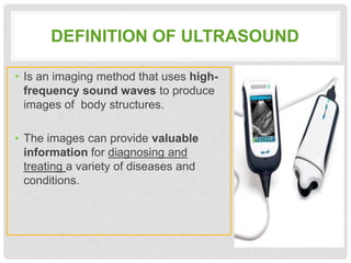
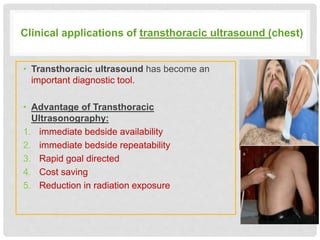
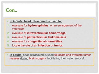
ULTRASONOGRAPHY
APPLICATIONS
Presented by :
Ghaida Al-Rashed
OUT LINE
• Definition of Ultrasound
• Ultrasound application
DEFINITION OF ULTRASOUND
• Is an imaging method that uses high-
frequency sound waves to produce
images of body structures.
• The images can provide valuable
information for diagnosing and
treating a variety of diseases and
conditions.
• Following the history and clinical examination,
ultrasound has become one of the investigations of
choice in patients with abdominal, chest or
Musculoskeletal problems.
• The reason is that ultrasound imaging is a cost
effective, quick, painless and non-invasive investigation
with no radiation hazards or parentral contrast reaction.
CLINICAL APPLICATIONS OF
ULTRASONOGRAPHY
Clinical applications of transthoracic ultrasound (chest)
• Transthoracic ultrasound has become an
important diagnostic tool.
• Advantage of Transthoracic
Ultrasonography:
1. immediate bedside availability
2. immediate bedside repeatability
3. Rapid goal directed
4. Cost saving
5. Reduction in radiation exposure
Con..
• Transthoracic Ultrasonography useful diagnostic
information for :
1. chest wall
2. pleural lesions
3. peripheral lung nodules
4. pulmonary consolidations
5. necrotizing pneumonias
6. lung abscesses
7. tumors with obstructive pneumonitis
8. mediastinal masses
9. peridiaphragmatic lesions
Clinical applications of Abdominal
ultrasound
• Abdominal ultrasound is performed to evaluate the:
1. kidneys
2. liver
3. gallbladder
4. pancreas
5. spleen
6. abdominal aorta and other blood vessels of the abdomen
Con..
• Ultrasound is used to help diagnose a
variety of conditions, such as:
1. abdominal pain or distention.
2. abnormal liver function.
3. enlarged abdominal organ.
4. stones in the gallbladder or kidney.
5. aneurysmin the aorta.
6. Trauma survey and follow up ( FAST)
7. Suspect acute cholecystitis or
pyelonephritis
Clinical applications of Musculoskeletal
ultrasound
• Ultrasound imaging uses to produce pictures of muscles, tendons,
ligaments and joints throughout the body.
• Ultrasound images are typically used to help diagnose:
1. tendon tears, or tendinitis
2. muscle tears, masses or fluid collections.
3. ligament sprains or tears.
4. inflammation or fluid (effusions) within the bursae and joints.
5. early changes of rheumatoid arthritis.
6. benign and malignant soft tissue tumors.
7. ganglion cysts.
8. dislocations of the hip in infants.
Clinical applications Head Ultrasound
• Ultrasound imaging uses to
produce pictures of the brain and
cerebrospinal fluid.
• It is most commonly performed
on infants, whose skulls have not
completely formed
Con..
• In infants, head ultrasound is used to:
1. evaluate for hydrocephalus, or an enlargement of the
ventricles
2. evaluate of intraventricular hemorrhage
3. evaluate of periventricular leukomalacia
4. evaluate for congenital abnormalities.
5. locate the site of an infection or tumor.
• In adults, head ultrasound is used to locate and evaluate tumor
masses during brain surgery, facilitating their safe removal.
SUMMARY
Ultrasound
application
(abdomine)
Ultrasound
application
(chest)
(muscularskl
eton)
Ultrasound
(definition)
REFERENCES
• https://www.radiologyinfo.org/en/info.cfm?pg=ultrasound-
cranial#common-uses
• https://www.radiologyinfo.org/en/info.cfm?pg=musculous
• https://www.radiologyinfo.org/en/info.cfm?pg=abdominus
Ultrasonography applications

Ultrasonography applications

  • 1.
  • 2.
    OUT LINE • Definitionof Ultrasound • Ultrasound application
  • 3.
    DEFINITION OF ULTRASOUND •Is an imaging method that uses high- frequency sound waves to produce images of body structures. • The images can provide valuable information for diagnosing and treating a variety of diseases and conditions.
  • 4.
    • Following thehistory and clinical examination, ultrasound has become one of the investigations of choice in patients with abdominal, chest or Musculoskeletal problems. • The reason is that ultrasound imaging is a cost effective, quick, painless and non-invasive investigation with no radiation hazards or parentral contrast reaction.
  • 5.
  • 6.
    Clinical applications oftransthoracic ultrasound (chest) • Transthoracic ultrasound has become an important diagnostic tool. • Advantage of Transthoracic Ultrasonography: 1. immediate bedside availability 2. immediate bedside repeatability 3. Rapid goal directed 4. Cost saving 5. Reduction in radiation exposure
  • 7.
    Con.. • Transthoracic Ultrasonographyuseful diagnostic information for : 1. chest wall 2. pleural lesions 3. peripheral lung nodules 4. pulmonary consolidations 5. necrotizing pneumonias 6. lung abscesses 7. tumors with obstructive pneumonitis 8. mediastinal masses 9. peridiaphragmatic lesions
  • 8.
    Clinical applications ofAbdominal ultrasound • Abdominal ultrasound is performed to evaluate the: 1. kidneys 2. liver 3. gallbladder 4. pancreas 5. spleen 6. abdominal aorta and other blood vessels of the abdomen
  • 9.
    Con.. • Ultrasound isused to help diagnose a variety of conditions, such as: 1. abdominal pain or distention. 2. abnormal liver function. 3. enlarged abdominal organ. 4. stones in the gallbladder or kidney. 5. aneurysmin the aorta. 6. Trauma survey and follow up ( FAST) 7. Suspect acute cholecystitis or pyelonephritis
  • 10.
    Clinical applications ofMusculoskeletal ultrasound • Ultrasound imaging uses to produce pictures of muscles, tendons, ligaments and joints throughout the body. • Ultrasound images are typically used to help diagnose: 1. tendon tears, or tendinitis 2. muscle tears, masses or fluid collections. 3. ligament sprains or tears. 4. inflammation or fluid (effusions) within the bursae and joints. 5. early changes of rheumatoid arthritis. 6. benign and malignant soft tissue tumors. 7. ganglion cysts. 8. dislocations of the hip in infants.
  • 11.
    Clinical applications HeadUltrasound • Ultrasound imaging uses to produce pictures of the brain and cerebrospinal fluid. • It is most commonly performed on infants, whose skulls have not completely formed
  • 12.
    Con.. • In infants,head ultrasound is used to: 1. evaluate for hydrocephalus, or an enlargement of the ventricles 2. evaluate of intraventricular hemorrhage 3. evaluate of periventricular leukomalacia 4. evaluate for congenital abnormalities. 5. locate the site of an infection or tumor. • In adults, head ultrasound is used to locate and evaluate tumor masses during brain surgery, facilitating their safe removal.
  • 13.
  • 14.

Editor's Notes

  • #5 The reason for choice US , the advantage of US
  • #12 tendon tears, or tendinitis of the rotator cuff in the shoulder, Achilles tendon in the ankle and other tendons throughout the body.
  • #14 detect bleeding within the brain tissue or the ventricles= intraventricular hemorrhage is damage to the white matter brain tissue surrounding the edges of the ventricles=periventricular leukomalacia (PVL).