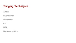
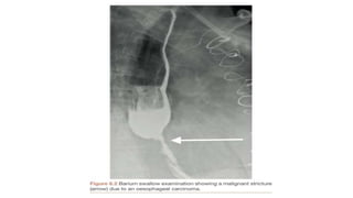
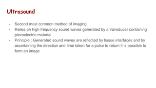
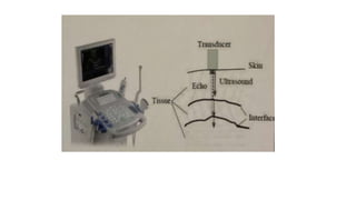
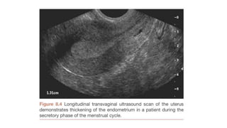
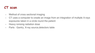
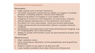
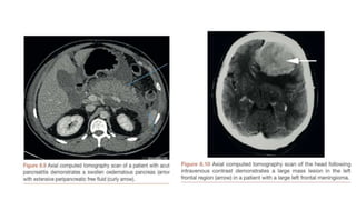
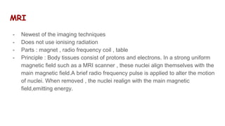
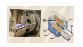
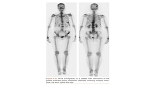
Imaging in Surgery
Introduction
-Imaging is a veritable tool in surgical patients.
-Radiological investigations remain vital to the diagnosis, preparation and follow
up of patients .
Uses
# To aid in diagnosis of a surgical disorder
# To guide a surgical procedure
# Monitoring
# Interventional radiological techniques
Imaging Techniques
X rays
Fluoroscopy
Ultrasound
CT
MRI
Nuclear medicine
Radiography
- Oldest and commonest imaging technique
- Uses ionising radiations
- Parts : X ray tube , collimator, table , film
- Principle : X-rays emitted from an X-ray source are absorbed to varying
degrees by different materials and tissues and therefore causes different
degree of darkening of radiographic films , resulting in an image
Advantages
- cheap
- Available
- Short developing time
- Good for assessing bone
Disadvantages
- ionising radiation
- Poor soft tissue contrast
Fluroscopy
- uses X-rays like in conventional radiographs. However, a fluorescent
screen,image intensifier and a video converter is used
- Allows for real time imaging
- Higher dose of radiation
- Extensive use in functional studies , interventional radiology and imagine
guidance
Ultrasound
- Second most common method of imaging
- Relies on high frequency sound waves generated by a transducer containing
piezoelectric material
- Principle : Generated sound waves are reflected by tissue interfaces and by
ascertaining the direction and time taken for a pulse to return it is possible to
form an image
CT scan
- Method of cross sectional imaging
- CT uses a computer to create an image from an integration of multiple X-rays
exposures taken in a circle round the patient
- Heavy ionising radiation dose
- Parts : Gantry, X-ray source,detectors table
MRI
- Newest of the imaging techniques
- Does not use ionising radiation
- Parts : magnet , radio frequency coil , table
- Principle : Body tissues consist of protons and electrons. In a strong uniform
magnetic field such as a MRI scanner , these nuclei align themselves with the
main magnetic field.A brief radio frequency pulse is applied to alter the motion
of nuclei. When removed , the nuclei realign with the main magnetic
field,emitting energy.
T1- fat, methemoglobin, mucinous
fluid appear white.
Important for delineating anatomy
T2- fat, water and most
pathological fluid appear white.
Highlights pathology better
Radionuclide scan
- A radioactive element (radionuclide) is administered as a radio
pharmaceutical agent and a detector ( gamma camera ) is used to record and
localise the emission ( gamma ray ) and thus form an image
- Agents : technetium,gallium,thallium,iodine
- These are usually labelled for organ specificity
PET scan
- An extension of radionuclide studies
- Same principle but uses substances that emit positron and not gamma rays
- Agent : 18F-2Fluoro-2deoxy-D glucose ( FDG)
Others
- CONTRAST STUDIES
- * positive , negative and combine
- * Radiocontrast
- * MRI contrast
- * USS contrast
- SPECT
Application in surgery
- Acute abdomen : Bowel obstruction, perforation , ischemia , GIT
haemorrhage , inflammatory lesions.
Modalities:- Chest X-ray , abdominal X-ray , USS , CT scan , MRI
- Trauma : USS , CT , MRI
- Masses : USS , Contrast studies , CT , MRI
THANK YOU

Imaging in Surgery Final Year MBBS .pptx

  • 1.
  • 2.
    Introduction -Imaging is averitable tool in surgical patients. -Radiological investigations remain vital to the diagnosis, preparation and follow up of patients .
  • 3.
    Uses # To aidin diagnosis of a surgical disorder # To guide a surgical procedure # Monitoring # Interventional radiological techniques
  • 4.
  • 5.
    Radiography - Oldest andcommonest imaging technique - Uses ionising radiations - Parts : X ray tube , collimator, table , film - Principle : X-rays emitted from an X-ray source are absorbed to varying degrees by different materials and tissues and therefore causes different degree of darkening of radiographic films , resulting in an image
  • 7.
    Advantages - cheap - Available -Short developing time - Good for assessing bone
  • 8.
    Disadvantages - ionising radiation -Poor soft tissue contrast
  • 10.
    Fluroscopy - uses X-rayslike in conventional radiographs. However, a fluorescent screen,image intensifier and a video converter is used - Allows for real time imaging - Higher dose of radiation - Extensive use in functional studies , interventional radiology and imagine guidance
  • 12.
    Ultrasound - Second mostcommon method of imaging - Relies on high frequency sound waves generated by a transducer containing piezoelectric material - Principle : Generated sound waves are reflected by tissue interfaces and by ascertaining the direction and time taken for a pulse to return it is possible to form an image
  • 16.
    CT scan - Methodof cross sectional imaging - CT uses a computer to create an image from an integration of multiple X-rays exposures taken in a circle round the patient - Heavy ionising radiation dose - Parts : Gantry, X-ray source,detectors table
  • 19.
    MRI - Newest ofthe imaging techniques - Does not use ionising radiation - Parts : magnet , radio frequency coil , table - Principle : Body tissues consist of protons and electrons. In a strong uniform magnetic field such as a MRI scanner , these nuclei align themselves with the main magnetic field.A brief radio frequency pulse is applied to alter the motion of nuclei. When removed , the nuclei realign with the main magnetic field,emitting energy.
  • 20.
    T1- fat, methemoglobin,mucinous fluid appear white. Important for delineating anatomy T2- fat, water and most pathological fluid appear white. Highlights pathology better
  • 24.
    Radionuclide scan - Aradioactive element (radionuclide) is administered as a radio pharmaceutical agent and a detector ( gamma camera ) is used to record and localise the emission ( gamma ray ) and thus form an image - Agents : technetium,gallium,thallium,iodine - These are usually labelled for organ specificity
  • 25.
    PET scan - Anextension of radionuclide studies - Same principle but uses substances that emit positron and not gamma rays - Agent : 18F-2Fluoro-2deoxy-D glucose ( FDG)
  • 28.
    Others - CONTRAST STUDIES -* positive , negative and combine - * Radiocontrast - * MRI contrast - * USS contrast - SPECT
  • 29.
    Application in surgery -Acute abdomen : Bowel obstruction, perforation , ischemia , GIT haemorrhage , inflammatory lesions. Modalities:- Chest X-ray , abdominal X-ray , USS , CT scan , MRI - Trauma : USS , CT , MRI - Masses : USS , Contrast studies , CT , MRI
  • 30.