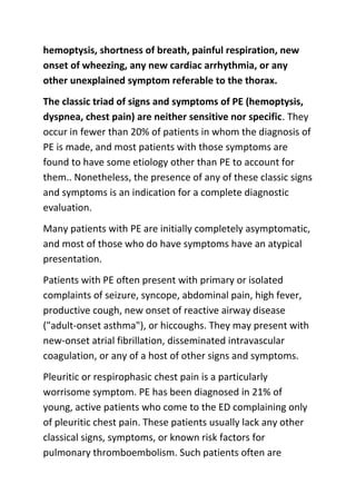
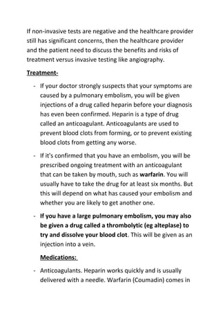
Premium Call Girls Cottonpet Whatsapp 7001035870 Independent Escort Service
Pulmonary embolism
1. Pulmonary embolism(PE) is a condition that occurs when
one or more arteries in your lungs become blocked. In most
cases, pulmonary embolism is caused by blood clots that
travel to your lungs from another part of your body — most
commonly, your legs.
Pulmonary embolism can occur in otherwise healthy people.
Signs and symptoms can vary from person to person, but
commonly include sudden and unexplained shortness of
breath, chest pain and a cough that may bring up bloodtinged sputum.
Pulmonary embolism can be life-threatening, but prompt
treatment with anti-clotting medications can greatly reduce
the risk of death. Taking measures to prevent blood clots in
your legs also can help protect you against pulmonary
embolism.
Pathophysiology- Pulmonary thromboembolism is not a
disease in and of itself. Rather, it is a complication of
underlying venous thrombosis. Under normal conditions,
microthrombi (tiny aggregates of red cells, platelets, and
fibrin) are formed and lysed continually within the venous
circulatory system. This dynamic equilibrium ensures local
hemostasis in response to injury without permitting
uncontrolled propagation of clot. Under pathological
conditions, microthrombi may escape the normal fibrinolytic
system to grow and propagate. Pulmonary embolism (PE)
2. occurs when these propagating clots break loose and
embolize to block pulmonary blood vessels.
Thrombosis in the veins is triggered by venostasis,
hypercoagulability, and vessel wall inflammation. These 3
underlying causes are known as the Virchow triad. All
known clinical risk factors for DVT ( deep vein thrombosis)
and PE have their basis in one or more elements of the triad.
Patients who have undergone gynecologic surgery, those
with major trauma, and those with indwelling venous
catheters may have DVTs that start in an area related to their
pathology. For other patients, venous thrombosis most often
involves the lower extremities and nearly always starts in the
calf veins, which are involved in virtually all cases of
symptomatic spontaneous lower extremity DVT. Although
DVT starts in the calf veins, in cases of pulmonary embolism,
it will usually propagate proximally to the popliteal vessels,
and from that area embolize.
Clinical
History- Pulmonary embolism (PE) is so common and so
lethal that the diagnosis should be sought actively in every
patient who presents with any chest symptoms that cannot
be proven to have another cause.
Symptoms that should provoke a suspicion of pulmonary
embolism must include chest pain, chest wall tenderness,
back pain, shoulder pain, upper abdominal pain, syncope,
3. hemoptysis, shortness of breath, painful respiration, new
onset of wheezing, any new cardiac arrhythmia, or any
other unexplained symptom referable to the thorax.
The classic triad of signs and symptoms of PE (hemoptysis,
dyspnea, chest pain) are neither sensitive nor specific. They
occur in fewer than 20% of patients in whom the diagnosis of
PE is made, and most patients with those symptoms are
found to have some etiology other than PE to account for
them.. Nonetheless, the presence of any of these classic signs
and symptoms is an indication for a complete diagnostic
evaluation.
Many patients with PE are initially completely asymptomatic,
and most of those who do have symptoms have an atypical
presentation.
Patients with PE often present with primary or isolated
complaints of seizure, syncope, abdominal pain, high fever,
productive cough, new onset of reactive airway disease
("adult-onset asthma"), or hiccoughs. They may present with
new-onset atrial fibrillation, disseminated intravascular
coagulation, or any of a host of other signs and symptoms.
Pleuritic or respirophasic chest pain is a particularly
worrisome symptom. PE has been diagnosed in 21% of
young, active patients who come to the ED complaining only
of pleuritic chest pain. These patients usually lack any other
classical signs, symptoms, or known risk factors for
pulmonary thromboembolism. Such patients often are
4. dismissed inappropriately with an inadequate workup and a
nonspecific diagnosis, such as musculoskeletal chest pain or
pleurisy.
Physical
Massive pulmonary embolism (PE) causes hypotension due
to acute cor pulmonale, but the physical examination
findings early in submassive PE may be completely normal.
After 24-72 hours, loss of pulmonary surfactant often
causes atelectasis and alveolar infiltrates that are
indistinguishable from pneumonia on clinical examination
and by radiography.
New wheezing may be appreciated. If pleural lung surfaces
are affected, a pulmonary rub may be heard.
In patients with recognized PE, the incidence of physical signs
has been reported as follows:
- tachypnea (respiratory rate >16/min)
- rales
- an accentuated second heart sound
- tachycardia (heart rate >100/min)
- fever (temperature >37.8°C)
- diaphoresis(sweating)
- an S 3 or S 4 gallop
5. - clinical signs and symptoms suggesting thrombophlebitis
- lower extremity edema
- cardiac murmur
- cyanosis
Causes- As stated in the Pathophysiology section, the
etiology of venous thrombosis and subsequent
thromboembolism results from a distortion in Virchow's
triad by venostasis, hypercoagulability, or vessel wall
inflammation. These risk factors for venous thrombosis and
pulmonary embolism can be broken down into hereditary
factors and acquired factors.
Hereditary factors (most result in a hypercoagulable state)
- Antithrombin III deficiency
- Protein C deficiency
- Protein S deficiency
- Factor V Leiden (most common genetic risk factor for
thrombophilia)
- Plasminogen abnormality
- Plasminogen activator abnormality
- Fibrinogen abnormality
- Resistance to activated protein C
6. Acquired factors (The most important clinically identifiable
risk factors for DVT and PE are a prior history of DVT or PE,
recent surgery or pregnancy, prolonged immobilization, or
underlying malignancy.)
- Reduced mobility- Fractures, Immobilization, Burns,
Obesity
- Old age
- Malignancy- Chemotherapy
- Acute medical illness- AIDS (lupus anticoagulant), Behçet
disease, Congestive heart failure (CHF), Myocardial
infarction, Polycythemia, Systemic lupus erythematosus,
Ulcerative colitis
- Trauma/major surgery- Spinal cord injury, Catheters
(indwelling venous infusion catheters), Postoperative
- Pregnancy- Postpartum period , Oral contraceptives,
Estrogen replacements (high dose only)
- Drug abuse (intravenous [IV] drugs)
- Drug-induced lupus anticoagulant
- Hemolytic anemias
- Heparin-associated thrombocytopenia
- Homocysteinemia
- Homocystinuria
7. - Hyperlipidemias
- Phenothiazines
- Thrombocytosis
- Varicose veins
- Venography
- Venous pacemakers
- Venous stasis
- Warfarin (first few days of therapy)
DiagnosisChest X-ray- This noninvasive test shows images of your
heart and lungs on film. Although X-rays can't diagnose
pulmonary embolism and may even appear normal when
pulmonary embolism exists, they can rule out conditions that
mimic the disease.
Ventilation-perfusion scans- This test, called a ventilationperfusion scan (V/Q scan), uses small amounts of radioactive
material to study airflow (ventilation) and blood flow
(perfusion) in your lungs.
For the first part of the test, you inhale a small amount of
radioactive material while a camera that's able to detect
radioactive substances takes pictures of the movement of air
in your lungs. Then a small amount of radioactive material is
injected into a vein in your arm, and pictures are taken of
8. blood flow in the blood vessels of your lungs. Comparing the
results of the two studies helps provide a more accurate
diagnosis of pulmonary embolism than does either study
alone. If a mismatch occurs, meaning that there is lung
tissue that has good air entry but no blood flow, it may be
indicative of a pulmonary embolus.
Spiral (helical) computerized tomography (CT) scan- Regular
CT scans take X-rays from many different angles and then
combine them to form images showing two-dimensional
"slices" of your internal structures. In a spiral or helical CT
scan, the scanner rotates around your body in a spiral — like
the stripe on a candy cane — to create three-dimensional
images. This type of CT can detect abnormalities with much
greater precision, and it's also much faster than are
conventional CT scans.
Pulmonary angiogram- This test provides a clear picture of
the blood flow in the arteries of your lungs. It's the most
accurate way to diagnose pulmonary embolism, but because
it requires a high degree of skill to administer and carries
potentially serious risks, it's usually performed when other
tests fail to provide a definitive diagnosis. It also has the
advantage of being able to measure the pressure in the right
side of your heart. It would be unusual to have normal
readings in the presence of pulmonary embolism.
In a pulmonary angiogram, a flexible tube (catheter) is
inserted into a large vein — usually in your groin — and
9. threaded through your heart into the pulmonary arteries. A
special dye is then injected into the catheter, and X-rays are
taken as the dye travels along the arteries in your lungs.
A risk of this procedure is a temporary change in your heart
rhythm. In addition, the dye may cause kidney damage in
people with decreased kidney function.
d-Dimer blood test- The d-Dimer blood test measures one of
the breakdown products of a blood clot. If this test is normal,
then the likelihood of a pulmonary embolism is very low.
Unfortunately, this test is not specific for blood clots in the
lung. It can be positive for a variety of reasons including
pregnancy, injury, recent surgery, or infection. Looking at the
list of deep vein thrombosis risk factors, one can imagine that
a d-Dimer blood test may not be helpful in those with
significant risk factors for deep vein thrombosis
Venous Doppler study- Ultrasound of the legs, also known as
venous Doppler studies, may be used to look for blood clots
in the legs of a patient suspected of having a pulmonary
embolus. If a deep vein thrombosis exists, it can be inferred
that chest pain and shortness of breath may be due to a
pulmonary embolism.
Echocardiography (EKG, ECG)- Echocardiography or
ultrasound of the heart may be helpful if it shows that there
is strain on the right side of the heart.
10. If non-invasive tests are negative and the healthcare provider
still has significant concerns, then the healthcare provider
and the patient need to discuss the benefits and risks of
treatment versus invasive testing like angiography.
Treatment- If your doctor strongly suspects that your symptoms are
caused by a pulmonary embolism, you will be given
injections of a drug called heparin before your diagnosis
has even been confirmed. Heparin is a type of drug
called an anticoagulant. Anticoagulants are used to
prevent blood clots from forming, or to prevent existing
blood clots from getting any worse.
- If it's confirmed that you have an embolism, you will be
prescribed ongoing treatment with an anticoagulant
that can be taken by mouth, such as warfarin. You will
usually have to take the drug for at least six months. But
this will depend on what has caused your embolism and
whether you are likely to get another one.
- If you have a large pulmonary embolism, you may also
be given a drug called a thrombolytic (eg alteplase) to
try and dissolve your blood clot. This will be given as an
injection into a vein.
Medications:
- Anticoagulants. Heparin works quickly and is usually
delivered with a needle. Warfarin (Coumadin) comes in
11. pill form. Both prevent new clots from forming, but it
takes a few days before warfarin begins to work. Risks
include bleeding gums and easy bruising.
- Clot dissolvers (thrombolytics). While clots usually
dissolve on their own, there are medications that can
dissolve clots quickly. Because these clot-busting drugs
can cause sudden and severe bleeding, they usually are
reserved for life-threatening situations.
Surgical and other procedures :
- Clot removal. If you have a very large clot in your lung,
your doctor may suggest removing it via a thin flexible
tube (catheter) threaded through your blood vessels.
- Vein filter. A catheter can also be used to position a
filter in the main vein — called the inferior vena cava —
that leads from your legs to the right side of your heart.
This filter can block clots from being carried into your
lungs. This procedure is typically reserved for people
who can't take anticoagulant drugs or when
anticoagulant drugs don't work well enough.
- Warfarin- - Initial dose: 5-15 mg/d PO qd
After initial anticoagulation obtained, adjust dose
according to desired INR
12. - Streptase- 2.5 lac IU loading dose over half to one hour,
followed by 1lac IU/hr for 24 hr
- Urokinase- 4400 IU/kg over 10 min i.v. followed by
4400IU/hr for 12 hr
- Alteplase- 100mg i.v infused over 2hr