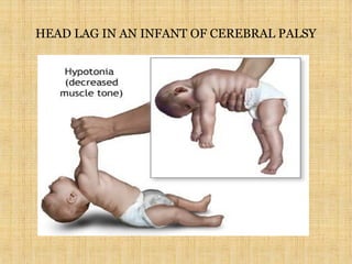
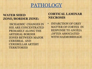
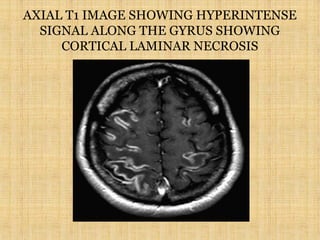
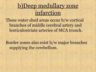
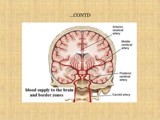
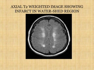
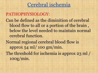
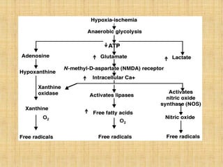
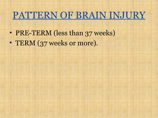
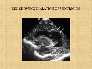
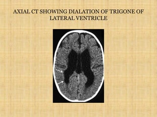
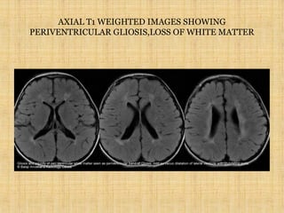
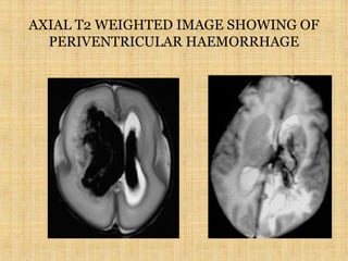
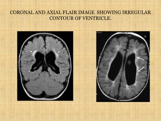
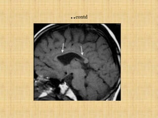
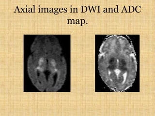
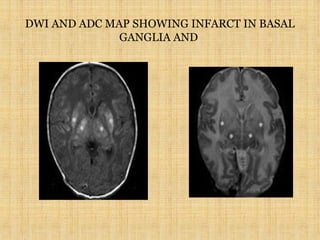
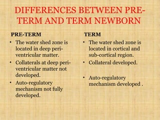
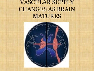
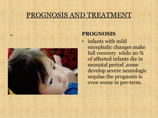
HYPOXIC ISCHEMIC
ENCEPHALOPATHY
DR NEHA NUPUR
JR -2
RIMS,RANCHI
DEFINITION
Hypoxic-Ischemic Encephalopathy:results
when there is global rather than focal
reduction in blood flow,oxygen or glucose
supply .
HIE Depends on:
1)Gestational age
2)Duration of insult
3)Collateral
ETIOLOGY

MATERNAL FACTORS
a) Hypotension
b) Cardiac arrest
c) In utero exposure to cocaine
d) Infection (chorio amnionitis)

UTERO- PLACENTAL FACTORS
a) uterine rupture
b) Umbilical cord-entanglement/prolapse
FETAL FACTORS*
a)Anaemia
b)Cardiomyopathy
c)Severe cardiac/circulatory failure
CLINICAL FEATURES
• 1- Delayed cry at birth.
• 2-drowsiness/lethargic
• 3-convulsions.
HEAD LAG IN AN INFANT OF CEREBRAL PALSY
PATHOLOGY
WATER SHED
ZONE/BORDER ZONE:
ISCHAEMIC CHANGES IN
HIE ARE CONCENTRATED
PRIMARILY ALONG THE
ARTERIAL BORDER
ZONES BETWEEN MAJOR
CEREBRAL AND
CEREBELLAR ARTERY
TERRITORIES
CORTICAL LAMINAR
NECROSIS
• INFARCTION OF GREY
MATTER OF CORTEX IN
RESPONSE TO ANOXIA
,OFTEN ASSOCIATED
WITH HAEMORRHAGE
AXIAL T1 IMAGE SHOWING HYPERINTENSE
SIGNAL ALONG THE GYRUS SHOWING
CORTICAL LAMINAR NECROSIS
WATER-SHED ZONES
BORDER OR WATER SHED ZONES ARE
THOSE AREAS BETWEEN THE
TERMINAL CAPILLARY BEDS OF
MAJOR TERRITORIAL ARTERIES.
a)Superficial border zone infarction:
ACA and MCA
MCA and PCA
b)Deep medullary zone
infarction
These water shed areas occur b/w cortical
branches of middle cerebral artery and
lenticulostriate arteries of MCA trunck.
Border zones also exist b/w major branches
supplying the cerebellum.
..CONTD
…CONTD
AXIAL T2 WEIGHTED IMAGE SHOWING
INFARCT IN WATER-SHED REGION
Cerebral ischemia
PATHOPHYSIOLOGY:
Can be defined as the diminition of cerebral
blood flow to all or a portion of the brain ,
below the level needed to maintain normal
cerebral function.
Normal regional cerebral blood flow is
approx 54 ml/ 100 gm/min.
The threshold for ischemia is approx 23 ml /
100g/min.
TYPES OF BRAIN EDEMA
• 1)VASOGENIC EDEMA
• 2)CYTOTOXIC EDEMA
• 3)INTERSTITIAL EDEMA
PATTERN OF BRAIN INJURY
• PRE-TERM (less than 37 weeks)
• TERM (37 weeks or more).
PRE-TERM:
MILD-TO MODERATE HYPOXIA:
The most common area to undergo ischaemic
injury in the pre-mature infant is the peri-
ventricular white matter,which in the
developing fetus is the vascular watershed
zone and has a relatively high metabolic
demand.
1 periventricular infarction(40%)
2 periventricular haemorrage(15%)-bleed may
occur in the region of germinal matrix which
may break through the ependymal lining
resulting into intra-ventricular bleed.
…contd
3) PERI-VENTRICULAR LEUKOMALACIA.
4)THINNING OF CORPUS CALLOSUM
5)DIALATION OF VENTRICLES
6)DEVELOPMENT OF PORENCEPHALIC CYST
GRADING OF GERMINAL MATRIX
HAEMORRHAGE
• DEFINITION:it is a loose network of highly vascularised tissue
with little supporting stroma containing primitive nerve cells.
• GRADE 1-haemorrhage confined to germinal matrix.
• GRADE 2-haemorrhage extending into ventricle or
subependymal region.
• GRADE 3-massive haemorrhage into ventricle causing
hydrocephalous.
IMAGING
• USG-to exclude dialation of ventricles or
haemorrhage.
• CT-to asses ventricular dilation and intra-ventricular
bleed.
• T1-intra ventricular bleed seen as hyperintense signal.
• T2-gliosis –seen as increased signal intensity in peri-
ventricular white matter.
• DWI-shows restriction in peri-ventricular region with
corresponding hypointense signal in ADC map
FLAIR
• Thinning of periventricular white matter.
• Thinning of corpus callosum.
• Development of porencephalic cyst.
• Ventriculomegaly.
USG SHOWING DIALATION OF VENTRICLES
AXIAL CT SHOWING DIALATION OF TRIGONE OF
LATERAL VENTRICLE
AXIAL T1 WEIGHTED IMAGES SHOWING
PERIVENTRICULAR GLIOSIS,LOSS OF WHITE MATTER
AXIAL T2 WEIGHTED IMAGE SHOWING OF
PERIVENTRICULAR HAEMORRHAGE
CORONAL AND AXIAL FLAIR IMAGE SHOWING IRREGULAR
CONTOUR OF VENTRICLE.
..contd
PRE-MATURE INFANTS WITH
PROFOUND ASPHYXIA
• CAUSE-hypotension or cardiac arrest
followed by resuscitation.
• IMAGING-infarction or bleed involving
thalami,basal ganglia and brainstem.
Axial images in DWI and ADC
map.
ISCHEMIC INJURY IN TERM
Less porfound asphyxia
• Cause-birth asphyxia
• Injury to cortical and sub-
cortical areas in a water –
shed distribution with
relative sparing of the
peri-ventricular white
matter.
Profound episodes of
asphyxia
• Cause-
hypotension/cardiac
• injury in the thalami and
basal ganglia.
• Cystic encephalomalacia-
TERM INFANTS WITH MILD TO MODERATE
HYPOXIA
• IMAGING
• 1)USG-for the assement of ventricular dilation .
• 2)CT-
• 24 hrs-hypodensity noted in bilateral cerebral hemisphere with
sparing of cerebellum and thalamus known as reversal sign.
• 2days-early calcific changes in bilateral basal ganglia.
• 5 days –ventricular dilation.
• 2 weeks –coarse calcification.
• 1 month-calcification resolves ,brain volume loss in cortical
and subcortical region.
…CONTD
3)T1- hypointense signal in the region of ischaemic grey
matter.
4)T2-hyperintense signal in the region of ischaemic grey
matter .
5)DWI AND ADC MAP-deep gery matter infarction
showing restriction.
AXIAL CT SHOWING REVERSAL SIGN
FEW CASES REPORTED IN DEPARTMENT OF RIMS
• . • .
IMAGING IN TERM INFANTS WITH
PROFOUND ISCHEMIA
1)CT-multicystic areas of fluid collection replacing brain
parenchyma.
2)T1-hypointensity primarily in grey matter.
3)T2-hyperintensity primarily in the grey matter.
4)DWI- high signal intensity in basal ganglia and
thalamus .
5)ADC-hypointensity in basal ganglia and thalamus.
6)FLAIR- shows multicystic encephalomalacia.
AXIAL CT SHOWING MUTICYSTIC
ENCEPHALOMALACIA
AXIAL FLAIR IMAGE SHOWING MULTICYSTIC
ENCEPHALOMALACIA IN FRONTAL REGIONS.
DWI AND ADC MAP SHOWING INFARCT IN BASAL
GANGLIA AND
DIFFERENCES BETWEEN PRE-
TERM AND TERM NEWBORN
PRE-TERM
• The water shed zone is
located in deep peri-
ventricular matter.
• Collaterals at deep peri-
ventricular matter not
developed.
• Auto-regulatory
mechanism not fully
developed.
TERM
• The water shed zone is
located in cortical and
sub-cortical region.
• Collateral developed.
• Auto-regulatory
mechanism developed .
VASCULAR SUPPLY
CHANGES AS BRAIN
MATURES
PROGNOSIS AND TREATMENT
.. PROGNOSIS
• infants with mild
encephalic changes make
full recovery while 20 %
of affected infants die in
neonatal period ,some
develop severe neurologic
sequlae.the prognosis is
even worse in pre-term.
TREATMENT
• Maintainence of adequate ventilation
,avoidance of hypotension ,maintainence
of metabolic glucose ,fluid and nutritional
status ,control of seizures and control of
brain edema lie the main treatment
CONCLUSION
• HIE remains an important cause of
morbidity and mortality in the neonatal
period and cerebral palsy is a late
neurologic sequale in the post natal
period .intervention remains supportive
,imaging becomes important for optimal
management and rule out other causes of
encephalopathy
REFERENCES
1) CRANIAL MRI AND CT –LEE AND RAO.
2) MRI OF BRAIN AND SPINE –SCOTT.
3) DIAGNOSTIC NEURORADIOLOGY-OSBORN
THANKS

HYPOXIC ISCHEMIC ENCEPHALOPATHY-RADIOLOGICAL APPROACH

  • 1.
  • 2.
    DEFINITION Hypoxic-Ischemic Encephalopathy:results when thereis global rather than focal reduction in blood flow,oxygen or glucose supply . HIE Depends on: 1)Gestational age 2)Duration of insult 3)Collateral
  • 3.
    ETIOLOGY  MATERNAL FACTORS a) Hypotension b)Cardiac arrest c) In utero exposure to cocaine d) Infection (chorio amnionitis)  UTERO- PLACENTAL FACTORS a) uterine rupture b) Umbilical cord-entanglement/prolapse
  • 4.
  • 5.
    CLINICAL FEATURES • 1-Delayed cry at birth. • 2-drowsiness/lethargic • 3-convulsions.
  • 6.
    HEAD LAG INAN INFANT OF CEREBRAL PALSY
  • 7.
    PATHOLOGY WATER SHED ZONE/BORDER ZONE: ISCHAEMICCHANGES IN HIE ARE CONCENTRATED PRIMARILY ALONG THE ARTERIAL BORDER ZONES BETWEEN MAJOR CEREBRAL AND CEREBELLAR ARTERY TERRITORIES CORTICAL LAMINAR NECROSIS • INFARCTION OF GREY MATTER OF CORTEX IN RESPONSE TO ANOXIA ,OFTEN ASSOCIATED WITH HAEMORRHAGE
  • 8.
    AXIAL T1 IMAGESHOWING HYPERINTENSE SIGNAL ALONG THE GYRUS SHOWING CORTICAL LAMINAR NECROSIS
  • 9.
    WATER-SHED ZONES BORDER ORWATER SHED ZONES ARE THOSE AREAS BETWEEN THE TERMINAL CAPILLARY BEDS OF MAJOR TERRITORIAL ARTERIES. a)Superficial border zone infarction: ACA and MCA MCA and PCA
  • 10.
    b)Deep medullary zone infarction Thesewater shed areas occur b/w cortical branches of middle cerebral artery and lenticulostriate arteries of MCA trunck. Border zones also exist b/w major branches supplying the cerebellum.
  • 11.
  • 12.
  • 13.
    AXIAL T2 WEIGHTEDIMAGE SHOWING INFARCT IN WATER-SHED REGION
  • 14.
    Cerebral ischemia PATHOPHYSIOLOGY: Can bedefined as the diminition of cerebral blood flow to all or a portion of the brain , below the level needed to maintain normal cerebral function. Normal regional cerebral blood flow is approx 54 ml/ 100 gm/min. The threshold for ischemia is approx 23 ml / 100g/min.
  • 16.
    TYPES OF BRAINEDEMA • 1)VASOGENIC EDEMA • 2)CYTOTOXIC EDEMA • 3)INTERSTITIAL EDEMA
  • 17.
    PATTERN OF BRAININJURY • PRE-TERM (less than 37 weeks) • TERM (37 weeks or more).
  • 18.
    PRE-TERM: MILD-TO MODERATE HYPOXIA: Themost common area to undergo ischaemic injury in the pre-mature infant is the peri- ventricular white matter,which in the developing fetus is the vascular watershed zone and has a relatively high metabolic demand. 1 periventricular infarction(40%) 2 periventricular haemorrage(15%)-bleed may occur in the region of germinal matrix which may break through the ependymal lining resulting into intra-ventricular bleed.
  • 19.
    …contd 3) PERI-VENTRICULAR LEUKOMALACIA. 4)THINNINGOF CORPUS CALLOSUM 5)DIALATION OF VENTRICLES 6)DEVELOPMENT OF PORENCEPHALIC CYST
  • 20.
    GRADING OF GERMINALMATRIX HAEMORRHAGE • DEFINITION:it is a loose network of highly vascularised tissue with little supporting stroma containing primitive nerve cells. • GRADE 1-haemorrhage confined to germinal matrix. • GRADE 2-haemorrhage extending into ventricle or subependymal region. • GRADE 3-massive haemorrhage into ventricle causing hydrocephalous.
  • 21.
    IMAGING • USG-to excludedialation of ventricles or haemorrhage. • CT-to asses ventricular dilation and intra-ventricular bleed. • T1-intra ventricular bleed seen as hyperintense signal. • T2-gliosis –seen as increased signal intensity in peri- ventricular white matter. • DWI-shows restriction in peri-ventricular region with corresponding hypointense signal in ADC map
  • 22.
    FLAIR • Thinning ofperiventricular white matter. • Thinning of corpus callosum. • Development of porencephalic cyst. • Ventriculomegaly.
  • 23.
    USG SHOWING DIALATIONOF VENTRICLES
  • 24.
    AXIAL CT SHOWINGDIALATION OF TRIGONE OF LATERAL VENTRICLE
  • 25.
    AXIAL T1 WEIGHTEDIMAGES SHOWING PERIVENTRICULAR GLIOSIS,LOSS OF WHITE MATTER
  • 26.
    AXIAL T2 WEIGHTEDIMAGE SHOWING OF PERIVENTRICULAR HAEMORRHAGE
  • 27.
    CORONAL AND AXIALFLAIR IMAGE SHOWING IRREGULAR CONTOUR OF VENTRICLE.
  • 28.
  • 29.
    PRE-MATURE INFANTS WITH PROFOUNDASPHYXIA • CAUSE-hypotension or cardiac arrest followed by resuscitation. • IMAGING-infarction or bleed involving thalami,basal ganglia and brainstem.
  • 30.
    Axial images inDWI and ADC map.
  • 31.
    ISCHEMIC INJURY INTERM Less porfound asphyxia • Cause-birth asphyxia • Injury to cortical and sub- cortical areas in a water – shed distribution with relative sparing of the peri-ventricular white matter. Profound episodes of asphyxia • Cause- hypotension/cardiac • injury in the thalami and basal ganglia. • Cystic encephalomalacia-
  • 32.
    TERM INFANTS WITHMILD TO MODERATE HYPOXIA • IMAGING • 1)USG-for the assement of ventricular dilation . • 2)CT- • 24 hrs-hypodensity noted in bilateral cerebral hemisphere with sparing of cerebellum and thalamus known as reversal sign. • 2days-early calcific changes in bilateral basal ganglia. • 5 days –ventricular dilation. • 2 weeks –coarse calcification. • 1 month-calcification resolves ,brain volume loss in cortical and subcortical region.
  • 33.
    …CONTD 3)T1- hypointense signalin the region of ischaemic grey matter. 4)T2-hyperintense signal in the region of ischaemic grey matter . 5)DWI AND ADC MAP-deep gery matter infarction showing restriction.
  • 34.
    AXIAL CT SHOWINGREVERSAL SIGN
  • 35.
    FEW CASES REPORTEDIN DEPARTMENT OF RIMS • . • .
  • 36.
    IMAGING IN TERMINFANTS WITH PROFOUND ISCHEMIA 1)CT-multicystic areas of fluid collection replacing brain parenchyma. 2)T1-hypointensity primarily in grey matter. 3)T2-hyperintensity primarily in the grey matter. 4)DWI- high signal intensity in basal ganglia and thalamus . 5)ADC-hypointensity in basal ganglia and thalamus. 6)FLAIR- shows multicystic encephalomalacia.
  • 37.
    AXIAL CT SHOWINGMUTICYSTIC ENCEPHALOMALACIA
  • 38.
    AXIAL FLAIR IMAGESHOWING MULTICYSTIC ENCEPHALOMALACIA IN FRONTAL REGIONS.
  • 39.
    DWI AND ADCMAP SHOWING INFARCT IN BASAL GANGLIA AND
  • 40.
    DIFFERENCES BETWEEN PRE- TERMAND TERM NEWBORN PRE-TERM • The water shed zone is located in deep peri- ventricular matter. • Collaterals at deep peri- ventricular matter not developed. • Auto-regulatory mechanism not fully developed. TERM • The water shed zone is located in cortical and sub-cortical region. • Collateral developed. • Auto-regulatory mechanism developed .
  • 41.
  • 42.
    PROGNOSIS AND TREATMENT ..PROGNOSIS • infants with mild encephalic changes make full recovery while 20 % of affected infants die in neonatal period ,some develop severe neurologic sequlae.the prognosis is even worse in pre-term.
  • 43.
    TREATMENT • Maintainence ofadequate ventilation ,avoidance of hypotension ,maintainence of metabolic glucose ,fluid and nutritional status ,control of seizures and control of brain edema lie the main treatment
  • 44.
    CONCLUSION • HIE remainsan important cause of morbidity and mortality in the neonatal period and cerebral palsy is a late neurologic sequale in the post natal period .intervention remains supportive ,imaging becomes important for optimal management and rule out other causes of encephalopathy
  • 45.
    REFERENCES 1) CRANIAL MRIAND CT –LEE AND RAO. 2) MRI OF BRAIN AND SPINE –SCOTT. 3) DIAGNOSTIC NEURORADIOLOGY-OSBORN
  • 46.