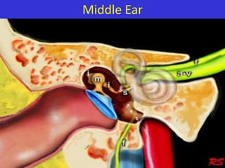
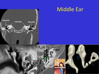
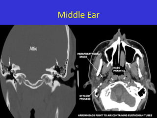
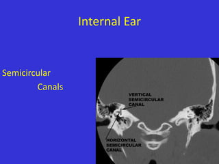
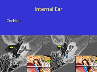
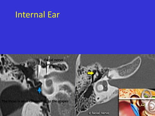
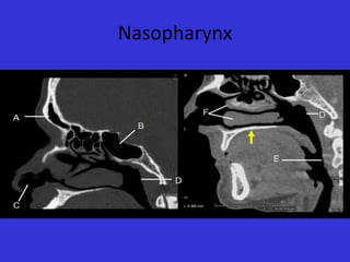
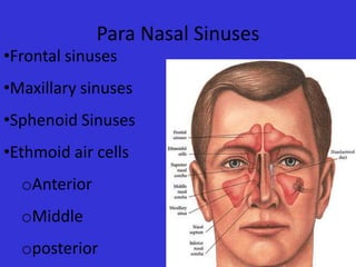
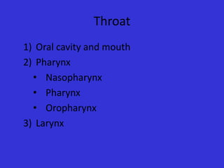
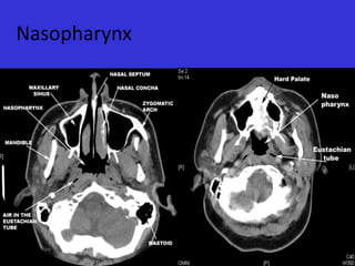
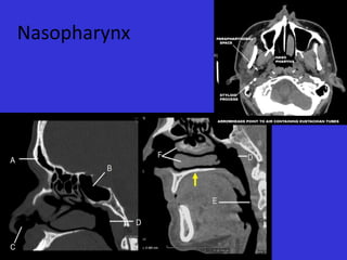
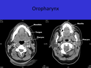
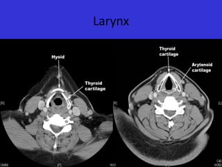
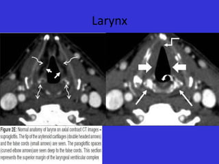
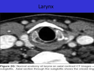
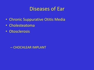
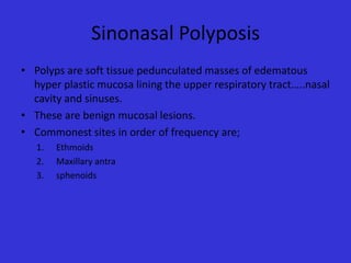
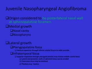
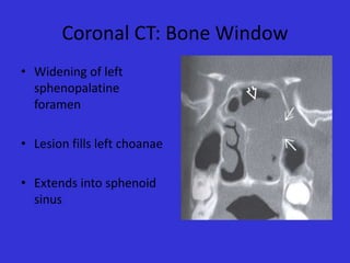
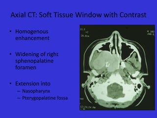
Imaging in ENT
Muhammad Bin Zulfiqar
PGR II SIMS/SHL
New Radiology Department
BASIC ANATOMY
•

EAR
1) External
2) Middle
3) Inner
• NOSE AND PARANASAL SINUSES
•

THROAT
1) Oral cavity and mouth
2) Pharynx
• Nasopharynx
• Oropharynx
• Pharynx
3) Larynx
Imaging modalities
•
•
•
•
•

Plain Radiography(main)
Computerized tomography(main)
Magnetic resonance Imaging
Ultrasound
Barium swallow
Anatomy of EAR
• External
• Middle
• Inner
Tympanic
Membrane
Separates external ear from
middle ear.
Middle Ear
Middle Ear

Stapes
Middle Ear
Middle Ear
Internal Ear
Internal Ear

Semicircular
Canals
Internal Ear
Cochlea
Internal Ear

Facial nerve
canal

The incus is seen connecting to the stapes
Internal Ear
Geniculate ganglion

The incus is seen connecting to the stapes
Nose and Para nasal Sinuses
• Nasal Bones
• Nasal Soft Tissue
• Nasopharynx
Nasal Anatomy
NASAL ANATOMY
Nasopharynx
Para Nasal Sinuses

•Frontal sinuses

•Maxillary sinuses
•Sphenoid Sinuses
•Ethmoid air cells
oAnterior

oMiddle
oposterior
Para Nasal Sinuses
Para Nasal Sinuses
A.
B.
C.
D.
E.
F.

Frontal Sinus
Sphenoid Sinus
Nostril
Posterior Nasopharyngeal wall
Posterior esophageal wall
Nasal turbinate
Osteomeatal Complex
Throat
1) Oral cavity and mouth
2) Pharynx
• Nasopharynx
• Pharynx
• Oropharynx
3) Larynx
•
•
•
•

Nasopharynx
Oropharynx
Pharynx
Larynx
Nasopharynx
Nasopharynx
Nasopharynx
Oropharynx
Pharynx
Pharynx
Oropharynx
Larynx
Larynx
Larynx
Larynx
Larynx
Larynx
Diseases of Ear
• Chronic Suppurative Otitis Media
• Cholesteatoma
• Otosclerosis

– CHOCHLEAR IMPLANT
Chronic Suppurative Otitis Media
• Poorly pneumatized mastoid air cells.
• Ear drum is thickened.
• Soft tissue seen around ossicles without
erosion.
• Calcification of ear drum, tendon of stapedius.
• Almost complete opacification of middle ear
cavity.
Chronic Suppurative Otitis Media
Loss of
pneumatization
Normal

Eardrum calcification

Stapes calcification

Eardrum calcification
Chronic Suppurative Otitis Media
Opacification of tympanic cavity
Cholesteatoma
• Present as soft tissue mass with associated
erosions.
• Auditory ossicles, especially the long process
and lenticular processes of the incus as well as
the head of the stapes
• Wall of the lateral semicircular canal
• Lateral epitympanic wall (the scutum)
Cholesteatoma
Cholesteatoma: 20-year old woman with
recurrent Otitis. Granulations on left ear
drum. Soft tissue mass between ossicular
chain and lateral tympanic wall, which is
eroded. Right side for comparison.

Granulations on left ear drum. Soft tissue mass
between ossicular chain and lateral tympanic
wall, which is eroded. Right side for comparison.
Cholesteatoma

Automastoidectomy due to a large cholesteatoma

Cholesteatoma with erosion of the wall of
the lateral semicircular canal

There is a soft tissue mass with erosion of
the long process of the incus.
Cholesteatoma

The examination shows a mass with mixed intensity on sagittal T1 and high
intensity on transverse T2 weighted images. It has a high intensity on
diffusion weighted images, which indicates restricted diffusion. (arrows)
Otosclerosis
• Otosclerosis is a genetically mediated metabolic
bone disease of unknown etiology.
• conductive hearing loss and is considered to be the
hallmark of the disease.
• The process starts in the region of the oval window,
classically at the fistula ante fenestram, i.e. in front of
the oval window (fenestral otosclerosis).

• It can also occur around the cochlea
(retrofenestral otosclerosis).
Otosclerosis
There is a lucency anterior to the oval window
(arrow) and between the cochlea and the
internal auditory canal.
This is combined fenestral and retrofenestral
otosclerosis.

Otosclerosis anteriorly to the oval window
(arrow)
Otosclerosis

Bilateral otosclerosis
Cochlear Implant
• Cochlear implantation is performed in patients
with sensorineural deafness due to
degeneration of the organ of Corti.
• The electrode is inserted into the scala
tympani of the cochlea via the round window
or via a drill hole directly into the basal turn.
• Post-operatively its position can be evaluated
with plain films or with CT.
Cochlear Implant

Status after cochlear implantation
Nose And Para Nasal Sinuses
•
•
•
•
•

Deviated Nasal Septum
Nasal Bone Fracture
Enlarged Adenoids
Sino nasal Polyposis
Angiofibroma
Deviated Nasal Septum
• Nasal septum deviation is a common physical
disorder of nose involving a displacement of
nasal septum.
• Trauma is a frequent cause.
• Can be congenital.
• Poor drainage of sinuses.
Deviated Nasal Septum

Waters view (close-up view of the
patient in the previous image) shows a
deviated nasal septum, quadrangular
cartilage displaced from the maxillary
crest, and a nasal root deviated to the
right
Fracture of Nasal bone
Enlarged Adenoids
• The adenoids are sections of soft tissue found
at the back of the nasal cavities where they
meet the pharynx.
• Like tonsils, adenoids help to stop harmful
bacteria and airborne pathogens from
entering the airways and causing infections.
• Multiple sinus infections, snoring and
worsened breathing esp. in children.
Enlarged Adenoids
Sinonasal Polyposis
• Polyps are soft tissue pedunculated masses of edematous
hyper plastic mucosa lining the upper respiratory tract…..nasal
cavity and sinuses.
• These are benign mucosal lesions.
• Commonest sites in order of frequency are;
1.
2.
3.

Ethmoids
Maxillary antra
sphenoids
Causes of Sinonasal Polyposis
1.
2.
3.
4.
5.
6.

Allergic rhinitis
Asthma
Cystic fibrosis(child)
Kartagener syndrome
Nickel exposure
Nonneoplastic hyperplastic hyperplasia of
inflammed mucous membranes.
Sinonasal Polyposis
Juvenile Nasopharyngeal Angiofibroma
• Benign highly vascular tumor
• Locally invasive, submucosal spread
• Vascular supply most commonly from internal
maxillary artery
– Also: internal carotid, external carotid, common
carotid, ascending pharyngeal
• Occurring almost exclusively in males
• Peak age of onset = 13-15 years old
• Intracranial Extension between 10-20%
• Recurrence Rates as high as 50%
Juvenile Nasopharyngeal Angiofibroma
Origen considered to be posterlateral nasal wall
at sphenopalatine foramen.
Medial growth
Nasal cavity
Nasopharynx

Lateral growth
Pterygopalatine fossa
 Vertical expansion through inferior orbital fissure to orbit possible

Infratemporal fossa
 Superior expansion through pterygoid process may involve middle cranial fossa
 Lateral and posterior walls of sphenoid sinus can be eroded
 Cavernous sinus may be involved
 Pituitary may involve.
Coronal CT: Bone Window
• Widening of left
sphenopalatine
foramen
• Lesion fills left choanae
• Extends into sphenoid
sinus
Axial CT: Soft Tissue Window with Contrast
• Homogenous
enhancement
• Widening of left
sphenopalatine
foramen
• Extension into
– Nasopharynx
– Pterygopalatine fossa
Axial CT: Soft Tissue Window with Contrast
• Homogenous
enhancement
• Widening of right
sphenopalatine
foramen
• Extension into
– Nasopharynx
– Pterygopalatine fossa
Axial MRI: T1
• Heterogeneous
intermediate signal
• Flow voids represent
enlarged vessels
• Extension into
– Nasopharynx
– Masticator space
Coronal MRI: T1 with Contrast
• Diffuse intense
enhancement
• Multiple flow voids
within hypervascular
mass
• Extension into
– Nasopharynx
– Pterygopalatine fossa
Axial MRI: T2
• Heterogeneous
intermediate to high signal
enhancement
• Multiple flow voids within
hypervascular mass
• Extension into
– Nasopharynx
– Pterygopalatine fossa
THROAT Diseases
• Enlarged adenoids
• Pharyngitis
• CROUP(Laryngotracheobronchitis)
Pharyngitis and Tonsillitis
• Bacterial
• Viral
• Fungal
Croup (acute laryngotracheobronchitis)
• Croup, also called acute laryngotracheobronchitis
is caused by viral infection of the upper airway
usually parainfluenza virus or respiratory
syncytial virus (RSV).
• It is common and has a peak incidence before
the age of 1 year (typically between 3 and 6
months of age).
• It is presented Clinically by protracted barking
cough and inspiratory strider due to tracheal
narrowing that is caused by mucosal edema .
Croup (acute laryngotracheobronchitis)

distension of the hypopharynx due to the patient's
attempt at decreasing airway resistance

steeple sign
Croup (acute laryngotracheobronchitis)

Steeple Sign
QUESTIONS ?
THANK YOU

Imaging in ent

  • 1.
    Imaging in ENT MuhammadBin Zulfiqar PGR II SIMS/SHL New Radiology Department
  • 2.
    BASIC ANATOMY • EAR 1) External 2)Middle 3) Inner • NOSE AND PARANASAL SINUSES • THROAT 1) Oral cavity and mouth 2) Pharynx • Nasopharynx • Oropharynx • Pharynx 3) Larynx
  • 3.
    Imaging modalities • • • • • Plain Radiography(main) Computerizedtomography(main) Magnetic resonance Imaging Ultrasound Barium swallow
  • 4.
    Anatomy of EAR •External • Middle • Inner
  • 6.
  • 7.
  • 8.
  • 9.
  • 10.
  • 11.
  • 12.
  • 13.
  • 14.
    Internal Ear Facial nerve canal Theincus is seen connecting to the stapes
  • 15.
    Internal Ear Geniculate ganglion Theincus is seen connecting to the stapes
  • 16.
    Nose and Paranasal Sinuses • Nasal Bones • Nasal Soft Tissue • Nasopharynx
  • 17.
  • 18.
  • 19.
  • 21.
    Para Nasal Sinuses •Frontalsinuses •Maxillary sinuses •Sphenoid Sinuses •Ethmoid air cells oAnterior oMiddle oposterior
  • 22.
  • 23.
    Para Nasal Sinuses A. B. C. D. E. F. FrontalSinus Sphenoid Sinus Nostril Posterior Nasopharyngeal wall Posterior esophageal wall Nasal turbinate
  • 24.
  • 25.
    Throat 1) Oral cavityand mouth 2) Pharynx • Nasopharynx • Pharynx • Oropharynx 3) Larynx
  • 26.
  • 27.
  • 28.
  • 29.
  • 30.
  • 31.
  • 32.
  • 33.
  • 34.
  • 35.
  • 36.
  • 37.
  • 38.
  • 39.
  • 40.
    Diseases of Ear •Chronic Suppurative Otitis Media • Cholesteatoma • Otosclerosis – CHOCHLEAR IMPLANT
  • 41.
    Chronic Suppurative OtitisMedia • Poorly pneumatized mastoid air cells. • Ear drum is thickened. • Soft tissue seen around ossicles without erosion. • Calcification of ear drum, tendon of stapedius. • Almost complete opacification of middle ear cavity.
  • 42.
    Chronic Suppurative OtitisMedia Loss of pneumatization Normal Eardrum calcification Stapes calcification Eardrum calcification
  • 43.
    Chronic Suppurative OtitisMedia Opacification of tympanic cavity
  • 44.
    Cholesteatoma • Present assoft tissue mass with associated erosions. • Auditory ossicles, especially the long process and lenticular processes of the incus as well as the head of the stapes • Wall of the lateral semicircular canal • Lateral epitympanic wall (the scutum)
  • 45.
    Cholesteatoma Cholesteatoma: 20-year oldwoman with recurrent Otitis. Granulations on left ear drum. Soft tissue mass between ossicular chain and lateral tympanic wall, which is eroded. Right side for comparison. Granulations on left ear drum. Soft tissue mass between ossicular chain and lateral tympanic wall, which is eroded. Right side for comparison.
  • 46.
    Cholesteatoma Automastoidectomy due toa large cholesteatoma Cholesteatoma with erosion of the wall of the lateral semicircular canal There is a soft tissue mass with erosion of the long process of the incus.
  • 47.
    Cholesteatoma The examination showsa mass with mixed intensity on sagittal T1 and high intensity on transverse T2 weighted images. It has a high intensity on diffusion weighted images, which indicates restricted diffusion. (arrows)
  • 48.
    Otosclerosis • Otosclerosis isa genetically mediated metabolic bone disease of unknown etiology. • conductive hearing loss and is considered to be the hallmark of the disease. • The process starts in the region of the oval window, classically at the fistula ante fenestram, i.e. in front of the oval window (fenestral otosclerosis). • It can also occur around the cochlea (retrofenestral otosclerosis).
  • 49.
    Otosclerosis There is alucency anterior to the oval window (arrow) and between the cochlea and the internal auditory canal. This is combined fenestral and retrofenestral otosclerosis. Otosclerosis anteriorly to the oval window (arrow)
  • 50.
  • 51.
    Cochlear Implant • Cochlearimplantation is performed in patients with sensorineural deafness due to degeneration of the organ of Corti. • The electrode is inserted into the scala tympani of the cochlea via the round window or via a drill hole directly into the basal turn. • Post-operatively its position can be evaluated with plain films or with CT.
  • 52.
    Cochlear Implant Status aftercochlear implantation
  • 53.
    Nose And ParaNasal Sinuses • • • • • Deviated Nasal Septum Nasal Bone Fracture Enlarged Adenoids Sino nasal Polyposis Angiofibroma
  • 54.
    Deviated Nasal Septum •Nasal septum deviation is a common physical disorder of nose involving a displacement of nasal septum. • Trauma is a frequent cause. • Can be congenital. • Poor drainage of sinuses.
  • 55.
    Deviated Nasal Septum Watersview (close-up view of the patient in the previous image) shows a deviated nasal septum, quadrangular cartilage displaced from the maxillary crest, and a nasal root deviated to the right
  • 56.
  • 57.
  • 58.
    Enlarged Adenoids • Theadenoids are sections of soft tissue found at the back of the nasal cavities where they meet the pharynx. • Like tonsils, adenoids help to stop harmful bacteria and airborne pathogens from entering the airways and causing infections. • Multiple sinus infections, snoring and worsened breathing esp. in children.
  • 59.
  • 60.
    Sinonasal Polyposis • Polypsare soft tissue pedunculated masses of edematous hyper plastic mucosa lining the upper respiratory tract…..nasal cavity and sinuses. • These are benign mucosal lesions. • Commonest sites in order of frequency are; 1. 2. 3. Ethmoids Maxillary antra sphenoids
  • 61.
    Causes of SinonasalPolyposis 1. 2. 3. 4. 5. 6. Allergic rhinitis Asthma Cystic fibrosis(child) Kartagener syndrome Nickel exposure Nonneoplastic hyperplastic hyperplasia of inflammed mucous membranes.
  • 62.
  • 63.
    Juvenile Nasopharyngeal Angiofibroma •Benign highly vascular tumor • Locally invasive, submucosal spread • Vascular supply most commonly from internal maxillary artery – Also: internal carotid, external carotid, common carotid, ascending pharyngeal • Occurring almost exclusively in males • Peak age of onset = 13-15 years old • Intracranial Extension between 10-20% • Recurrence Rates as high as 50%
  • 64.
    Juvenile Nasopharyngeal Angiofibroma Origenconsidered to be posterlateral nasal wall at sphenopalatine foramen. Medial growth Nasal cavity Nasopharynx Lateral growth Pterygopalatine fossa  Vertical expansion through inferior orbital fissure to orbit possible Infratemporal fossa  Superior expansion through pterygoid process may involve middle cranial fossa  Lateral and posterior walls of sphenoid sinus can be eroded  Cavernous sinus may be involved  Pituitary may involve.
  • 65.
    Coronal CT: BoneWindow • Widening of left sphenopalatine foramen • Lesion fills left choanae • Extends into sphenoid sinus
  • 66.
    Axial CT: SoftTissue Window with Contrast • Homogenous enhancement • Widening of left sphenopalatine foramen • Extension into – Nasopharynx – Pterygopalatine fossa
  • 67.
    Axial CT: SoftTissue Window with Contrast • Homogenous enhancement • Widening of right sphenopalatine foramen • Extension into – Nasopharynx – Pterygopalatine fossa
  • 68.
    Axial MRI: T1 •Heterogeneous intermediate signal • Flow voids represent enlarged vessels • Extension into – Nasopharynx – Masticator space
  • 69.
    Coronal MRI: T1with Contrast • Diffuse intense enhancement • Multiple flow voids within hypervascular mass • Extension into – Nasopharynx – Pterygopalatine fossa
  • 70.
    Axial MRI: T2 •Heterogeneous intermediate to high signal enhancement • Multiple flow voids within hypervascular mass • Extension into – Nasopharynx – Pterygopalatine fossa
  • 71.
    THROAT Diseases • Enlargedadenoids • Pharyngitis • CROUP(Laryngotracheobronchitis)
  • 72.
    Pharyngitis and Tonsillitis •Bacterial • Viral • Fungal
  • 73.
    Croup (acute laryngotracheobronchitis) •Croup, also called acute laryngotracheobronchitis is caused by viral infection of the upper airway usually parainfluenza virus or respiratory syncytial virus (RSV). • It is common and has a peak incidence before the age of 1 year (typically between 3 and 6 months of age). • It is presented Clinically by protracted barking cough and inspiratory strider due to tracheal narrowing that is caused by mucosal edema .
  • 74.
    Croup (acute laryngotracheobronchitis) distensionof the hypopharynx due to the patient's attempt at decreasing airway resistance steeple sign
  • 75.
  • 76.
  • 77.