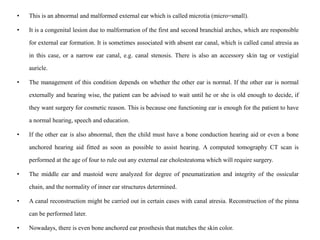
Download to read offline

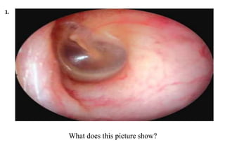

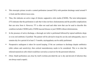
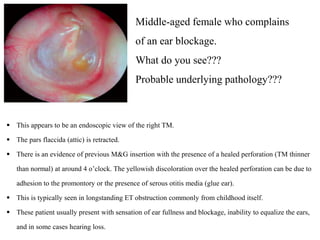
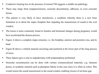
![ The normal translucent color of the TM is not present; instead it looks yellowish due to the
accumulation of fluid in the middle ear space.
The TM is retracted (handle of malleus is more acute than usual 45°) and the Prussak’s space
is retracted (a potential space just above the lateral process of the malleus).
This is consistent with middle ear effusion (MEE) or serous otitis media.
The middle ear space is an air-filled cavity that extends posteriorly to the mastoid antrum via
the aditus and is connected to the nasopharynx via the Eustachian tube (ET). The ET functions
to equalize pressure in the middle ear space and the atmospheric pressure outside.
Malfunctions occur most commonly due to mechanical obstruction, which causes a gradual
negative pressure development in the middle ear that is eventually filled with fluid.
The causes of MEE include ET dysfunction (abnormal ET or muscular movements of ET, e.g.
cleft palate, craniofacial abnormalities, mechanical obstruction, e.g. adenoids, tumors [e.g.
nasopharyngeal carcinoma (CA), very common cancer in Southeast Asia], allergic rhinitis,
and rhinosinusitis, etc.](https://image.slidesharecdn.com/earmcqppt-220831150956-db20f066/85/Image-based-ear-MCQ-pptx-5-320.jpg)
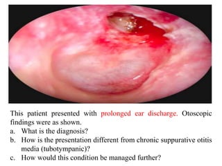
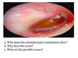
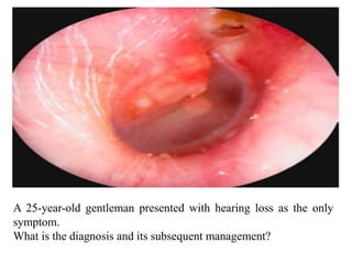
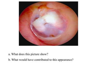
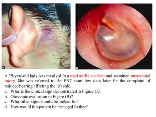
![ There is an attic (area above the anterior and posterior malleolar ligament or pars flaccida and scutum)
pocket with white flaky debris characteristic of cholesteatoma. The pars tensa appears normal.
The discharge in these patients tends to be persistent, scanty, and foul-smelling as compared to intermittent,
copious, mucopurulent, odorless discharge of CSOM (TT). Thus, the diagnosis is CSOM [atticoantral
(AA)].
Atticoantral indicates that the perforation is unsafe; that it does have cholesteatoma and the possible
complications that can arise from it. However, AA is often not used and other terms that can be used to
describe this condition include CSOM (unsafe), CSOM epithelial disease or just CSOM with
cholesteatoma.
The management of this condition is essential to prevent the possible complications if it is allowed to
expand; namely brain abscess, labyrinthitis, facial palsy, subperiosteal abscess and sigmoid sinus
thrombosis. In a small cavity and a medically unfit patient, regular cleaning will be adequate if all the
boundaries of the sac or cavity can be visualized and easily assessed with the help of a microscope.
If this is not possible, mastoid exploration and exteriorization of cholesteatoma should be carried out.
This can be divided into an atticotomy, modified radical mastoidectomy (MRM) or combined approach
mastoidectomy depending on the severity and intraoperative findings.](https://image.slidesharecdn.com/earmcqppt-220831150956-db20f066/85/Image-based-ear-MCQ-pptx-29-320.jpg)
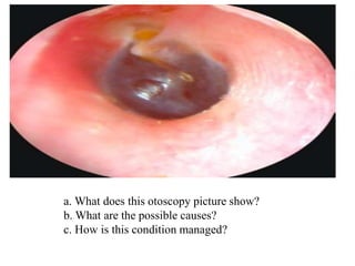
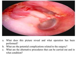
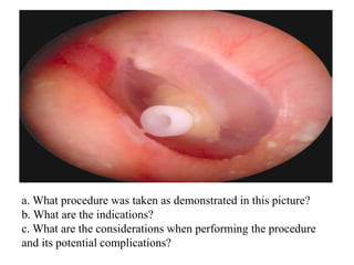
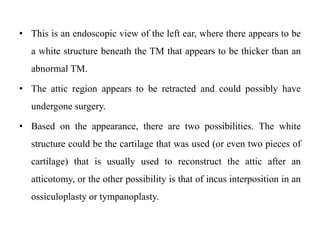
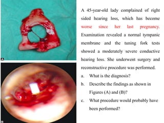
![• This picture reveals a post MRM cavity where mastoid, attic and the external ear were operated and joined together to form a single
chamber.
• It is usually performed to exteriorize the cholesteatoma to render the ‘unsafe’ ear into ‘safe ear’. The middle ear cavity is usually closed
off by laying the TM over the medial aspect of middle ear space since the scutum has been removed.
• This is important as an open middle ear cavity with its respiratory mucosa (goblets cells and other mucous secreting glands) will cause a
discharging mastoid cavity. In
• MRM, the facial ridge is lowered sufficiently to prevent ‘sump’ effect in the mastoid bowl.
• Complications of this surgery include injury to the dura [with or without cerebrospinal fluid (CSF) leak], labyrinth, facial nerve and
sigmoid sinus.
• There can also be a profound hearing loss that is believed due to the drills used during the surgery.
• Other procedures include atticotomy and combined approach tympanoplasty. In atticotomy, disease that is limited to the attic is removed
by removing the scutum and exteriorizing the attic pocket. This can be reconstructed with a cartilage grafting the same sitting if the
surgeon is confident that all the cholesteatoma has been removed or perform later in the second sitting. Combined approach
tympanoplasty or canal wall up procedure is usually performed in children, with well-pneumatized mastoid and in patients, who
complaint and understand that this is a two-stage procedure.
• This procedure prevents a mastoid cavity and its complications (discharging ear, regular ear toileting for life, vertigo and imbalance in
cold winds) and retains a normal external ear canal. It can be technically more challenging as a posterior tympanotomy (boundary: facial
nerve, chorda tympani and buttress) is performed together with the elevation of a tympanomeatal flap to remove the disease.
• As recurrence rate is high, a second look is vital.](https://image.slidesharecdn.com/earmcqppt-220831150956-db20f066/85/Image-based-ear-MCQ-pptx-33-320.jpg)

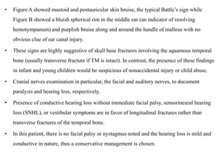


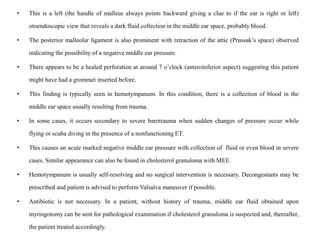
This endoscopic view shows a normal left ear canal and tympanic membrane. The ear canal is free from earwax due to outward epithelial migration from the umbo. The tympanic membrane appears pearly and translucent with the malleus handle at a 45 degree angle. This indicates a healthy, self-cleansing ear canal without need for cotton buds.


