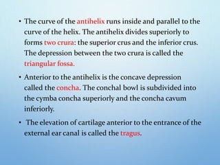
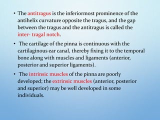
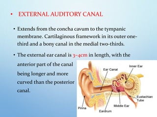
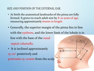
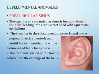
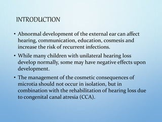
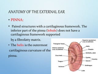
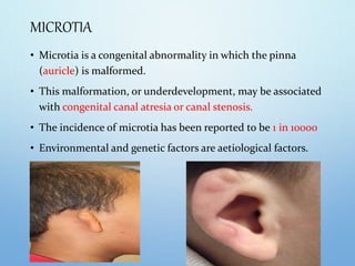
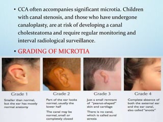
SlideShare a Scribd company logo
SCOTT BROWN LEARNING 2020
MICROTIA AND EXTERNAL EAR ABNORMALITIES
VOLUME-II
CHAPTER- 16
PAGES- 165-172
MODERATOR: Dr. Arul sundaresh kumar MS
PRESENTER: Dr. E.Selvapriya MS PG
INTRODUCTION
• Abnormal development of the external ear can affect
hearing, communication, education, cosmesis and
increase the risk of recurrent infections.
• While many children with unilateral hearing loss
develop normally, some may have negative effects upon
development.
• The management of the cosmetic consequences of
microtia should not occur in isolation, but in
combination with the rehabilitation of hearing loss due
to congenital canal atresia (CCA).
ANATOMY OF THE EXTERNAL EAR
• PINNA:
• Paired structures with a cartilaginous framework. The
inferior part of the pinna (lobule) does not have a
cartilaginous framework supported
by a fibrofatty matrix.
• The helix is the outermost
cartilaginous curvature of the
pinna.
• The curve of the antihelix runs inside and parallel to the
curve of the helix. The antihelix divides superiorly to
forms two crura: the superior crus and the inferior crus.
The depression between the two crura is called the
triangular fossa.
• Anterior to the antihelix is the concave depression
called the concha. The conchal bowl is subdivided into
the cymba concha superiorly and the concha cavum
inferiorly.
• The elevation of cartilage anterior to the entrance of the
external ear canal is called the tragus.
• The antitragus is the inferiormost prominence of the
antihelix curvature opposite the tragus, and the gap
between the tragus and the antitragus is called the
inter- tragal notch.
• The cartilage of the pinna is continuous with the
cartilaginous ear canal, thereby fixing it to the temporal
bone along with muscles and ligaments (anterior,
posterior and superior ligaments).
• The intrinsic muscles of the pinna are poorly
developed; the extrinsic muscles (anterior, posterior
and superior) may be well developed in some
individuals.
• EXTERNAL AUDITORY CANAL
• Extends from the concha cavum to the tympanic
membrane. Cartilaginous framework in its outer one-
third and a bony canal in the medial two-thirds.
• The external ear canal is 3–4cm in length, with the
anterior part of the canal
being longer and more
curved than the posterior
canal.
SIZE AND POSITION OF THE EXTERNAL EAR
• At birth the anatomical landmarks of the pinna are fully
formed. It grows to reach adult size by 8–10 years of age,
measuring approximately 60mm in length.
• Generally, the superior margin of the pinna lies in line
with the eyebrow, and the lower limit of the lobule is in
line with the base of the nasal
septal columella.
• It is inclined approximately
15–20° posteriorly and
protrudes 15–20mm from the scalp.
DEVELOPMENTAL ANOMALIES
• PREAURICULAR SINUS
• The opening of a preauricular sinus is found in front of
the helix, leading into a sinus tract lined with squamous
epithelium .
• The tract lies in the subcutaneous tissues lateral to the
temporalis fascia superiorly and
parotid fascia inferiorly, and with a
tortuous and branching course.
• The terminal portion of the tract is
adherent to the cartilage of the helix.
• Preauricular sinuses are postulated to develop from
defective or incomplete fusion of the hillocks of His
during auricular embryogenesis.
• An alternative theory suggests that isolated or localized
folding of ectoderm during auricular embryogenesis is
the cause of preauricular sinus formation.
• Preauricular sinuses may be associated with several
syndromes, most notably branchio-oto-renal syndrome.
• These patients often investigated using renal ultrasound
to detect concurrent renal abnormalities.
LEFT PREAURICULAR SINUS WITH OVERLYING INFLAMMATION
OF THE SKIN
INDICATIONS FOR SURGERY
• Frequency and severity of infective episodes,
• Chronicity of sinus discharge
• Development of unsightly overlying skin inflammation
• Incision and drainage should be avoided in acute
infections due to the risk of sinus disruption and
seeding, and less likely facial nerve injury.
• Acute infections are best treated with intravenous
antibiotics and in severe cases needle aspiration with
microbiology of the aspirate.
SURGICAL TECHNIQUE
• Several techniques have been suggested like curettage,
microdissection and wide local excision.
• When selecting a surgical technique, the fact that the
sinus may branch extensively and be adherent to the
pinna cartilage must be taken into consideration.
• Curettage is no longer routinely performed due to high
recurrence rates and unsightly scarring.
• Microdissection techniques rely upon identification of
the extent of the sinus and its branches, with lacrimal
probes and methylene blue being used.
• The ‘supra-auricular’ approach involves identification of
the plane of the temporalis fascia and dissection of the
soft tissue between this plane and the helix of the pinna,
remaining posterior to the parotid fascia, and without a
formal attempt to identify the extent of the sinus and its
branches.
• The resultant wide local excision is considered to give
the lowest recurrence rate.
• Care must be taken to avoid facial nerve injury in
revision cases and when skin and soft-tissue
inflammation is extensive.
• PREAURICULAR APPENDAGES
• Preauricular appendages result from abnormalities of
embryogenesis of the external ear and may be unilateral
or bilateral, solitary or multiple.
• Fibrofatty core and often contain a cartilaginous
component. They are usually found along a line drawn
from the tragus to the angle of the mandible, reflecting
origin from the first branchial arch.
• An increased risk of associated permanent hearing loss
has been suggested in infants with preauricular
appendages.
INDICATIONS FOR SURGERY
• To improve cosmesis and facial symmetry.
• Usually undertaken after 1 year of age.
• Preauricular appendages associated with microtia may
be operated upon to improve facial symmetry while
waiting for the child to reach an age when ear
reconstruction is considered.
SURGICAL TECHNIQUE
• Appendages may contain a deeply extending
cartilaginous core that may be more extensive than
apparent from examination of the external component.
• Care must therefore be taken when resecting the deep
components of the appendage, due to the potential for
inadvertent facial nerve injury.
• Often complete excision of the skin and soft- tissue
components is undertaken with partial resection of the
superficial part of the cartilaginous core.
MICROTIA
• Microtia is a congenital abnormality in which the pinna
(auricle) is malformed.
• This malformation, or underdevelopment, may be associated
with congenital canal atresia or canal stenosis.
• The incidence of microtia has been reported to be 1 in 10000
• Environmental and genetic factors are aetiological factors.
• CCA often accompanies significant microtia. Children
with canal stenosis, and those who have undergone
canaloplasty, are at risk of developing a canal
cholesteatoma and require regular monitoring and
interval radiological surveillance.
• GRADING OF MICROTIA
CLINICAL ASSESSMENT
• Unilateral or bilateral microtia
• Size of the microtic ear and its location
• Development of the contralateral pinna in unilateral microtia
• Hairline
• Site and size of a remnant lobule
• Presence or absence of normal skin separating the remnants
of the microtic ear – this may suggest a superficial course of
the facial nerve and caution is exercised during
reconstruction to avoid damage to the superficially placed
facial nerve
• Presence of a stenotic ear canal
• Growth and development of the mastoid bone
• Space between the temporomandibular joint (TMJ) and
the mastoid tip
• Presence or absence of facial asymmetry (e.g,
Hemifacial microsomia in goldenhar syndrome)
• Facial nerve function.
AUDIOLOGICAL ASSESSMENT
• Children with microtia and canal stenosis/atresia will
usually have a conductive hearing loss (CHL).
• A small proportion of these children may also have an
underlying sensorineural hearing loss (SNHL).
• Management of bilateral CHL in children with bilateral
CCA is of fundamental importance to normal speech
and language development.
MANAGEMENT OF SIGNIFICANT MICROTIA
• The multidisciplinary team delivering care should be able to
offer both hearing rehabilitation and all types of pinna
reconstruction.
• Most commonly, children with microtia have three options
regarding cosmesis:
No intervention,
Autologous ear reconstruction using cartilage,
Bone-anchored auricular prosthesis (BAAP).
• A proportion of children and young people with significant
dysplasia will prefer to keep their ‘special ear’.
AUTOLOGOUS EAR RECONSTRUCTION
• Construction of a cartilaginous framework,
• Soft-tissue cover and projection of the reconstructed pinna.
• Autologous rib cartilage is harvested and used to carve the
new cartilage framework.
• Timing of surgery will depend on availability of requisite
amount of cartilage (8–10 years), and the child’s ability to
cooperate with the demands of surgery.
• The risk of resorption and extrusion is lower in autologous
cartilaginous frameworks as compared to frameworks made
from artificial materials.
BONE-ANCHORED AURICULAR PROSTHESIS (BAAP)
• A BAAP necessitates removal of the vestigial ear (including
the lobule), therefore precluding subsequent autologous ear
reconstruction if the recipient was dissatisfied with the
BAAP. The surgeon and prosthetist must agree the position of
the two osseointegrating fixtures.
• Reasons for opting for a BAAP include
• Patient preference,
• Failed autologous ear reconstruction and
• Significant comorbidities precluding autologous ear
reconstruction.
• The cosmetic outcome of an auricular prosthesis is
closely linked to the experience of the prosthetist, with
an experienced prosthetist able to add fine contouring
and closely match skin colour.
• However, despite this, some patients prefer a
reconstructed pinna that is made from their own
tissues.
• Recurrent soft-tissue inflammation ,
traumatic fixture loss may complicate
the use of percutaneous abutments,
as encountered in percutaneous BAHA.
TREACHER COLLIN
SYNDROME
GOLDENHAR
SYNDROME
CHARGE SYNDROME
THANK YOU!

More Related Content

What's hot

Phonosurgery
PhonosurgeryPhonosurgery
Phonosurgery
Vaibhav Lahane
 
Phonosurgery
PhonosurgeryPhonosurgery
Phonosurgery
Yousuf Choudhury
 
Canal Wall Down Mastoidectomy(MRM)
Canal Wall Down Mastoidectomy(MRM)Canal Wall Down Mastoidectomy(MRM)
Canal Wall Down Mastoidectomy(MRM)
Kanu Saha
 
Biofilms IN ENT
Biofilms IN ENTBiofilms IN ENT
Biofilms IN ENT
Utpal Sarmah
 
Hadad.bassagasteguy flap
Hadad.bassagasteguy flap Hadad.bassagasteguy flap
Hadad.bassagasteguy flap
Chandra Veer Suryavanshi
 
Approach to a patient with cholesteatoma
Approach to a patient with cholesteatoma Approach to a patient with cholesteatoma
Approach to a patient with cholesteatoma
Dr Safika Zaman
 
Physiology Of Voice Production Its Disorders And Management.pptx
Physiology Of Voice Production Its Disorders And Management.pptxPhysiology Of Voice Production Its Disorders And Management.pptx
Physiology Of Voice Production Its Disorders And Management.pptx
Kunal Jha
 
surgical approaches to frontal sinus ppt
surgical approaches to frontal sinus pptsurgical approaches to frontal sinus ppt
surgical approaches to frontal sinus ppt
Vaibhav Lahane
 
Mucosal folds of the middle ear
Mucosal folds of the middle earMucosal folds of the middle ear
Mucosal folds of the middle ear
Dʀ Smruti Ranjan Samal
 
SUBGLOTTIC STENOSIS.pptx
SUBGLOTTIC STENOSIS.pptxSUBGLOTTIC STENOSIS.pptx
SUBGLOTTIC STENOSIS.pptx
shankarnaikvarthya
 
MIDDLE EAR ANOMALIES
MIDDLE EAR ANOMALIESMIDDLE EAR ANOMALIES
MIDDLE EAR ANOMALIES
JINORAJ RAJAN
 
Glomus Tumour and its Approaches
Glomus Tumour and its ApproachesGlomus Tumour and its Approaches
Glomus Tumour and its Approaches
Lady Hardinge Medical College
 
Iatrogenic facial nerve injury
Iatrogenic facial nerve injury Iatrogenic facial nerve injury
Iatrogenic facial nerve injury
Mamoon Ameen
 
Inner ear malformations and Implantation
Inner ear malformations and ImplantationInner ear malformations and Implantation
Inner ear malformations and Implantation
Utkal Mishra
 
Spaces of middle ear and their surgical importance
Spaces of middle ear  and their surgical importanceSpaces of middle ear  and their surgical importance
Spaces of middle ear and their surgical importance
Dr Soumya Singh
 
Laryngeal framework surgery
Laryngeal framework  surgeryLaryngeal framework  surgery
Laryngeal framework surgery
Dr Safika Zaman
 
Coblation in ent
Coblation in entCoblation in ent
Coblation in ent
Dr. Pruthvi Raj S
 
Superior Semicircular Canal Dehiscence Syndrome
Superior Semicircular Canal Dehiscence SyndromeSuperior Semicircular Canal Dehiscence Syndrome
Superior Semicircular Canal Dehiscence Syndrome
Ade Wijaya
 
Flaps in otolaryngology
Flaps in otolaryngology Flaps in otolaryngology
Flaps in otolaryngology
shivjee Prashant
 

What's hot (20)

Phonosurgery
PhonosurgeryPhonosurgery
Phonosurgery
 
Phonosurgery
PhonosurgeryPhonosurgery
Phonosurgery
 
Canal Wall Down Mastoidectomy(MRM)
Canal Wall Down Mastoidectomy(MRM)Canal Wall Down Mastoidectomy(MRM)
Canal Wall Down Mastoidectomy(MRM)
 
Biofilms IN ENT
Biofilms IN ENTBiofilms IN ENT
Biofilms IN ENT
 
Hadad.bassagasteguy flap
Hadad.bassagasteguy flap Hadad.bassagasteguy flap
Hadad.bassagasteguy flap
 
Microtia
MicrotiaMicrotia
Microtia
 
Approach to a patient with cholesteatoma
Approach to a patient with cholesteatoma Approach to a patient with cholesteatoma
Approach to a patient with cholesteatoma
 
Physiology Of Voice Production Its Disorders And Management.pptx
Physiology Of Voice Production Its Disorders And Management.pptxPhysiology Of Voice Production Its Disorders And Management.pptx
Physiology Of Voice Production Its Disorders And Management.pptx
 
surgical approaches to frontal sinus ppt
surgical approaches to frontal sinus pptsurgical approaches to frontal sinus ppt
surgical approaches to frontal sinus ppt
 
Mucosal folds of the middle ear
Mucosal folds of the middle earMucosal folds of the middle ear
Mucosal folds of the middle ear
 
SUBGLOTTIC STENOSIS.pptx
SUBGLOTTIC STENOSIS.pptxSUBGLOTTIC STENOSIS.pptx
SUBGLOTTIC STENOSIS.pptx
 
MIDDLE EAR ANOMALIES
MIDDLE EAR ANOMALIESMIDDLE EAR ANOMALIES
MIDDLE EAR ANOMALIES
 
Glomus Tumour and its Approaches
Glomus Tumour and its ApproachesGlomus Tumour and its Approaches
Glomus Tumour and its Approaches
 
Iatrogenic facial nerve injury
Iatrogenic facial nerve injury Iatrogenic facial nerve injury
Iatrogenic facial nerve injury
 
Inner ear malformations and Implantation
Inner ear malformations and ImplantationInner ear malformations and Implantation
Inner ear malformations and Implantation
 
Spaces of middle ear and their surgical importance
Spaces of middle ear  and their surgical importanceSpaces of middle ear  and their surgical importance
Spaces of middle ear and their surgical importance
 
Laryngeal framework surgery
Laryngeal framework  surgeryLaryngeal framework  surgery
Laryngeal framework surgery
 
Coblation in ent
Coblation in entCoblation in ent
Coblation in ent
 
Superior Semicircular Canal Dehiscence Syndrome
Superior Semicircular Canal Dehiscence SyndromeSuperior Semicircular Canal Dehiscence Syndrome
Superior Semicircular Canal Dehiscence Syndrome
 
Flaps in otolaryngology
Flaps in otolaryngology Flaps in otolaryngology
Flaps in otolaryngology
 

Similar to Microtia and ear abnormalities final

Congenital malformation of external ear and it’s management
Congenital malformation of external ear and it’s managementCongenital malformation of external ear and it’s management
Congenital malformation of external ear and it’s management
Yousuf Choudhury
 
Ear Reconstruction_084051.pptx
Ear Reconstruction_084051.pptxEar Reconstruction_084051.pptx
Ear Reconstruction_084051.pptx
drazizsaleh94
 
Prominent ears otoplasty
Prominent ears otoplastyProminent ears otoplasty
Prominent ears otoplasty
Mohammed Aljodah
 
Pediatric facial injuries
Pediatric facial injuriesPediatric facial injuries
Pediatric facial injuries
Dr.Amit kumar choudhary
 
AURICULAR Reconstruction 1.pptx
AURICULAR Reconstruction 1.pptxAURICULAR Reconstruction 1.pptx
AURICULAR Reconstruction 1.pptx
Maheen Fatima
 
CONGENITAL MALFORATION OF EAR AND ITS MANAGEMENT
CONGENITAL MALFORATION OF EAR AND ITS MANAGEMENTCONGENITAL MALFORATION OF EAR AND ITS MANAGEMENT
CONGENITAL MALFORATION OF EAR AND ITS MANAGEMENT
abhijeet89singh
 
Lesions of the temporal bone & petrous ppt
Lesions of the  temporal bone & petrous  pptLesions of the  temporal bone & petrous  ppt
Lesions of the temporal bone & petrous ppt
DEBKUMAR BISWAS
 
maxillarysinus-170705134531 [Autosaved].pptx
maxillarysinus-170705134531 [Autosaved].pptxmaxillarysinus-170705134531 [Autosaved].pptx
maxillarysinus-170705134531 [Autosaved].pptx
AniketChoudhary65
 
Modified coronoid process grafts combined with sagittal split osteotomy for t...
Modified coronoid process grafts combined with sagittal split osteotomy for t...Modified coronoid process grafts combined with sagittal split osteotomy for t...
Modified coronoid process grafts combined with sagittal split osteotomy for t...Indian dental academy
 
Syndromic cranial synostosis
Syndromic cranial synostosisSyndromic cranial synostosis
Syndromic cranial synostosis
Jamil Kifayatullah
 
Choanal atresia
Choanal atresiaChoanal atresia
Choanal atresia
Brian Wells, MD, MS, MPH
 
Maxillary sinus
Maxillary sinusMaxillary sinus
Maxillary sinus
sauvik2014
 
Presentation
PresentationPresentation
Presentation
Mohammed Sayed
 
Pdf clcp
Pdf clcpPdf clcp
Pdf clcp
sourav chandra
 
Sinus Lift ppt about maxillary sinus lift
Sinus Lift ppt about maxillary sinus liftSinus Lift ppt about maxillary sinus lift
Sinus Lift ppt about maxillary sinus lift
senthilnathanhl
 
Case prsentation tmj ankylosis
Case prsentation tmj ankylosisCase prsentation tmj ankylosis
Case prsentation tmj ankylosis
Kanokporn Tungsakul
 
The Orbit
The OrbitThe Orbit
Craniofacial Microsomia and Hemifacial Atrophy
Craniofacial Microsomia and Hemifacial AtrophyCraniofacial Microsomia and Hemifacial Atrophy
Craniofacial Microsomia and Hemifacial Atrophy
Satish Kumar
 
Congenital Benign Neck masses
Congenital Benign Neck masses Congenital Benign Neck masses
Congenital Benign Neck masses
Haya Taha
 
CYSTS OF THE JAWS Part II
CYSTS OF THE JAWS Part IICYSTS OF THE JAWS Part II
CYSTS OF THE JAWS Part II
Abhishek PT
 

Similar to Microtia and ear abnormalities final (20)

Congenital malformation of external ear and it’s management
Congenital malformation of external ear and it’s managementCongenital malformation of external ear and it’s management
Congenital malformation of external ear and it’s management
 
Ear Reconstruction_084051.pptx
Ear Reconstruction_084051.pptxEar Reconstruction_084051.pptx
Ear Reconstruction_084051.pptx
 
Prominent ears otoplasty
Prominent ears otoplastyProminent ears otoplasty
Prominent ears otoplasty
 
Pediatric facial injuries
Pediatric facial injuriesPediatric facial injuries
Pediatric facial injuries
 
AURICULAR Reconstruction 1.pptx
AURICULAR Reconstruction 1.pptxAURICULAR Reconstruction 1.pptx
AURICULAR Reconstruction 1.pptx
 
CONGENITAL MALFORATION OF EAR AND ITS MANAGEMENT
CONGENITAL MALFORATION OF EAR AND ITS MANAGEMENTCONGENITAL MALFORATION OF EAR AND ITS MANAGEMENT
CONGENITAL MALFORATION OF EAR AND ITS MANAGEMENT
 
Lesions of the temporal bone & petrous ppt
Lesions of the  temporal bone & petrous  pptLesions of the  temporal bone & petrous  ppt
Lesions of the temporal bone & petrous ppt
 
maxillarysinus-170705134531 [Autosaved].pptx
maxillarysinus-170705134531 [Autosaved].pptxmaxillarysinus-170705134531 [Autosaved].pptx
maxillarysinus-170705134531 [Autosaved].pptx
 
Modified coronoid process grafts combined with sagittal split osteotomy for t...
Modified coronoid process grafts combined with sagittal split osteotomy for t...Modified coronoid process grafts combined with sagittal split osteotomy for t...
Modified coronoid process grafts combined with sagittal split osteotomy for t...
 
Syndromic cranial synostosis
Syndromic cranial synostosisSyndromic cranial synostosis
Syndromic cranial synostosis
 
Choanal atresia
Choanal atresiaChoanal atresia
Choanal atresia
 
Maxillary sinus
Maxillary sinusMaxillary sinus
Maxillary sinus
 
Presentation
PresentationPresentation
Presentation
 
Pdf clcp
Pdf clcpPdf clcp
Pdf clcp
 
Sinus Lift ppt about maxillary sinus lift
Sinus Lift ppt about maxillary sinus liftSinus Lift ppt about maxillary sinus lift
Sinus Lift ppt about maxillary sinus lift
 
Case prsentation tmj ankylosis
Case prsentation tmj ankylosisCase prsentation tmj ankylosis
Case prsentation tmj ankylosis
 
The Orbit
The OrbitThe Orbit
The Orbit
 
Craniofacial Microsomia and Hemifacial Atrophy
Craniofacial Microsomia and Hemifacial AtrophyCraniofacial Microsomia and Hemifacial Atrophy
Craniofacial Microsomia and Hemifacial Atrophy
 
Congenital Benign Neck masses
Congenital Benign Neck masses Congenital Benign Neck masses
Congenital Benign Neck masses
 
CYSTS OF THE JAWS Part II
CYSTS OF THE JAWS Part IICYSTS OF THE JAWS Part II
CYSTS OF THE JAWS Part II
 

More from Arul Lakshmanaperumal

Tinnitus
Tinnitus Tinnitus
Otitis media with effusion
Otitis media with effusion Otitis media with effusion
Otitis media with effusion
Arul Lakshmanaperumal
 
Jorrp
JorrpJorrp
Hearing tests in children
Hearing tests in children Hearing tests in children
Hearing tests in children
Arul Lakshmanaperumal
 
Issnhl final
Issnhl finalIssnhl final
Issnhl final
Arul Lakshmanaperumal
 
Ototoxicity
Ototoxicity Ototoxicity
Ototoxicity
Arul Lakshmanaperumal
 
Adenoids and adenoidectomy
Adenoids and adenoidectomyAdenoids and adenoidectomy
Adenoids and adenoidectomy
Arul Lakshmanaperumal
 
Acquired laryngotracheal stenosis
Acquired laryngotracheal stenosisAcquired laryngotracheal stenosis
Acquired laryngotracheal stenosis
Arul Lakshmanaperumal
 
Vestibular system
Vestibular system Vestibular system
Vestibular system
Arul Lakshmanaperumal
 
Neonatal nasal obstruction final
Neonatal nasal obstruction finalNeonatal nasal obstruction final
Neonatal nasal obstruction final
Arul Lakshmanaperumal
 
Acute larynx infections , congenital cause copy
Acute larynx infections , congenital cause copyAcute larynx infections , congenital cause copy
Acute larynx infections , congenital cause copy
Arul Lakshmanaperumal
 
Pediatric trachostomy
Pediatric trachostomyPediatric trachostomy
Pediatric trachostomy
Arul Lakshmanaperumal
 
Cervicofacial infection
Cervicofacial infection Cervicofacial infection
Cervicofacial infection
Arul Lakshmanaperumal
 
Aom,ome,com copy
Aom,ome,com copyAom,ome,com copy
Aom,ome,com copy
Arul Lakshmanaperumal
 
Perinatal airway management haemangiomas and vascular malformations
Perinatal   airway   management  haemangiomas   and   vascular   malformationsPerinatal   airway   management  haemangiomas   and   vascular   malformations
Perinatal airway management haemangiomas and vascular malformations
Arul Lakshmanaperumal
 
Lacrimal disorders in children
Lacrimal disorders in children Lacrimal disorders in children
Lacrimal disorders in children
Arul Lakshmanaperumal
 
Examination of ear.
Examination of ear. Examination of ear.
Examination of ear.
Arul Lakshmanaperumal
 
Acute otitis media final
Acute otitis media finalAcute otitis media final
Acute otitis media final
Arul Lakshmanaperumal
 
Vesti. migraine, neuronitis, ssc dehiscence
Vesti. migraine, neuronitis, ssc dehiscenceVesti. migraine, neuronitis, ssc dehiscence
Vesti. migraine, neuronitis, ssc dehiscence
Arul Lakshmanaperumal
 
Inner ear scott brown full
Inner ear scott brown fullInner ear scott brown full
Inner ear scott brown full
Arul Lakshmanaperumal
 

More from Arul Lakshmanaperumal (20)

Tinnitus
Tinnitus Tinnitus
Tinnitus
 
Otitis media with effusion
Otitis media with effusion Otitis media with effusion
Otitis media with effusion
 
Jorrp
JorrpJorrp
Jorrp
 
Hearing tests in children
Hearing tests in children Hearing tests in children
Hearing tests in children
 
Issnhl final
Issnhl finalIssnhl final
Issnhl final
 
Ototoxicity
Ototoxicity Ototoxicity
Ototoxicity
 
Adenoids and adenoidectomy
Adenoids and adenoidectomyAdenoids and adenoidectomy
Adenoids and adenoidectomy
 
Acquired laryngotracheal stenosis
Acquired laryngotracheal stenosisAcquired laryngotracheal stenosis
Acquired laryngotracheal stenosis
 
Vestibular system
Vestibular system Vestibular system
Vestibular system
 
Neonatal nasal obstruction final
Neonatal nasal obstruction finalNeonatal nasal obstruction final
Neonatal nasal obstruction final
 
Acute larynx infections , congenital cause copy
Acute larynx infections , congenital cause copyAcute larynx infections , congenital cause copy
Acute larynx infections , congenital cause copy
 
Pediatric trachostomy
Pediatric trachostomyPediatric trachostomy
Pediatric trachostomy
 
Cervicofacial infection
Cervicofacial infection Cervicofacial infection
Cervicofacial infection
 
Aom,ome,com copy
Aom,ome,com copyAom,ome,com copy
Aom,ome,com copy
 
Perinatal airway management haemangiomas and vascular malformations
Perinatal   airway   management  haemangiomas   and   vascular   malformationsPerinatal   airway   management  haemangiomas   and   vascular   malformations
Perinatal airway management haemangiomas and vascular malformations
 
Lacrimal disorders in children
Lacrimal disorders in children Lacrimal disorders in children
Lacrimal disorders in children
 
Examination of ear.
Examination of ear. Examination of ear.
Examination of ear.
 
Acute otitis media final
Acute otitis media finalAcute otitis media final
Acute otitis media final
 
Vesti. migraine, neuronitis, ssc dehiscence
Vesti. migraine, neuronitis, ssc dehiscenceVesti. migraine, neuronitis, ssc dehiscence
Vesti. migraine, neuronitis, ssc dehiscence
 
Inner ear scott brown full
Inner ear scott brown fullInner ear scott brown full
Inner ear scott brown full
 

Recently uploaded

Top 10 Best Ayurvedic Kidney Stone Syrups in India
Top 10 Best Ayurvedic Kidney Stone Syrups in IndiaTop 10 Best Ayurvedic Kidney Stone Syrups in India
Top 10 Best Ayurvedic Kidney Stone Syrups in India
Swastik Ayurveda
 
Top-Vitamin-Supplement-Brands-in-India List
Top-Vitamin-Supplement-Brands-in-India ListTop-Vitamin-Supplement-Brands-in-India List
Top-Vitamin-Supplement-Brands-in-India List
SwisschemDerma
 
Aortic Association CBL Pilot April 19 – 20 Bern
Aortic Association CBL Pilot April 19 – 20 BernAortic Association CBL Pilot April 19 – 20 Bern
Aortic Association CBL Pilot April 19 – 20 Bern
suvadeepdas911
 
Non-respiratory Functions of the Lungs.pdf
Non-respiratory Functions of the Lungs.pdfNon-respiratory Functions of the Lungs.pdf
Non-respiratory Functions of the Lungs.pdf
MedicoseAcademics
 
Physiology of Special Chemical Sensation of Taste
Physiology of Special Chemical Sensation of TastePhysiology of Special Chemical Sensation of Taste
Physiology of Special Chemical Sensation of Taste
MedicoseAcademics
 
Ozempic: Preoperative Management of Patients on GLP-1 Receptor Agonists
Ozempic: Preoperative Management of Patients on GLP-1 Receptor Agonists  Ozempic: Preoperative Management of Patients on GLP-1 Receptor Agonists
Ozempic: Preoperative Management of Patients on GLP-1 Receptor Agonists
Saeid Safari
 
Novas diretrizes da OMS para os cuidados perinatais de mais qualidade
Novas diretrizes da OMS para os cuidados perinatais de mais qualidadeNovas diretrizes da OMS para os cuidados perinatais de mais qualidade
Novas diretrizes da OMS para os cuidados perinatais de mais qualidade
Prof. Marcus Renato de Carvalho
 
Physiology of Chemical Sensation of smell.pdf
Physiology of Chemical Sensation of smell.pdfPhysiology of Chemical Sensation of smell.pdf
Physiology of Chemical Sensation of smell.pdf
MedicoseAcademics
 
Top Effective Soaps for Fungal Skin Infections in India
Top Effective Soaps for Fungal Skin Infections in IndiaTop Effective Soaps for Fungal Skin Infections in India
Top Effective Soaps for Fungal Skin Infections in India
SwisschemDerma
 
Thyroid Gland- Gross Anatomy by Dr. Rabia Inam Gandapore.pptx
Thyroid Gland- Gross Anatomy by Dr. Rabia Inam Gandapore.pptxThyroid Gland- Gross Anatomy by Dr. Rabia Inam Gandapore.pptx
Thyroid Gland- Gross Anatomy by Dr. Rabia Inam Gandapore.pptx
Dr. Rabia Inam Gandapore
 
Chapter 11 Nutrition and Chronic Diseases.pptx
Chapter 11 Nutrition and Chronic Diseases.pptxChapter 11 Nutrition and Chronic Diseases.pptx
Chapter 11 Nutrition and Chronic Diseases.pptx
Earlene McNair
 
Phone Us ❤8107221448❤ #ℂall #gIRLS In Dehradun By Dehradun @ℂall @Girls Hotel...
Phone Us ❤8107221448❤ #ℂall #gIRLS In Dehradun By Dehradun @ℂall @Girls Hotel...Phone Us ❤8107221448❤ #ℂall #gIRLS In Dehradun By Dehradun @ℂall @Girls Hotel...
Phone Us ❤8107221448❤ #ℂall #gIRLS In Dehradun By Dehradun @ℂall @Girls Hotel...
chandankumarsmartiso
 
A Classical Text Review on Basavarajeeyam
A Classical Text Review on BasavarajeeyamA Classical Text Review on Basavarajeeyam
A Classical Text Review on Basavarajeeyam
Dr. Jyothirmai Paindla
 
Superficial & Deep Fascia of the NECK.pptx
Superficial & Deep Fascia of the NECK.pptxSuperficial & Deep Fascia of the NECK.pptx
Superficial & Deep Fascia of the NECK.pptx
Dr. Rabia Inam Gandapore
 
Netter's Atlas of Human Anatomy 7.ed.pdf
Netter's Atlas of Human Anatomy 7.ed.pdfNetter's Atlas of Human Anatomy 7.ed.pdf
Netter's Atlas of Human Anatomy 7.ed.pdf
BrissaOrtiz3
 
Light House Retreats: Plant Medicine Retreat Europe
Light House Retreats: Plant Medicine Retreat EuropeLight House Retreats: Plant Medicine Retreat Europe
Light House Retreats: Plant Medicine Retreat Europe
Lighthouse Retreat
 
Basavarajeeyam - Ayurvedic heritage book of Andhra pradesh
Basavarajeeyam - Ayurvedic heritage book of Andhra pradeshBasavarajeeyam - Ayurvedic heritage book of Andhra pradesh
Basavarajeeyam - Ayurvedic heritage book of Andhra pradesh
Dr. Madduru Muni Haritha
 
Integrating Ayurveda into Parkinson’s Management: A Holistic Approach
Integrating Ayurveda into Parkinson’s Management: A Holistic ApproachIntegrating Ayurveda into Parkinson’s Management: A Holistic Approach
Integrating Ayurveda into Parkinson’s Management: A Holistic Approach
Ayurveda ForAll
 
Efficacy of Avartana Sneha in Ayurveda
Efficacy of Avartana Sneha in AyurvedaEfficacy of Avartana Sneha in Ayurveda
Efficacy of Avartana Sneha in Ayurveda
Dr. Jyothirmai Paindla
 
#cALL# #gIRLS# In Dehradun ꧁❤8107221448❤꧂#cALL# #gIRLS# Service In Dehradun W...
#cALL# #gIRLS# In Dehradun ꧁❤8107221448❤꧂#cALL# #gIRLS# Service In Dehradun W...#cALL# #gIRLS# In Dehradun ꧁❤8107221448❤꧂#cALL# #gIRLS# Service In Dehradun W...
#cALL# #gIRLS# In Dehradun ꧁❤8107221448❤꧂#cALL# #gIRLS# Service In Dehradun W...
chandankumarsmartiso
 

Recently uploaded (20)

Top 10 Best Ayurvedic Kidney Stone Syrups in India
Top 10 Best Ayurvedic Kidney Stone Syrups in IndiaTop 10 Best Ayurvedic Kidney Stone Syrups in India
Top 10 Best Ayurvedic Kidney Stone Syrups in India
 
Top-Vitamin-Supplement-Brands-in-India List
Top-Vitamin-Supplement-Brands-in-India ListTop-Vitamin-Supplement-Brands-in-India List
Top-Vitamin-Supplement-Brands-in-India List
 
Aortic Association CBL Pilot April 19 – 20 Bern
Aortic Association CBL Pilot April 19 – 20 BernAortic Association CBL Pilot April 19 – 20 Bern
Aortic Association CBL Pilot April 19 – 20 Bern
 
Non-respiratory Functions of the Lungs.pdf
Non-respiratory Functions of the Lungs.pdfNon-respiratory Functions of the Lungs.pdf
Non-respiratory Functions of the Lungs.pdf
 
Physiology of Special Chemical Sensation of Taste
Physiology of Special Chemical Sensation of TastePhysiology of Special Chemical Sensation of Taste
Physiology of Special Chemical Sensation of Taste
 
Ozempic: Preoperative Management of Patients on GLP-1 Receptor Agonists
Ozempic: Preoperative Management of Patients on GLP-1 Receptor Agonists  Ozempic: Preoperative Management of Patients on GLP-1 Receptor Agonists
Ozempic: Preoperative Management of Patients on GLP-1 Receptor Agonists
 
Novas diretrizes da OMS para os cuidados perinatais de mais qualidade
Novas diretrizes da OMS para os cuidados perinatais de mais qualidadeNovas diretrizes da OMS para os cuidados perinatais de mais qualidade
Novas diretrizes da OMS para os cuidados perinatais de mais qualidade
 
Physiology of Chemical Sensation of smell.pdf
Physiology of Chemical Sensation of smell.pdfPhysiology of Chemical Sensation of smell.pdf
Physiology of Chemical Sensation of smell.pdf
 
Top Effective Soaps for Fungal Skin Infections in India
Top Effective Soaps for Fungal Skin Infections in IndiaTop Effective Soaps for Fungal Skin Infections in India
Top Effective Soaps for Fungal Skin Infections in India
 
Thyroid Gland- Gross Anatomy by Dr. Rabia Inam Gandapore.pptx
Thyroid Gland- Gross Anatomy by Dr. Rabia Inam Gandapore.pptxThyroid Gland- Gross Anatomy by Dr. Rabia Inam Gandapore.pptx
Thyroid Gland- Gross Anatomy by Dr. Rabia Inam Gandapore.pptx
 
Chapter 11 Nutrition and Chronic Diseases.pptx
Chapter 11 Nutrition and Chronic Diseases.pptxChapter 11 Nutrition and Chronic Diseases.pptx
Chapter 11 Nutrition and Chronic Diseases.pptx
 
Phone Us ❤8107221448❤ #ℂall #gIRLS In Dehradun By Dehradun @ℂall @Girls Hotel...
Phone Us ❤8107221448❤ #ℂall #gIRLS In Dehradun By Dehradun @ℂall @Girls Hotel...Phone Us ❤8107221448❤ #ℂall #gIRLS In Dehradun By Dehradun @ℂall @Girls Hotel...
Phone Us ❤8107221448❤ #ℂall #gIRLS In Dehradun By Dehradun @ℂall @Girls Hotel...
 
A Classical Text Review on Basavarajeeyam
A Classical Text Review on BasavarajeeyamA Classical Text Review on Basavarajeeyam
A Classical Text Review on Basavarajeeyam
 
Superficial & Deep Fascia of the NECK.pptx
Superficial & Deep Fascia of the NECK.pptxSuperficial & Deep Fascia of the NECK.pptx
Superficial & Deep Fascia of the NECK.pptx
 
Netter's Atlas of Human Anatomy 7.ed.pdf
Netter's Atlas of Human Anatomy 7.ed.pdfNetter's Atlas of Human Anatomy 7.ed.pdf
Netter's Atlas of Human Anatomy 7.ed.pdf
 
Light House Retreats: Plant Medicine Retreat Europe
Light House Retreats: Plant Medicine Retreat EuropeLight House Retreats: Plant Medicine Retreat Europe
Light House Retreats: Plant Medicine Retreat Europe
 
Basavarajeeyam - Ayurvedic heritage book of Andhra pradesh
Basavarajeeyam - Ayurvedic heritage book of Andhra pradeshBasavarajeeyam - Ayurvedic heritage book of Andhra pradesh
Basavarajeeyam - Ayurvedic heritage book of Andhra pradesh
 
Integrating Ayurveda into Parkinson’s Management: A Holistic Approach
Integrating Ayurveda into Parkinson’s Management: A Holistic ApproachIntegrating Ayurveda into Parkinson’s Management: A Holistic Approach
Integrating Ayurveda into Parkinson’s Management: A Holistic Approach
 
Efficacy of Avartana Sneha in Ayurveda
Efficacy of Avartana Sneha in AyurvedaEfficacy of Avartana Sneha in Ayurveda
Efficacy of Avartana Sneha in Ayurveda
 
#cALL# #gIRLS# In Dehradun ꧁❤8107221448❤꧂#cALL# #gIRLS# Service In Dehradun W...
#cALL# #gIRLS# In Dehradun ꧁❤8107221448❤꧂#cALL# #gIRLS# Service In Dehradun W...#cALL# #gIRLS# In Dehradun ꧁❤8107221448❤꧂#cALL# #gIRLS# Service In Dehradun W...
#cALL# #gIRLS# In Dehradun ꧁❤8107221448❤꧂#cALL# #gIRLS# Service In Dehradun W...
 

Microtia and ear abnormalities final

  • 1. SCOTT BROWN LEARNING 2020 MICROTIA AND EXTERNAL EAR ABNORMALITIES VOLUME-II CHAPTER- 16 PAGES- 165-172 MODERATOR: Dr. Arul sundaresh kumar MS PRESENTER: Dr. E.Selvapriya MS PG
  • 2. INTRODUCTION • Abnormal development of the external ear can affect hearing, communication, education, cosmesis and increase the risk of recurrent infections. • While many children with unilateral hearing loss develop normally, some may have negative effects upon development. • The management of the cosmetic consequences of microtia should not occur in isolation, but in combination with the rehabilitation of hearing loss due to congenital canal atresia (CCA).
  • 3. ANATOMY OF THE EXTERNAL EAR • PINNA: • Paired structures with a cartilaginous framework. The inferior part of the pinna (lobule) does not have a cartilaginous framework supported by a fibrofatty matrix. • The helix is the outermost cartilaginous curvature of the pinna.
  • 4. • The curve of the antihelix runs inside and parallel to the curve of the helix. The antihelix divides superiorly to forms two crura: the superior crus and the inferior crus. The depression between the two crura is called the triangular fossa. • Anterior to the antihelix is the concave depression called the concha. The conchal bowl is subdivided into the cymba concha superiorly and the concha cavum inferiorly. • The elevation of cartilage anterior to the entrance of the external ear canal is called the tragus.
  • 5. • The antitragus is the inferiormost prominence of the antihelix curvature opposite the tragus, and the gap between the tragus and the antitragus is called the inter- tragal notch. • The cartilage of the pinna is continuous with the cartilaginous ear canal, thereby fixing it to the temporal bone along with muscles and ligaments (anterior, posterior and superior ligaments). • The intrinsic muscles of the pinna are poorly developed; the extrinsic muscles (anterior, posterior and superior) may be well developed in some individuals.
  • 6. • EXTERNAL AUDITORY CANAL • Extends from the concha cavum to the tympanic membrane. Cartilaginous framework in its outer one- third and a bony canal in the medial two-thirds. • The external ear canal is 3–4cm in length, with the anterior part of the canal being longer and more curved than the posterior canal.
  • 7. SIZE AND POSITION OF THE EXTERNAL EAR • At birth the anatomical landmarks of the pinna are fully formed. It grows to reach adult size by 8–10 years of age, measuring approximately 60mm in length. • Generally, the superior margin of the pinna lies in line with the eyebrow, and the lower limit of the lobule is in line with the base of the nasal septal columella. • It is inclined approximately 15–20° posteriorly and protrudes 15–20mm from the scalp.
  • 8. DEVELOPMENTAL ANOMALIES • PREAURICULAR SINUS • The opening of a preauricular sinus is found in front of the helix, leading into a sinus tract lined with squamous epithelium . • The tract lies in the subcutaneous tissues lateral to the temporalis fascia superiorly and parotid fascia inferiorly, and with a tortuous and branching course. • The terminal portion of the tract is adherent to the cartilage of the helix.
  • 9. • Preauricular sinuses are postulated to develop from defective or incomplete fusion of the hillocks of His during auricular embryogenesis. • An alternative theory suggests that isolated or localized folding of ectoderm during auricular embryogenesis is the cause of preauricular sinus formation. • Preauricular sinuses may be associated with several syndromes, most notably branchio-oto-renal syndrome. • These patients often investigated using renal ultrasound to detect concurrent renal abnormalities.
  • 10. LEFT PREAURICULAR SINUS WITH OVERLYING INFLAMMATION OF THE SKIN
  • 11. INDICATIONS FOR SURGERY • Frequency and severity of infective episodes, • Chronicity of sinus discharge • Development of unsightly overlying skin inflammation • Incision and drainage should be avoided in acute infections due to the risk of sinus disruption and seeding, and less likely facial nerve injury. • Acute infections are best treated with intravenous antibiotics and in severe cases needle aspiration with microbiology of the aspirate.
  • 12. SURGICAL TECHNIQUE • Several techniques have been suggested like curettage, microdissection and wide local excision. • When selecting a surgical technique, the fact that the sinus may branch extensively and be adherent to the pinna cartilage must be taken into consideration. • Curettage is no longer routinely performed due to high recurrence rates and unsightly scarring. • Microdissection techniques rely upon identification of the extent of the sinus and its branches, with lacrimal probes and methylene blue being used.
  • 13. • The ‘supra-auricular’ approach involves identification of the plane of the temporalis fascia and dissection of the soft tissue between this plane and the helix of the pinna, remaining posterior to the parotid fascia, and without a formal attempt to identify the extent of the sinus and its branches. • The resultant wide local excision is considered to give the lowest recurrence rate. • Care must be taken to avoid facial nerve injury in revision cases and when skin and soft-tissue inflammation is extensive.
  • 14. • PREAURICULAR APPENDAGES • Preauricular appendages result from abnormalities of embryogenesis of the external ear and may be unilateral or bilateral, solitary or multiple. • Fibrofatty core and often contain a cartilaginous component. They are usually found along a line drawn from the tragus to the angle of the mandible, reflecting origin from the first branchial arch. • An increased risk of associated permanent hearing loss has been suggested in infants with preauricular appendages.
  • 15.
  • 16. INDICATIONS FOR SURGERY • To improve cosmesis and facial symmetry. • Usually undertaken after 1 year of age. • Preauricular appendages associated with microtia may be operated upon to improve facial symmetry while waiting for the child to reach an age when ear reconstruction is considered.
  • 17. SURGICAL TECHNIQUE • Appendages may contain a deeply extending cartilaginous core that may be more extensive than apparent from examination of the external component. • Care must therefore be taken when resecting the deep components of the appendage, due to the potential for inadvertent facial nerve injury. • Often complete excision of the skin and soft- tissue components is undertaken with partial resection of the superficial part of the cartilaginous core.
  • 18. MICROTIA • Microtia is a congenital abnormality in which the pinna (auricle) is malformed. • This malformation, or underdevelopment, may be associated with congenital canal atresia or canal stenosis. • The incidence of microtia has been reported to be 1 in 10000 • Environmental and genetic factors are aetiological factors.
  • 19. • CCA often accompanies significant microtia. Children with canal stenosis, and those who have undergone canaloplasty, are at risk of developing a canal cholesteatoma and require regular monitoring and interval radiological surveillance. • GRADING OF MICROTIA
  • 20.
  • 21. CLINICAL ASSESSMENT • Unilateral or bilateral microtia • Size of the microtic ear and its location • Development of the contralateral pinna in unilateral microtia • Hairline • Site and size of a remnant lobule • Presence or absence of normal skin separating the remnants of the microtic ear – this may suggest a superficial course of the facial nerve and caution is exercised during reconstruction to avoid damage to the superficially placed facial nerve
  • 22. • Presence of a stenotic ear canal • Growth and development of the mastoid bone • Space between the temporomandibular joint (TMJ) and the mastoid tip • Presence or absence of facial asymmetry (e.g, Hemifacial microsomia in goldenhar syndrome) • Facial nerve function.
  • 23. AUDIOLOGICAL ASSESSMENT • Children with microtia and canal stenosis/atresia will usually have a conductive hearing loss (CHL). • A small proportion of these children may also have an underlying sensorineural hearing loss (SNHL). • Management of bilateral CHL in children with bilateral CCA is of fundamental importance to normal speech and language development.
  • 24. MANAGEMENT OF SIGNIFICANT MICROTIA • The multidisciplinary team delivering care should be able to offer both hearing rehabilitation and all types of pinna reconstruction. • Most commonly, children with microtia have three options regarding cosmesis: No intervention, Autologous ear reconstruction using cartilage, Bone-anchored auricular prosthesis (BAAP). • A proportion of children and young people with significant dysplasia will prefer to keep their ‘special ear’.
  • 25. AUTOLOGOUS EAR RECONSTRUCTION • Construction of a cartilaginous framework, • Soft-tissue cover and projection of the reconstructed pinna. • Autologous rib cartilage is harvested and used to carve the new cartilage framework. • Timing of surgery will depend on availability of requisite amount of cartilage (8–10 years), and the child’s ability to cooperate with the demands of surgery. • The risk of resorption and extrusion is lower in autologous cartilaginous frameworks as compared to frameworks made from artificial materials.
  • 26.
  • 27. BONE-ANCHORED AURICULAR PROSTHESIS (BAAP) • A BAAP necessitates removal of the vestigial ear (including the lobule), therefore precluding subsequent autologous ear reconstruction if the recipient was dissatisfied with the BAAP. The surgeon and prosthetist must agree the position of the two osseointegrating fixtures. • Reasons for opting for a BAAP include • Patient preference, • Failed autologous ear reconstruction and • Significant comorbidities precluding autologous ear reconstruction.
  • 28. • The cosmetic outcome of an auricular prosthesis is closely linked to the experience of the prosthetist, with an experienced prosthetist able to add fine contouring and closely match skin colour. • However, despite this, some patients prefer a reconstructed pinna that is made from their own tissues. • Recurrent soft-tissue inflammation , traumatic fixture loss may complicate the use of percutaneous abutments, as encountered in percutaneous BAHA.
  • 29.