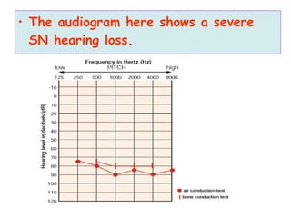
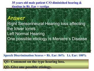
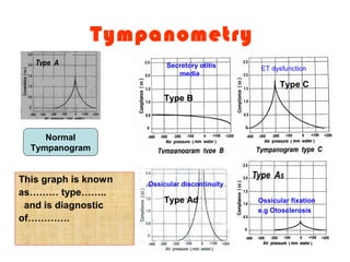
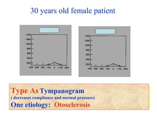
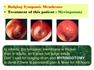
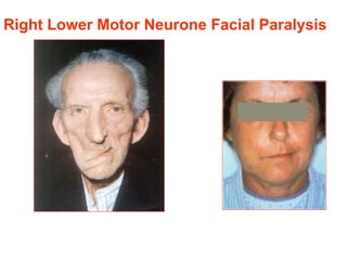
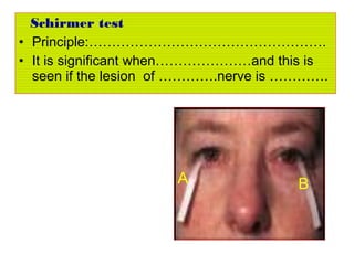
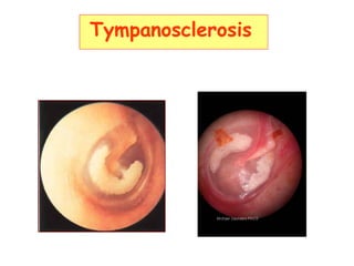
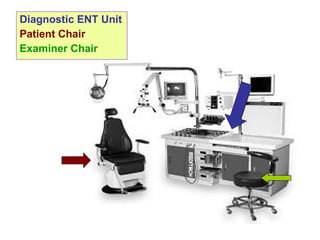
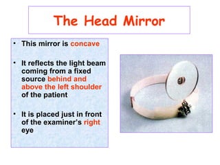
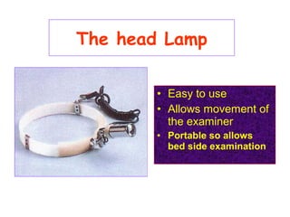
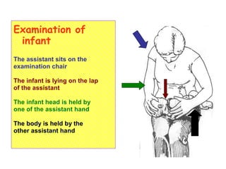
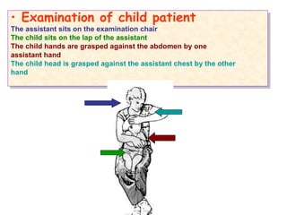
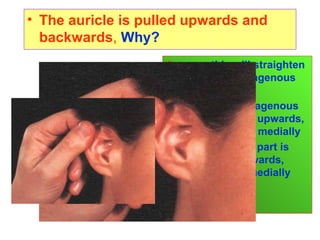
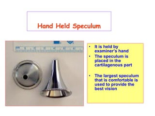
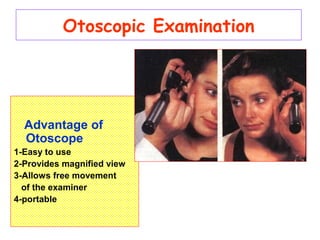
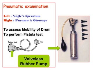
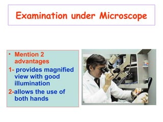
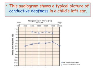
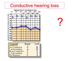
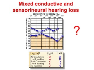
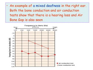
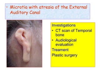
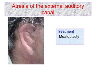
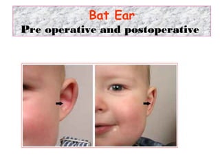
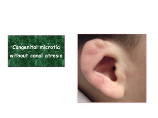
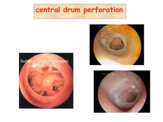
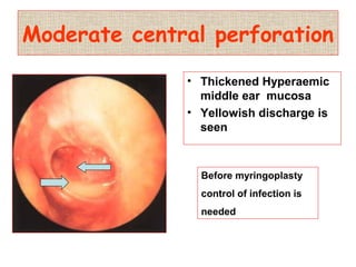
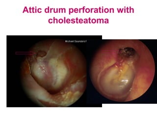
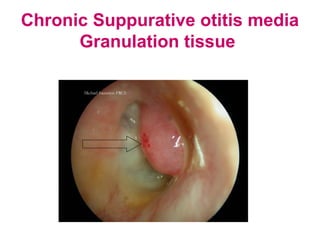
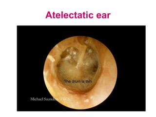
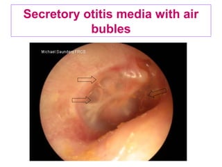
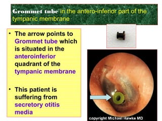
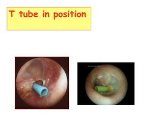
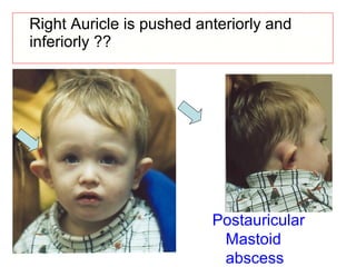
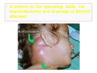
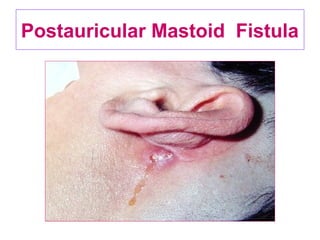
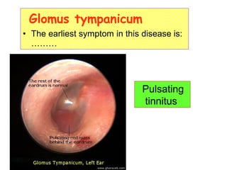
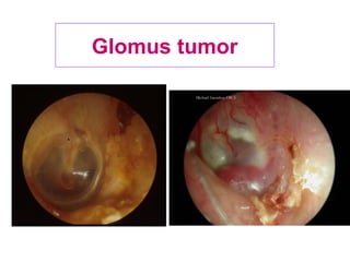
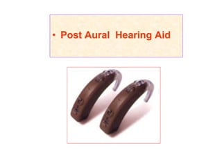
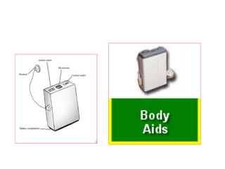
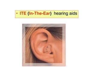
Oto-rhino-laryngologic
illustrations
Diagnostic ENT Unit
Patient Chair
Examiner Chair
• This mirror is concave
• It reflects the light beam
coming from a fixed
source behind and
above the left shoulder
of the patient
• It is placed just in front
of the examiner’s right
eye
The Head Mirror
The head Lamp
• Easy to use
• Allows movement of
the examiner
• Portable so allows
bed side examination
Examination of
infant
The assistant sits on the
examination chair
The infant is lying on the lap
of the assistant
The infant head is held by
one of the assistant hand
The body is held by the
other assistant hand
• Examination of child patient
The assistant sits on the examination chair
The child sits on the lap of the assistant
The child hands are grasped against the abdomen by one
assistant hand
The child head is grasped against the assistant chest by the other
hand
• Examination of child patient
The assistant sits on the examination chair
The child sits on the lap of the assistant
The child hands are grasped against the abdomen by one
assistant hand
The child head is grasped against the assistant chest by the other
hand
• The auricle is pulled upwards and
backwards, Why?
Answer: this will straighten
the outer cartilagenous
part because:
• The outer cartilagenous
part is directed upwards,
backwards and medially
• The inner bony part is
directed downwards,
forwards and medially
Hand Held Speculum
• It is held by
examiner’s hand
• The speculum is
placed in the
cartilagenous part
• The largest speculum
that is comfortable is
used to provide the
best vision
Otoscopic Examination
Advantage of
Otoscope
1-Easy to use
2-Provides magnified view
3-Allows free movement
of the examiner
4-portable
Pneumatic examination
Left : Seigle’s Speculum
Right : Pneumatic Otoscope
To assess Mobility of Drum
To perform Fistula test
Valveless
Rubber Pump
Examination under Microscope
• Mention 2
advantages
1- provides magnified
view with good
illumination
2-allows the use of
both hands
Enumerate2 tests done by
this instrument
Mention the principle of
every test
Mention the results of
each test in normal case
and in conductive and
sensorineural hearing
loss
Tunning fork
•Pure Tone Audiometry
•Value :
Usefull for diagnosis of:
1- Degree of Hearing
Loss ( mild, moderate
severe or profound)
2-Type of Hearing loss,
( CD, SNHL Or Mixed)
Degree of Hearing Loss
• This audiogram shows normal hearing.
• This audiogram shows a typical picture of
conductive deafness in a child’s left ear.
Conductive hearing loss
?
Mixed conductive and
sensorineural hearing loss
?
• An example of a mixed deafness in the right ear.
Both the bone conduction and air conduction
tests show that there is a hearing loss and Air
Bone Gap is also seen
• The audiogram here shows a severe
SN hearing loss.
35 years old male patient C/O diminished hearing &
tinnitus in Rt. Ear + vertigo
250 500 1000 2000 4000 8000 Hz
Speech Discrimination Scores = Rt. Ear: 84% Lt. Ear: 100%
250 500 1000 2000 4000 8000
Hz
0
10
20
30
40
50
60
70
80
90
100
110
120
0
10
20
30
40
50
60
70
80
90
100
110
120
dB
Q1- Comment on the type hearing loss.
Q2- Give one possible etiology.
dB
Answer
Right Sensorineural Hearing loss affecting
the lower tones
Left Normal Hearing
One possible etiology is Meniere’s Disease
Tympanometry
This graph is known
as……… type……..
and is diagnostic
of………….
Normal
Tympanogram
Type Ad
Type B
Type C
Secretory otitis
media
ET dysfunction
Ossicular discontinuity
Ossicular fixation
e.g Otosclerosis
30 years old female patient
Q1 – Comment on the type of tympanograms.
Q2 – Give one possible etiology.
Type AsTympanogram
( decreases compliance and normal pressure)
One etiology: Otosclerosis
• On application of a
loud sound to an
infant (below 6
month) the infant
responds by a reflex
movement
• This movement may
be a jerk of the whole
body called Moro or
Startle reflex
The Moro reflex
• This test is known as Positional Test
• One disease diagnosed by this test is
Benign Paroxysmal Positional Vertigo
(BPPV)
1- The Handel of Malleus
2-The Cone of Light
3-lateral process of
Malleus
4-Pars Tensa
5-Pars Flaccida
Normal Tympanic membrane
1
2
3
4
5
• The bone number…… is that one included in
otosclerosis
• The handle of the bone number…….. is
foreshortened when the drum is retracted
1 2 3
• The bone number 3 is that one included in
otosclerosis
• The handle of the bone number 1 is
foreshortened when the drum is retracted
Bat earBat ear
• Accessory auricle
Bifid lobule
• Microtia with atresia of the External
Auditory Canal
Investigations
• CT scan of Temporal
bone
• Audiological
evaluation
Treament
Plastic surgery
Atresia of the external auditory
canal
Treatment
Meatoplasty
Bat Ear
Pre operative and postoperative
Congenital microtia
without canal atresia
Preauricular
sinus
• Infected Preauricular sinus
• Infection is clear (left)
Investigations:
• Culture and
sensitivity of the
discharge
Treatment:
- Systemic antibiotic
- Analgesic
antipyretic
- Incision for drainage
of pus
- Later on: excision
Foreign body (FB)in the
external auditory canal
- On the left side it is animate FB (insect), the
treatment is to kill the insect by alcohol or oil
before removal
- On the left side it is animate FB (insect), the
treatment is to kill the insect by alcohol or oil
before removal
Cerumen impaction
• Treatment
- Removal by ear wash or instruments
- If wax is hard, it should be softened by glycrine
bicarbonate or hydrogen peroxide before ear wash
A brownish mass in the external canal
• Hearing loss
& Tinnitus
Wax accumulation
Q- Symptoms in
this patient are:
1-…………..
2-…………..
Complications ?
• Perichondritis
• Cauliflower ear
Auricular hematoma
• Diagnosis :………..
• It is a complication of
1- …………..
2-……………
• Diagnosis
Cauliflower ear
• It is a complication of
1- hematoma auris
• 2- perichondrtitis
• Painless, slowly growing, solid tumor that distorts the auricle
and changes the normal architecture. It resembles a
cauliflower ear, but it is much harder and cannot be
compressed by squeezing it with the fingers
Chondroma of the Auricle
•Keloid
• A 64-year-old man presented with a 7-day history
of pain and increased swelling behind his left ear.
This 2-cm fluctuant, mass represented
………………………….., that required incision
and drainage
Infected sebaceous cyst
• Severe perichondritis of the left auricle
with abscess formation
• Squamous cell carcinoma (left)
• Basal cell carcinoma (right)
• Describe the character of each ulcer seen
Everted edge Inverted beaded
edge
Necrotic floor
• Right Facial paralysis + vesicular eruptions
of the concha
• Diagnosis ?
Ramsay Hunt Syndrome
Frunculosis of the
external auditory canal
Otomycosis
• Right: whitish massa filling the external canal
( Candida Albicans)
• Left: black mass with hyphae and spores
(asprigillus Niger)
What is the main symptom that patient in patient ? • Itching
Otomycosis
Tympanic Membrane
Spores are well seen
Exostoses
Traumatic drum perforation
Central perforation in the pars tensa elliptical in shape
with irregular hyperaemic sharp edge
• In this pateint:
1-surgical treatment is
mandatory
2- conservative treatment is not
successful in most cases
3- The use of antibiotic ear
drops is helpful
4- surgical treatment is indicated
when the perforation fails to
heal after 3 months
Answer: 4
•Retracted Tympanic
Membrane
• What are the signs of Retracted Drum seen in this
case?
• Prominent lateral
process of malleus
• Fore-shortened
handel of malleus
• Distorted cone of
light
Hyperaemic tympanic membrane
Notice the hyperaemia at the periphery of the
tympanic membrane
• Bulging Tympanic Membrane
• Treatment of this patient : Myringotomy
In infants, the tympanic membrane is thicker
than in adults, so it does not bulge easily
Don’ t wait for bulging drum and MYRINGOTOMY
is done if there is persistent pain & fever for 48 hours
Right Lower Motor Neurone Facial Paralysis
Schirmer test
• Principle:…………………………………………….
• It is significant when…………………and this is
seen if the lesion of ………….nerve is ………….
A B
Tympanosclerosis
central drum perforation
Subtotal Drum Perforation
Moderate central perforation
• Thickened Hyperaemic
middle ear mucosa
• Yellowish discharge is
seen
Before myringoplasty
control of infection is
needed
Attic drum perforation with
cholesteatoma
Chronic Suppurative otitis media
Granulation tissue
Atelectatic ear
The drum is thin
Secretory otitis media with air
bubles
Grommet tube in the antero-inferior part of the
tympanic membrane
• The arrow points to
………….. which is
situated in
the…………quadrant
of the ………..
• This patient is
suffering from
………….
• The arrow points to
Grommet tube which
is situated in the
anteroinferior
quadrant of the
tympanic membrane
• This patient is
suffering from
secretory otitis
media
T tube in position
Right Auricle is pushed anteriorly and
inferiorly ??
Postauricular
Mastoid
abscess
• A patient on the operating table for
mastoidectomy and drainage of Bezold
abscess
Postauricular Mastoid Fistula
- Positive Kernig sign
inability to extend the
knee completely when
the hip is flexed
This sign is positive in
MENINGITIS
Poaitive Brudzniski sign
Reflex flexion of the
hip and knees when
the neck is flexed
This sign is positive in
MENINGITIS
Glomus tympanicum
• The earliest symptom in this disease is:
………
Pulsating
tinnitus
Glomus tumor
• Post Aural Hearing Aid
• ITE (In-The-Ear) hearing aids

Ear

  • 1.
  • 3.
    Diagnostic ENT Unit PatientChair Examiner Chair
  • 4.
    • This mirroris concave • It reflects the light beam coming from a fixed source behind and above the left shoulder of the patient • It is placed just in front of the examiner’s right eye The Head Mirror
  • 5.
    The head Lamp •Easy to use • Allows movement of the examiner • Portable so allows bed side examination
  • 6.
    Examination of infant The assistantsits on the examination chair The infant is lying on the lap of the assistant The infant head is held by one of the assistant hand The body is held by the other assistant hand
  • 7.
    • Examination ofchild patient The assistant sits on the examination chair The child sits on the lap of the assistant The child hands are grasped against the abdomen by one assistant hand The child head is grasped against the assistant chest by the other hand • Examination of child patient The assistant sits on the examination chair The child sits on the lap of the assistant The child hands are grasped against the abdomen by one assistant hand The child head is grasped against the assistant chest by the other hand
  • 8.
    • The auricleis pulled upwards and backwards, Why? Answer: this will straighten the outer cartilagenous part because: • The outer cartilagenous part is directed upwards, backwards and medially • The inner bony part is directed downwards, forwards and medially
  • 9.
    Hand Held Speculum •It is held by examiner’s hand • The speculum is placed in the cartilagenous part • The largest speculum that is comfortable is used to provide the best vision
  • 10.
    Otoscopic Examination Advantage of Otoscope 1-Easyto use 2-Provides magnified view 3-Allows free movement of the examiner 4-portable
  • 11.
    Pneumatic examination Left :Seigle’s Speculum Right : Pneumatic Otoscope To assess Mobility of Drum To perform Fistula test Valveless Rubber Pump
  • 12.
    Examination under Microscope •Mention 2 advantages 1- provides magnified view with good illumination 2-allows the use of both hands
  • 13.
    Enumerate2 tests doneby this instrument Mention the principle of every test Mention the results of each test in normal case and in conductive and sensorineural hearing loss Tunning fork
  • 14.
    •Pure Tone Audiometry •Value: Usefull for diagnosis of: 1- Degree of Hearing Loss ( mild, moderate severe or profound) 2-Type of Hearing loss, ( CD, SNHL Or Mixed)
  • 15.
  • 16.
    • This audiogramshows normal hearing.
  • 17.
    • This audiogramshows a typical picture of conductive deafness in a child’s left ear.
  • 18.
  • 19.
  • 20.
    • An exampleof a mixed deafness in the right ear. Both the bone conduction and air conduction tests show that there is a hearing loss and Air Bone Gap is also seen
  • 21.
    • The audiogramhere shows a severe SN hearing loss.
  • 22.
    35 years oldmale patient C/O diminished hearing & tinnitus in Rt. Ear + vertigo 250 500 1000 2000 4000 8000 Hz Speech Discrimination Scores = Rt. Ear: 84% Lt. Ear: 100% 250 500 1000 2000 4000 8000 Hz 0 10 20 30 40 50 60 70 80 90 100 110 120 0 10 20 30 40 50 60 70 80 90 100 110 120 dB Q1- Comment on the type hearing loss. Q2- Give one possible etiology. dB Answer Right Sensorineural Hearing loss affecting the lower tones Left Normal Hearing One possible etiology is Meniere’s Disease
  • 23.
    Tympanometry This graph isknown as……… type…….. and is diagnostic of…………. Normal Tympanogram Type Ad Type B Type C Secretory otitis media ET dysfunction Ossicular discontinuity Ossicular fixation e.g Otosclerosis
  • 24.
    30 years oldfemale patient Q1 – Comment on the type of tympanograms. Q2 – Give one possible etiology. Type AsTympanogram ( decreases compliance and normal pressure) One etiology: Otosclerosis
  • 25.
    • On applicationof a loud sound to an infant (below 6 month) the infant responds by a reflex movement • This movement may be a jerk of the whole body called Moro or Startle reflex The Moro reflex
  • 26.
    • This testis known as Positional Test • One disease diagnosed by this test is Benign Paroxysmal Positional Vertigo (BPPV)
  • 28.
    1- The Handelof Malleus 2-The Cone of Light 3-lateral process of Malleus 4-Pars Tensa 5-Pars Flaccida Normal Tympanic membrane 1 2 3 4 5
  • 29.
    • The bonenumber…… is that one included in otosclerosis • The handle of the bone number…….. is foreshortened when the drum is retracted 1 2 3 • The bone number 3 is that one included in otosclerosis • The handle of the bone number 1 is foreshortened when the drum is retracted
  • 30.
  • 31.
  • 32.
  • 33.
    • Microtia withatresia of the External Auditory Canal Investigations • CT scan of Temporal bone • Audiological evaluation Treament Plastic surgery
  • 34.
    Atresia of theexternal auditory canal Treatment Meatoplasty
  • 35.
    Bat Ear Pre operativeand postoperative
  • 36.
  • 37.
  • 38.
    • Infected Preauricularsinus • Infection is clear (left) Investigations: • Culture and sensitivity of the discharge Treatment: - Systemic antibiotic - Analgesic antipyretic - Incision for drainage of pus - Later on: excision
  • 39.
    Foreign body (FB)inthe external auditory canal - On the left side it is animate FB (insect), the treatment is to kill the insect by alcohol or oil before removal - On the left side it is animate FB (insect), the treatment is to kill the insect by alcohol or oil before removal
  • 40.
    Cerumen impaction • Treatment -Removal by ear wash or instruments - If wax is hard, it should be softened by glycrine bicarbonate or hydrogen peroxide before ear wash
  • 41.
    A brownish massin the external canal • Hearing loss & Tinnitus Wax accumulation Q- Symptoms in this patient are: 1-………….. 2-…………..
  • 42.
    Complications ? • Perichondritis •Cauliflower ear Auricular hematoma
  • 43.
    • Diagnosis :……….. •It is a complication of 1- ………….. 2-…………… • Diagnosis Cauliflower ear • It is a complication of 1- hematoma auris • 2- perichondrtitis
  • 44.
    • Painless, slowlygrowing, solid tumor that distorts the auricle and changes the normal architecture. It resembles a cauliflower ear, but it is much harder and cannot be compressed by squeezing it with the fingers Chondroma of the Auricle
  • 45.
  • 46.
    • A 64-year-oldman presented with a 7-day history of pain and increased swelling behind his left ear. This 2-cm fluctuant, mass represented ………………………….., that required incision and drainage Infected sebaceous cyst
  • 47.
    • Severe perichondritisof the left auricle with abscess formation
  • 48.
    • Squamous cellcarcinoma (left) • Basal cell carcinoma (right) • Describe the character of each ulcer seen Everted edge Inverted beaded edge Necrotic floor
  • 49.
    • Right Facialparalysis + vesicular eruptions of the concha • Diagnosis ? Ramsay Hunt Syndrome
  • 50.
  • 51.
    Otomycosis • Right: whitishmassa filling the external canal ( Candida Albicans) • Left: black mass with hyphae and spores (asprigillus Niger) What is the main symptom that patient in patient ? • Itching
  • 52.
  • 53.
  • 54.
    Traumatic drum perforation Centralperforation in the pars tensa elliptical in shape with irregular hyperaemic sharp edge • In this pateint: 1-surgical treatment is mandatory 2- conservative treatment is not successful in most cases 3- The use of antibiotic ear drops is helpful 4- surgical treatment is indicated when the perforation fails to heal after 3 months Answer: 4
  • 55.
    •Retracted Tympanic Membrane • Whatare the signs of Retracted Drum seen in this case? • Prominent lateral process of malleus • Fore-shortened handel of malleus • Distorted cone of light
  • 56.
    Hyperaemic tympanic membrane Noticethe hyperaemia at the periphery of the tympanic membrane
  • 57.
    • Bulging TympanicMembrane • Treatment of this patient : Myringotomy In infants, the tympanic membrane is thicker than in adults, so it does not bulge easily Don’ t wait for bulging drum and MYRINGOTOMY is done if there is persistent pain & fever for 48 hours
  • 58.
    Right Lower MotorNeurone Facial Paralysis
  • 59.
    Schirmer test • Principle:……………………………………………. •It is significant when…………………and this is seen if the lesion of ………….nerve is …………. A B
  • 60.
  • 61.
  • 62.
    Moderate central perforation •Thickened Hyperaemic middle ear mucosa • Yellowish discharge is seen Before myringoplasty control of infection is needed
  • 63.
    Attic drum perforationwith cholesteatoma
  • 64.
    Chronic Suppurative otitismedia Granulation tissue
  • 65.
  • 66.
    Secretory otitis mediawith air bubles
  • 67.
    Grommet tube inthe antero-inferior part of the tympanic membrane • The arrow points to ………….. which is situated in the…………quadrant of the ……….. • This patient is suffering from …………. • The arrow points to Grommet tube which is situated in the anteroinferior quadrant of the tympanic membrane • This patient is suffering from secretory otitis media
  • 68.
    T tube inposition
  • 69.
    Right Auricle ispushed anteriorly and inferiorly ?? Postauricular Mastoid abscess
  • 70.
    • A patienton the operating table for mastoidectomy and drainage of Bezold abscess
  • 71.
  • 72.
    - Positive Kernigsign inability to extend the knee completely when the hip is flexed This sign is positive in MENINGITIS
  • 73.
    Poaitive Brudzniski sign Reflexflexion of the hip and knees when the neck is flexed This sign is positive in MENINGITIS
  • 74.
    Glomus tympanicum • Theearliest symptom in this disease is: ……… Pulsating tinnitus
  • 75.
  • 76.
    • Post AuralHearing Aid
  • 78.