Downloaded 18 times


![Risk factors
• Risk factors for atherosclerosis are generally risk factors for
myocardial infarction:
• Diabetes (with or without insulin resistance) - the single most
important risk factor for ischaemic heart disease (IHD)
• Tobacco smoking
• Hypercholesterolemia (more accurately hyperlipoproteinemia,
especially high low density lipoprotein and low
high density lipoprotein)
• High blood pressure
• Family history of ischaemic heart disease (IHD)
• Obesity[29] (defined by a body mass index of more than 30 kg/m², or
alternatively by waist circumference or waist-hip ratio).](https://image.slidesharecdn.com/ihd-141017074345-conversion-gate01/75/Ihd-9-2048.jpg)
![Risk factors
• Age: Men acquire an independent risk factor at age 45, Women acquire an
independent risk factor at age 55; in addition individuals acquire another
independent risk factor if they have a first-degree male relative (brother, father)
who suffered a coronary vascular event at or before age 55. Another independent
risk factor is acquired if one has a first-degree female relative (mother, sister) who
suffered a coronary vascular event at age 65 or younger.
• Hyperhomocysteinemia (high homocysteine, a toxic blood amino acid that is
elevated when intakes of vitamins B2, B6, B12 and folic acid are insufficient)
• Stress (occupations with high stress index are known to have susceptibility
for atherosclerosis)
• Alcohol Studies show that prolonged exposure to high quantities of alcohol can
increase the risk of heart attack
• Males are more at risk than females.[20]
• Many of these risk factors are modifiable, so many heart attacks can be prevented
by maintaining a healthier lifestyle. Physical activity, for example, is associated
with a lower risk p](https://image.slidesharecdn.com/ihd-141017074345-conversion-gate01/75/Ihd-10-2048.jpg)
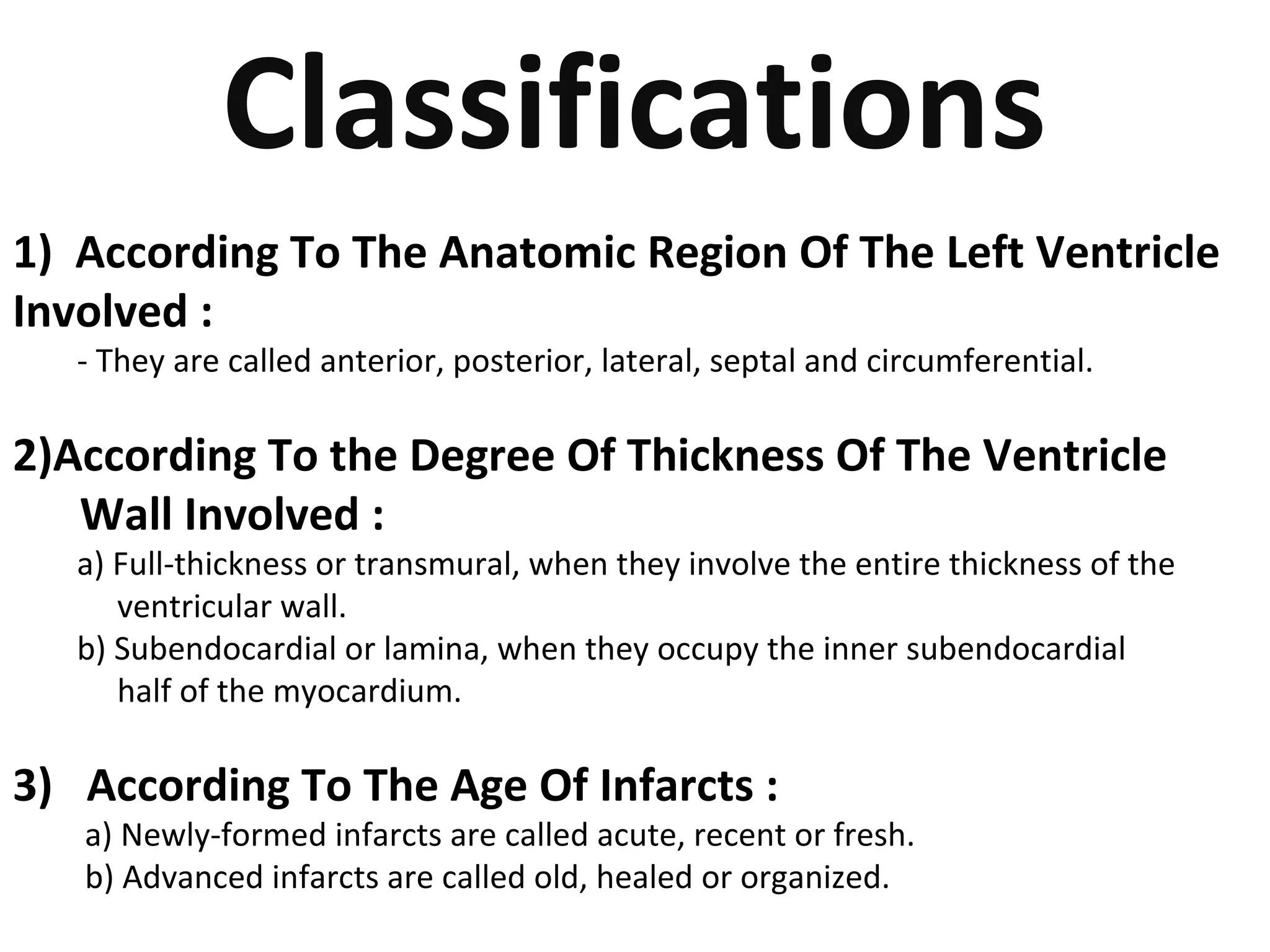


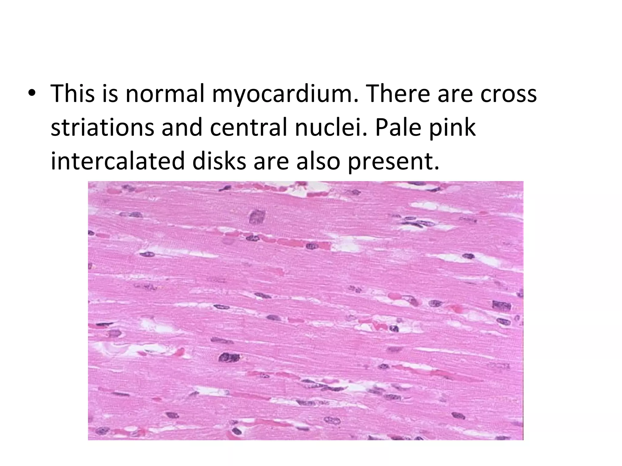
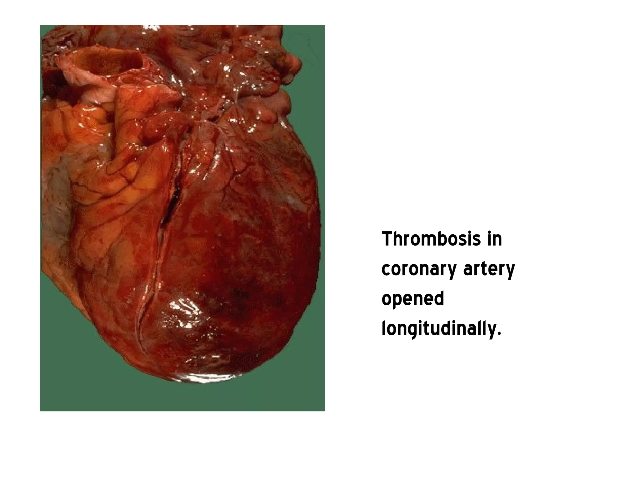
The document discusses diseases of the heart, specifically chronic ischemic heart disease and myocardial infarction. It describes the blood supply to the heart from the coronary arteries and the risks factors, causes, classifications, signs and symptoms, progression, complications and treatment of myocardial infarction. Key points include that myocardial infarction is mainly caused by blockages in the coronary arteries from atherosclerosis, and can lead to complications like arrhythmias, heart failure, cardiogenic shock or cardiac rupture if not properly treated.


