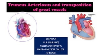
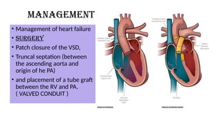
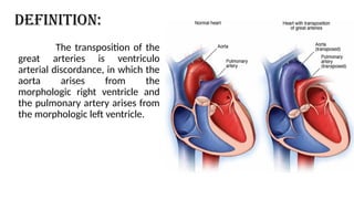
Truncus Arteriosus and transposition
of great vessels
DEEPIKA.R
M.Sc (NURSING)
COLLEGE OF NURSING
MADRAS MEDICAL COLLEGE
CHENNAI
DEFINITION
• A rare congenital heart disease characterized by a single blood vessel
arising from the right and left ventricles, instead of the pulmonary artery
and aorta.
• It occurs when the two large arteries carrying blood away from the heart
don’t form properly and one large artery is present instead. This single-
great vessel carries blood both to the body and to the lungs.
• truncus sits over a large opening or hole in the wall between the two
pumping chambers (ventricular septal defect).
CAUSES
• Some congenital heart defects may have a genetic link, either
occurring due to
• A defect in a gene,
• A chromosome abnormality, or
• Environmental exposure,
• Causing heart problems to occur more often in certain families.
• Other times this heart defect occurs sporadically (by chance),
• With no clear reason for its development.
RISK FACTORS
• A mother who had rubella (German measles) or another viral illness
during early pregnancy
• A parent who had a congenital heart defect
• Excessive alcohol consumption during pregnancy
• A mother who has diabetes
• Taking some types of medications during pregnancy
TYPES
TYPE
IV
TYPE
III
TYPE
II
TYPE
1
Types
• In type I, the main
pulmonary artery (PA) arises
from the truncus and then
divides into the right (RPA)
and left pulmonary artery
(LPA) branches. (there is
short MPA)
Type II
•In type II, the pulmonary
arteries arise from the
posterior aspect of the
truncus (very close to each other )
Type III
•In type III, the
pulmonary arteries
arise from the
lateral aspects of
the truncus.
• Two orifices of the PAs are more distant
from the truncal valve .
• more distant from each other as
compared with type II.
Type IV
• In type IV, or pseudotruncus
arteriosus, arteries arising
from the descending aorta
(AO) supply the lungs.
types
Pathophysiology
Blood eject from
both right and left
ventricles
Enter into common
trunk
Mixing pulmonary
and systemic
circulation
According to
resistances blood
flow is distributed to
the pulmonary and
systemic circulation
Resistance to
pulmonary blood
flow is less than
systemic vascular
resistance
Preferential blood
flow to the lungs
CLINICAL MANIFESTATION
• Tachypnea
• Costosternal retraction
• Cyanosis
• Signs of CHF
• Bounding peripheral pulses
• Systolic murmur at left sternal
border
• Diastolic murmur of aortic
regurgitation
• Prominent ejection click at the
apex or left sternal border
Diagnostic studies
• ECG- RT or LT Ventricular
hypertrophy, with left or
combined hypertrophy
• Chest radiography-
cardiomegaly, pulmonary
vasculature increased may in the
right aortic arch
• ECHO- Overriding truncal root
• Cardiac catheterization-
anatomic information
• Management of heart failure
• SURGERY
• Patch closure of the VSD,
• Truncal septation (between
the ascending aorta and
origin of he PA)
• and placement of a tube graft
between the RV and PA.
( VALVED CONDUIT )
MANAGEMENT
TRANSPOSITION OF GREAT
ARTERIES
DEFINITION:
The transposition of the
great arteries is ventriculo
arterial discordance, in which the
aorta arises from the
morphologic right ventricle and
the pulmonary artery arises from
the morphologic left ventricle.
INCIDENCE:
• Transposition of the great arteries (TGA) is the most common cyanotic
congenital heart lesion that presents in neonates.
• This lesion presents in 5-7% of all patients with congenital heart
disease.
• The overall annual incidence is 20-30 per 100,000 live births.
ETIOLOGY:
• unknown and is presumed to be multifactorial.
• This congenital heart defect is more common in infants of diabetic
mothers.
Pathophysiology
Due to etiological factors
Transposition of great arteries
Oxygenated pulmonary venous blood returns to
the left atrium and left ventricle
It was recirculated to the pulmonary vascular
bed via the abnormal pulmonary arterial
connection to the left ventricle.
Deoxygenated systemic venous blood returns to the right atrium and
right ventricle
pumped to the systemic circulation, effectively bypassing the lungs
deficient oxygen supply to the tissues and an excessive right and left
ventricular workload
It is incompatible with prolonged survival unless mixing of
oxygenated and deoxygenated blood occurs at some anatomic level
like
• ASD
• VSD
• PDA
Clinical manifestations:
• Prominent and progressive cyanosis within the first 24 hours of life is
the usual finding in infants.
• Tachypnea
• Tachycardia
• Diaphoresis
• Failure to gain weight
HEART SOUND:
• A single or narrowly split, diminished second heart sound
• Systolic ejection murmur may be present.
DIAGNOSTIC EVALUATION:
• History collection
• Physical examination
• ABG analysis
• Echocardiography
• Cardiac catheterization
• Chest radiography
demonstrate
the classic "egg on a
string"
appearance in
approximately one
third of patients.
Medical management:
• Initial treatment consists of maintaining ductal patency with
continuous intravenous (IV) prostaglandin E1 infusion to promote
pulmonary blood flow.
• Antibiotic Prophylactic Regimens for Endocarditis.
• Fluid replacement
• Bicarbonate administration- Acidosis
• Mechanical ventilation
Surgical management:
• Rastelli-type procedure the main
pulmonary artery and place an
aortopulmonary shunt during the
newborn period to restrict
pulmonary blood flow.
• Arterial switch procedure
Arterial switch procedure:
Complications:
• Congestive heart failure
• Arrhythmia
• Eisenmenger syndrome (irreversible and progressive pulmonary
vascular obstructive disease)
Prognosis:
• The overall survival rate following arterial switch operation is 90%.
• The overall mortality rate following an atrial level switch is low;
however, long-term morbidity associated with systemic (right)
ventricular dilatation and failure, systemic atrioventricular (tricuspid)
valve regurgitation, and atrial bradyarrhythmia's and
tachyarrhythmias is significant.
NURSING
MANAGEMENT
Relieving
Respiratory
Distress
Improving
cardiac
output
Improving
oxygenatio
n
Nutrition
Preventing
infection
Health
education
Truncus Arteriosus.cardiothoracic pptx ..

Truncus Arteriosus.cardiothoracic pptx ..

  • 1.
    Truncus Arteriosus andtransposition of great vessels DEEPIKA.R M.Sc (NURSING) COLLEGE OF NURSING MADRAS MEDICAL COLLEGE CHENNAI
  • 2.
    DEFINITION • A rarecongenital heart disease characterized by a single blood vessel arising from the right and left ventricles, instead of the pulmonary artery and aorta. • It occurs when the two large arteries carrying blood away from the heart don’t form properly and one large artery is present instead. This single- great vessel carries blood both to the body and to the lungs. • truncus sits over a large opening or hole in the wall between the two pumping chambers (ventricular septal defect).
  • 3.
    CAUSES • Some congenitalheart defects may have a genetic link, either occurring due to • A defect in a gene, • A chromosome abnormality, or • Environmental exposure, • Causing heart problems to occur more often in certain families. • Other times this heart defect occurs sporadically (by chance), • With no clear reason for its development.
  • 4.
    RISK FACTORS • Amother who had rubella (German measles) or another viral illness during early pregnancy • A parent who had a congenital heart defect • Excessive alcohol consumption during pregnancy • A mother who has diabetes • Taking some types of medications during pregnancy
  • 5.
  • 6.
    Types • In typeI, the main pulmonary artery (PA) arises from the truncus and then divides into the right (RPA) and left pulmonary artery (LPA) branches. (there is short MPA)
  • 7.
    Type II •In typeII, the pulmonary arteries arise from the posterior aspect of the truncus (very close to each other )
  • 8.
    Type III •In typeIII, the pulmonary arteries arise from the lateral aspects of the truncus. • Two orifices of the PAs are more distant from the truncal valve . • more distant from each other as compared with type II.
  • 9.
    Type IV • Intype IV, or pseudotruncus arteriosus, arteries arising from the descending aorta (AO) supply the lungs.
  • 10.
  • 11.
    Pathophysiology Blood eject from bothright and left ventricles Enter into common trunk Mixing pulmonary and systemic circulation According to resistances blood flow is distributed to the pulmonary and systemic circulation Resistance to pulmonary blood flow is less than systemic vascular resistance Preferential blood flow to the lungs
  • 12.
    CLINICAL MANIFESTATION • Tachypnea •Costosternal retraction • Cyanosis • Signs of CHF • Bounding peripheral pulses • Systolic murmur at left sternal border • Diastolic murmur of aortic regurgitation • Prominent ejection click at the apex or left sternal border
  • 13.
    Diagnostic studies • ECG-RT or LT Ventricular hypertrophy, with left or combined hypertrophy • Chest radiography- cardiomegaly, pulmonary vasculature increased may in the right aortic arch • ECHO- Overriding truncal root • Cardiac catheterization- anatomic information
  • 14.
    • Management ofheart failure • SURGERY • Patch closure of the VSD, • Truncal septation (between the ascending aorta and origin of he PA) • and placement of a tube graft between the RV and PA. ( VALVED CONDUIT ) MANAGEMENT
  • 15.
  • 16.
    DEFINITION: The transposition ofthe great arteries is ventriculo arterial discordance, in which the aorta arises from the morphologic right ventricle and the pulmonary artery arises from the morphologic left ventricle.
  • 17.
    INCIDENCE: • Transposition ofthe great arteries (TGA) is the most common cyanotic congenital heart lesion that presents in neonates. • This lesion presents in 5-7% of all patients with congenital heart disease. • The overall annual incidence is 20-30 per 100,000 live births.
  • 18.
    ETIOLOGY: • unknown andis presumed to be multifactorial. • This congenital heart defect is more common in infants of diabetic mothers.
  • 19.
    Pathophysiology Due to etiologicalfactors Transposition of great arteries Oxygenated pulmonary venous blood returns to the left atrium and left ventricle It was recirculated to the pulmonary vascular bed via the abnormal pulmonary arterial connection to the left ventricle.
  • 20.
    Deoxygenated systemic venousblood returns to the right atrium and right ventricle pumped to the systemic circulation, effectively bypassing the lungs deficient oxygen supply to the tissues and an excessive right and left ventricular workload
  • 21.
    It is incompatiblewith prolonged survival unless mixing of oxygenated and deoxygenated blood occurs at some anatomic level like • ASD • VSD • PDA
  • 22.
    Clinical manifestations: • Prominentand progressive cyanosis within the first 24 hours of life is the usual finding in infants. • Tachypnea • Tachycardia • Diaphoresis • Failure to gain weight
  • 23.
    HEART SOUND: • Asingle or narrowly split, diminished second heart sound • Systolic ejection murmur may be present.
  • 24.
    DIAGNOSTIC EVALUATION: • Historycollection • Physical examination • ABG analysis • Echocardiography • Cardiac catheterization
  • 25.
    • Chest radiography demonstrate theclassic "egg on a string" appearance in approximately one third of patients.
  • 26.
    Medical management: • Initialtreatment consists of maintaining ductal patency with continuous intravenous (IV) prostaglandin E1 infusion to promote pulmonary blood flow. • Antibiotic Prophylactic Regimens for Endocarditis. • Fluid replacement • Bicarbonate administration- Acidosis • Mechanical ventilation
  • 27.
    Surgical management: • Rastelli-typeprocedure the main pulmonary artery and place an aortopulmonary shunt during the newborn period to restrict pulmonary blood flow. • Arterial switch procedure
  • 28.
  • 29.
    Complications: • Congestive heartfailure • Arrhythmia • Eisenmenger syndrome (irreversible and progressive pulmonary vascular obstructive disease)
  • 30.
    Prognosis: • The overallsurvival rate following arterial switch operation is 90%. • The overall mortality rate following an atrial level switch is low; however, long-term morbidity associated with systemic (right) ventricular dilatation and failure, systemic atrioventricular (tricuspid) valve regurgitation, and atrial bradyarrhythmia's and tachyarrhythmias is significant.
  • 31.