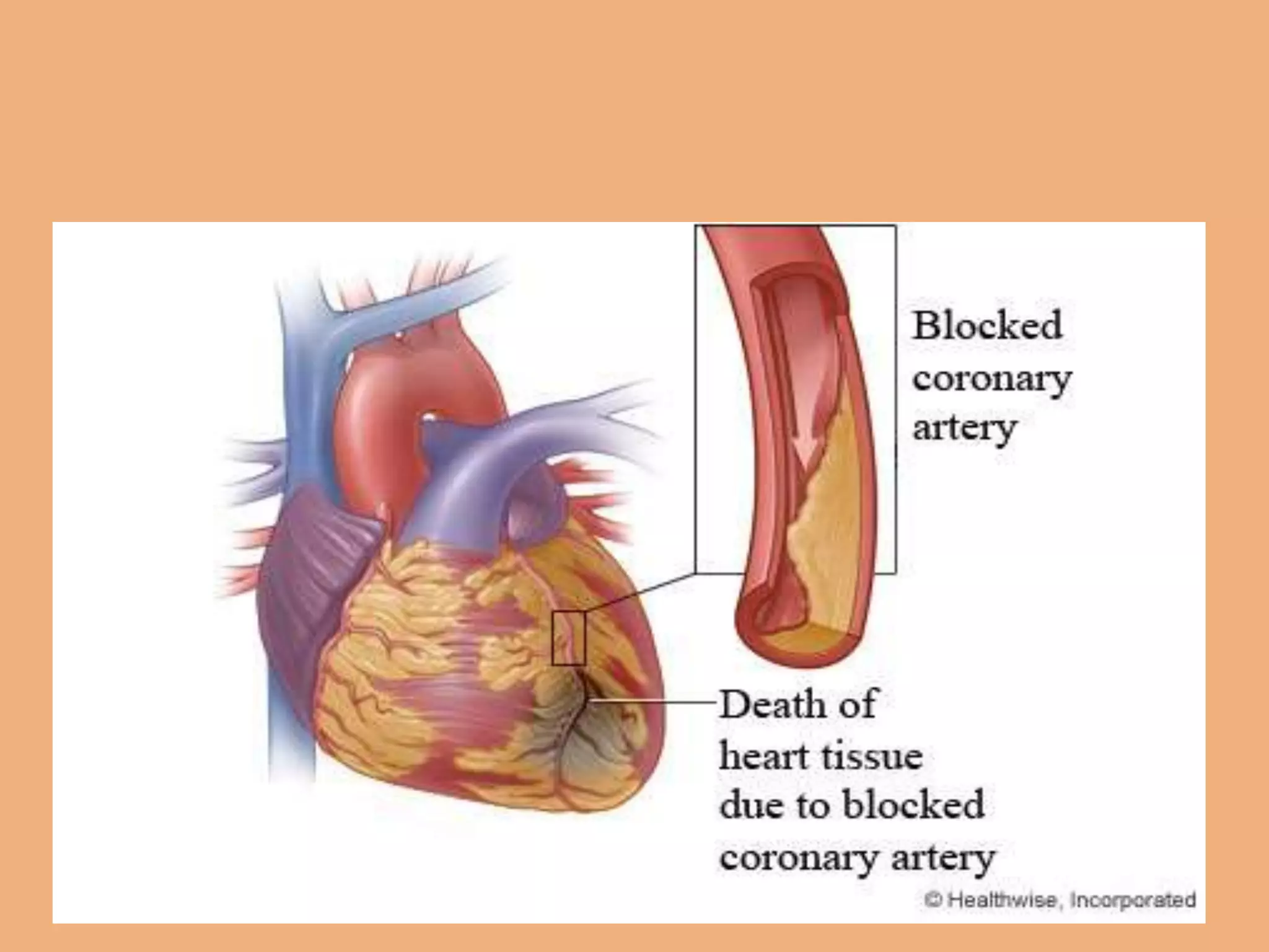
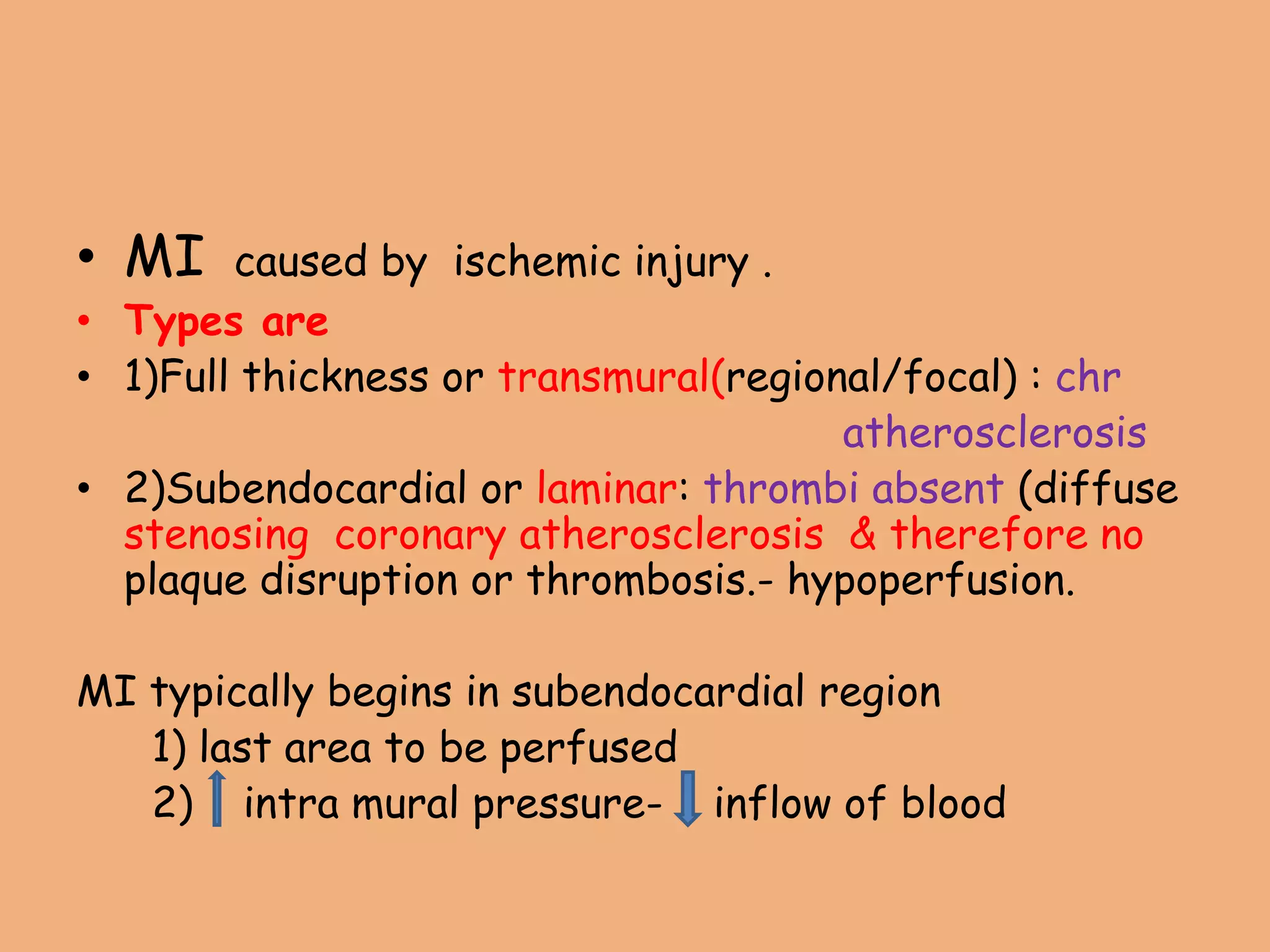
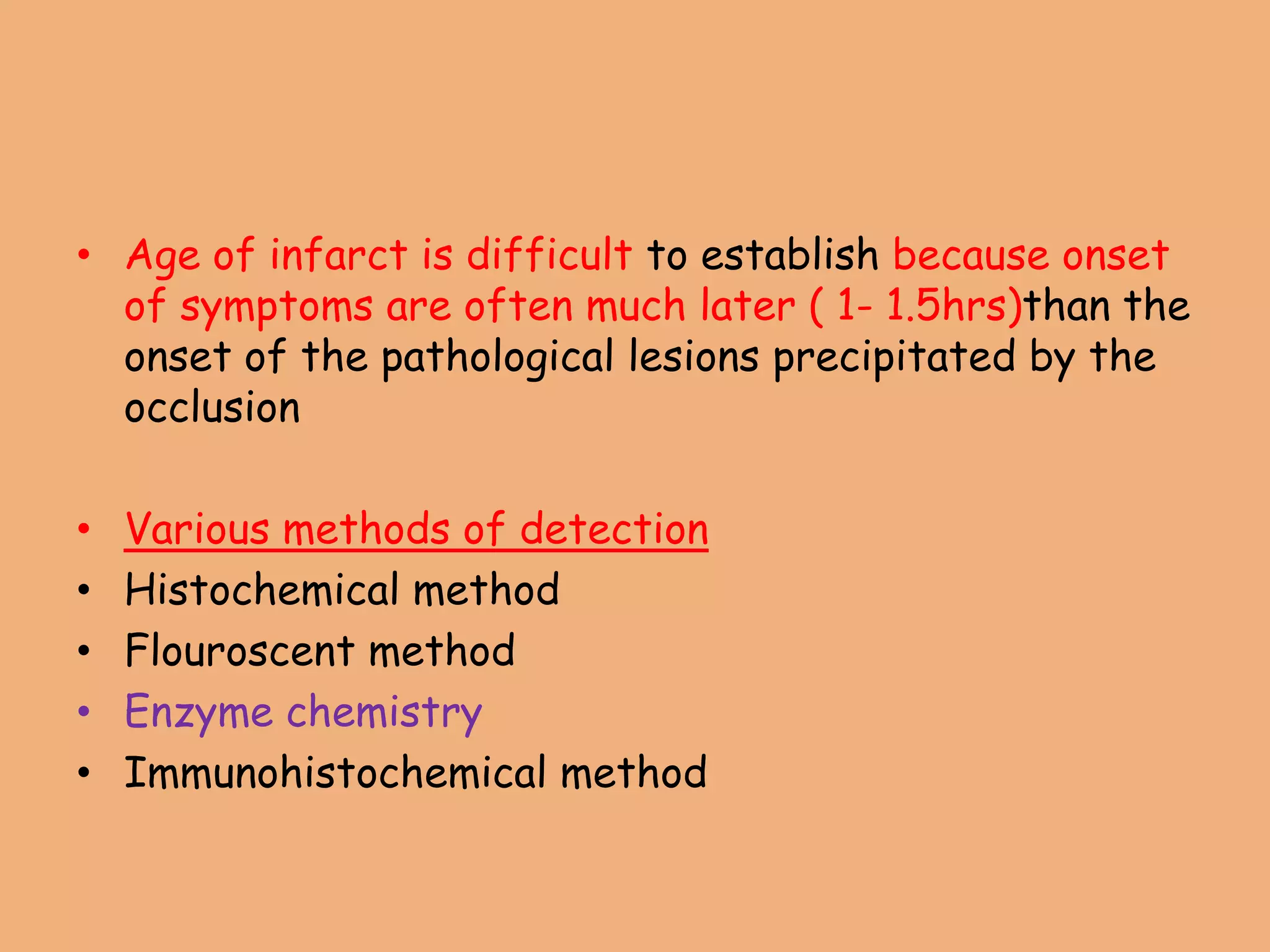
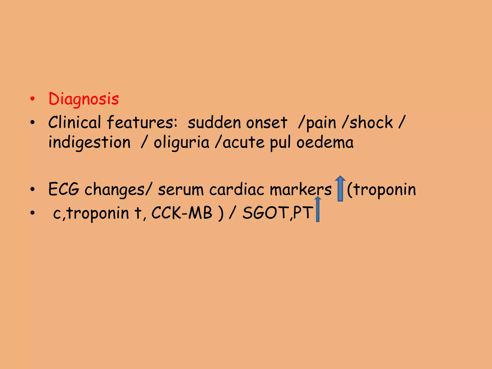
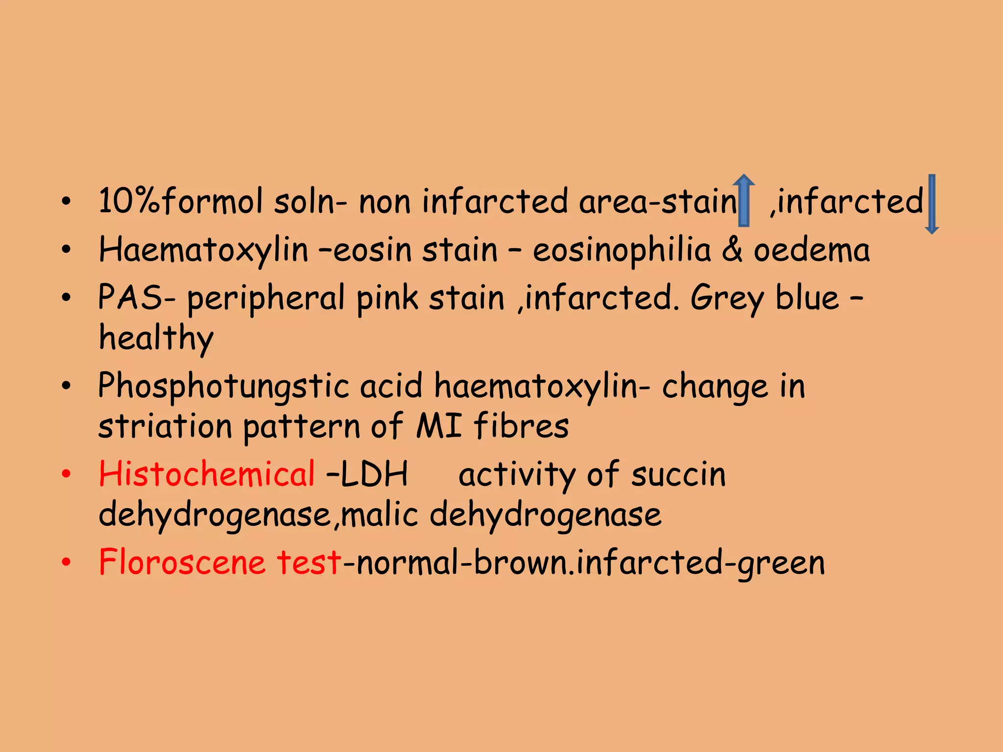
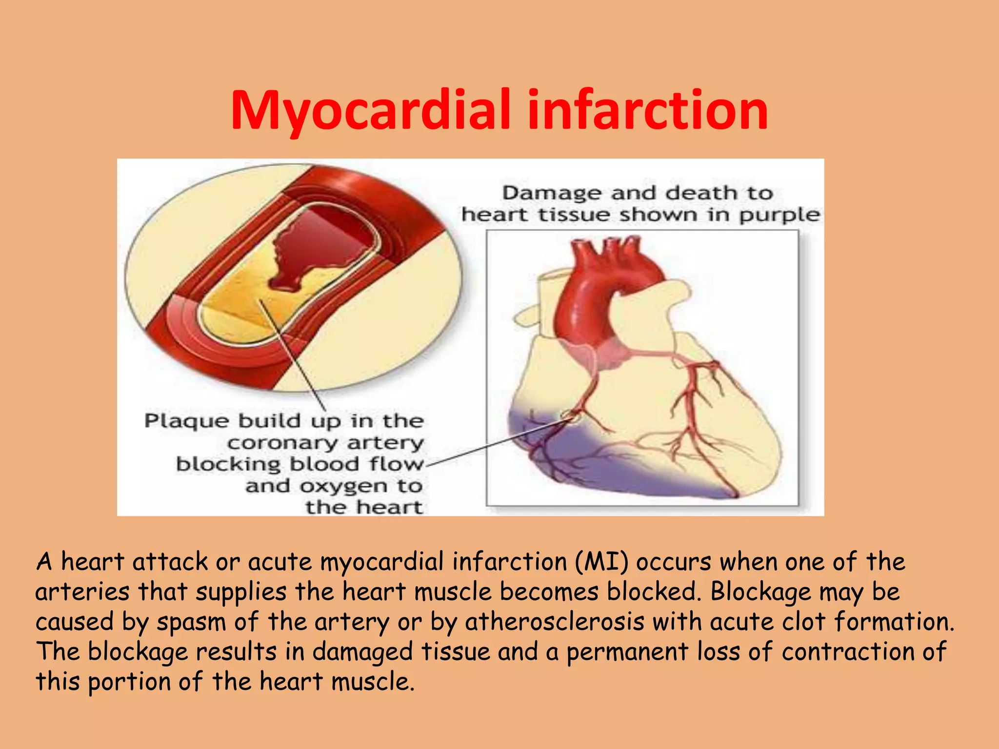
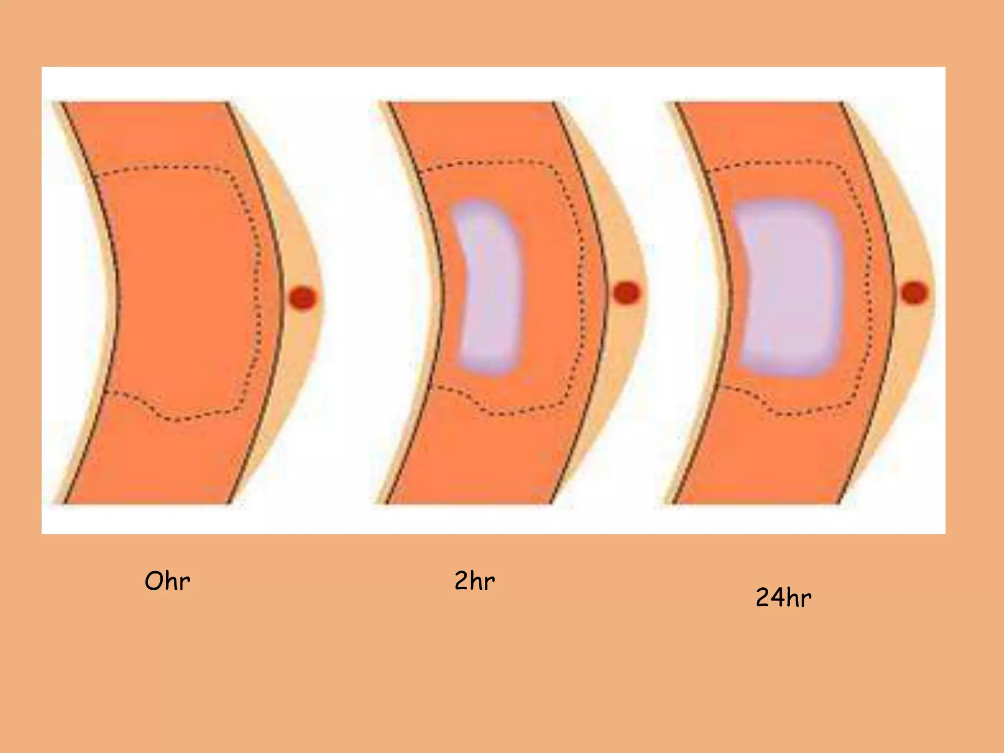
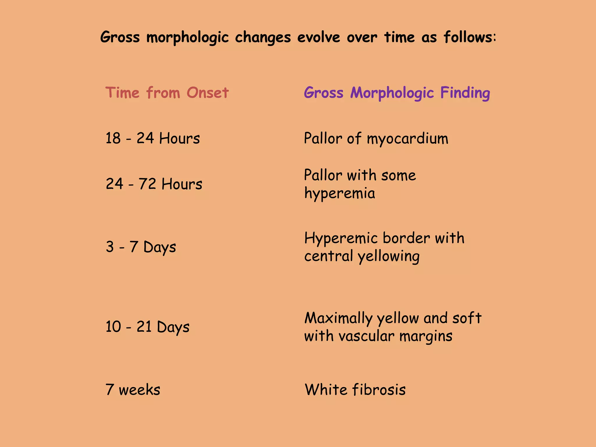
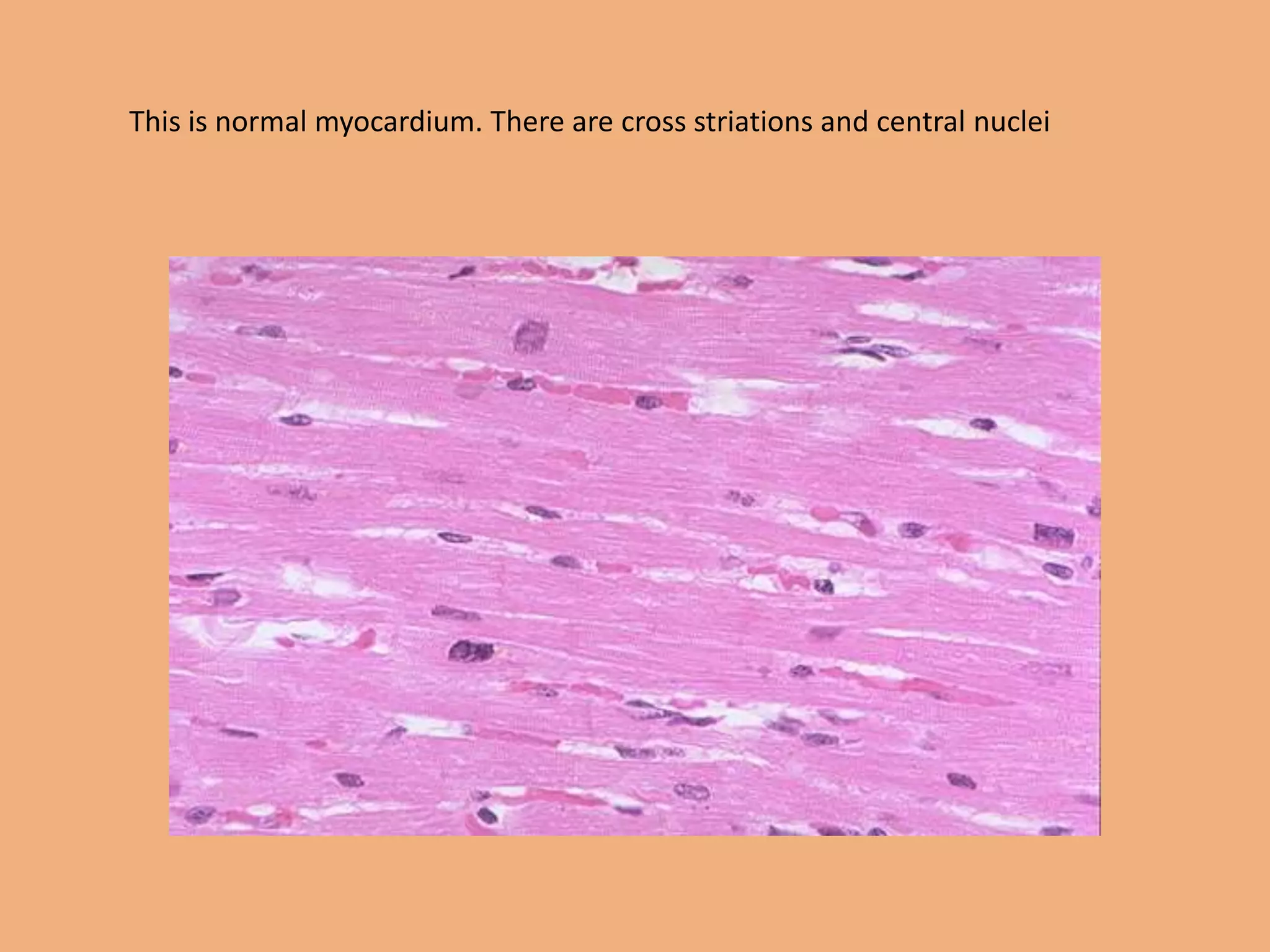
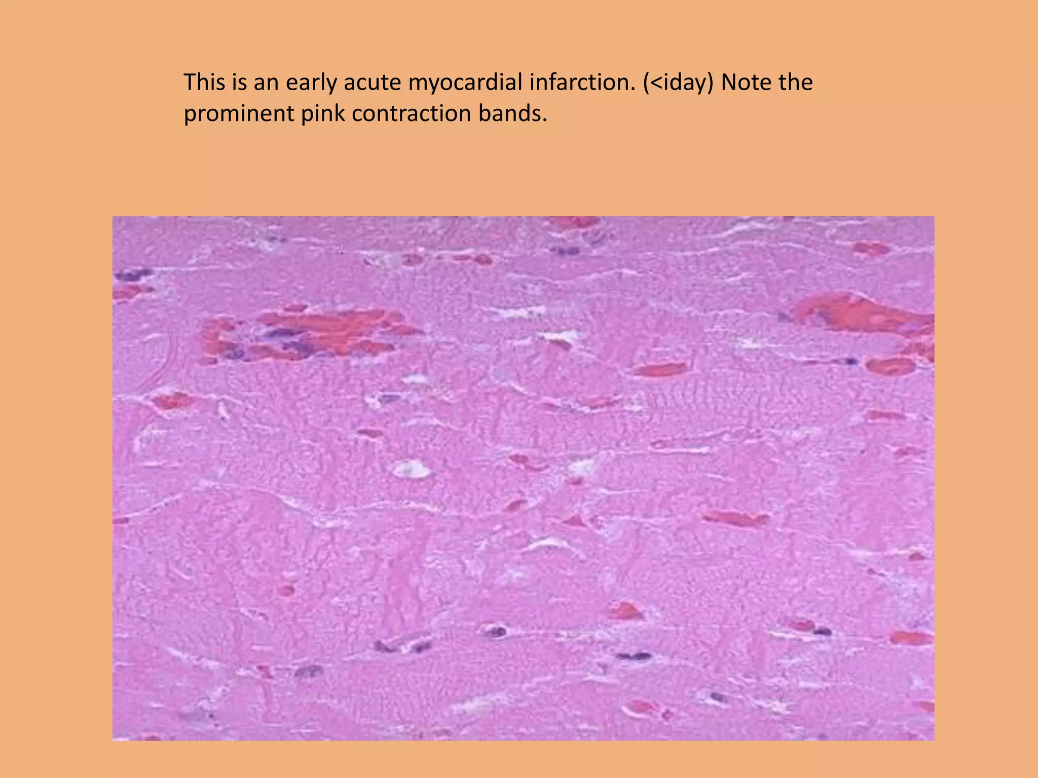
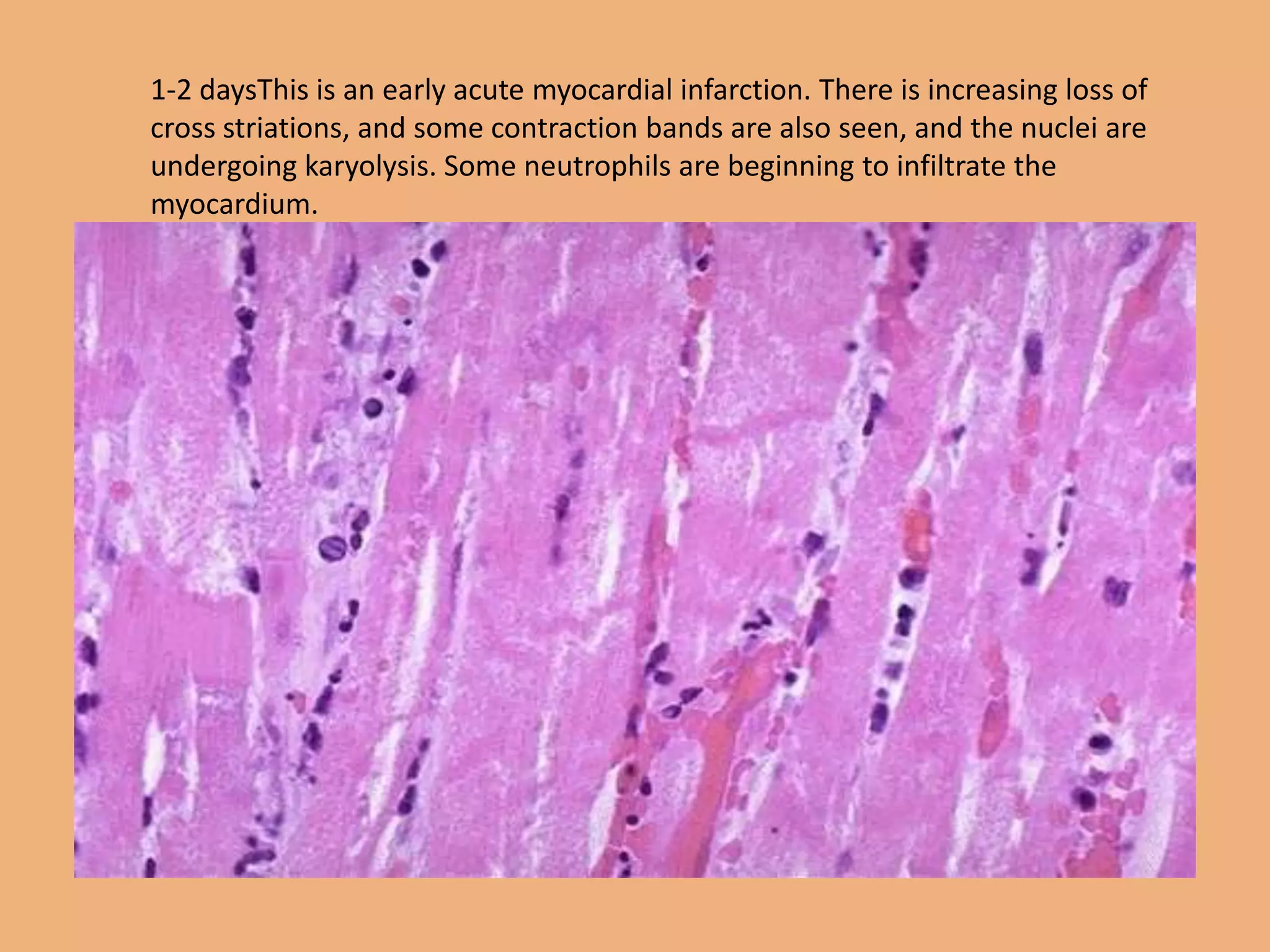
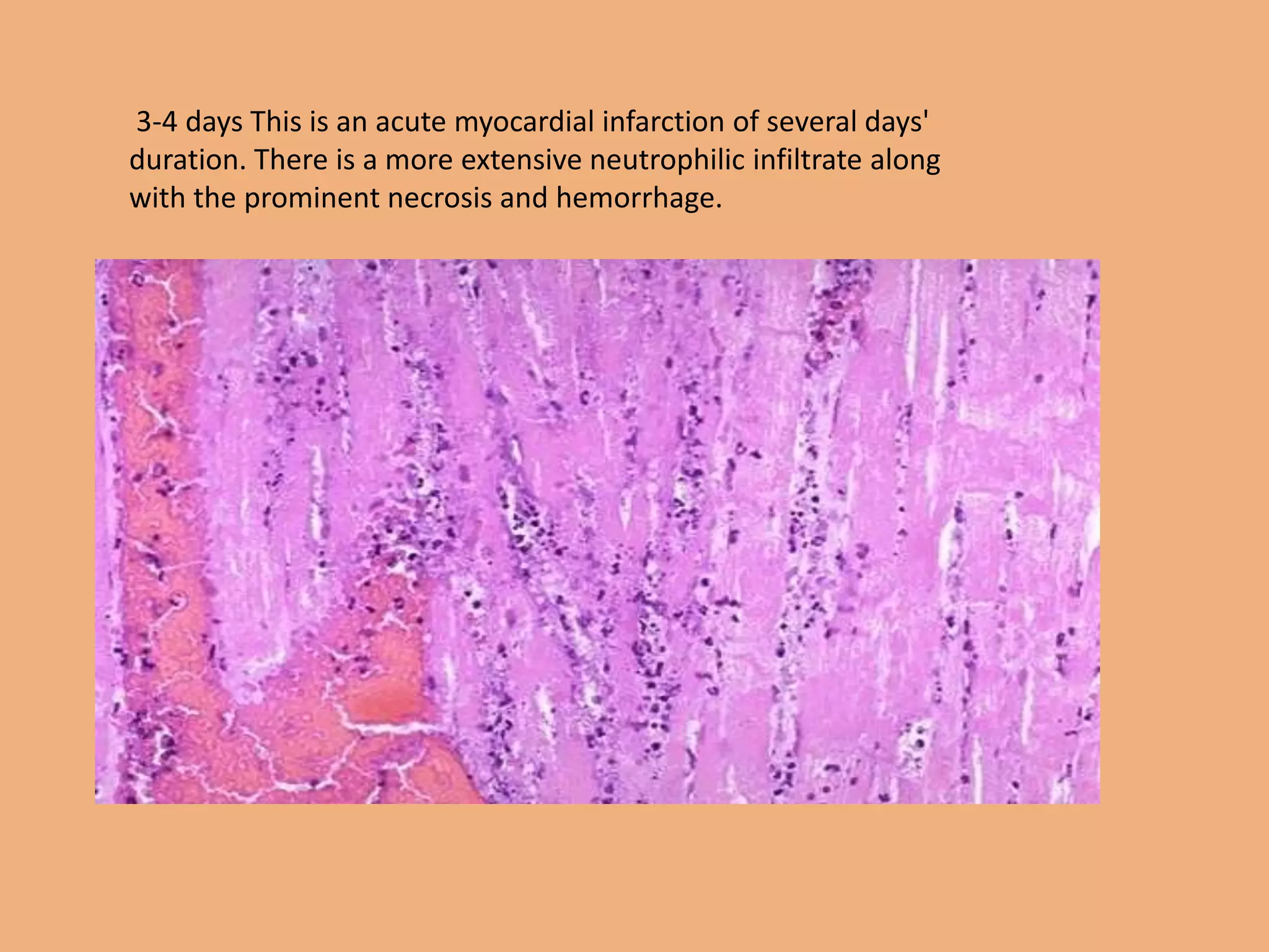
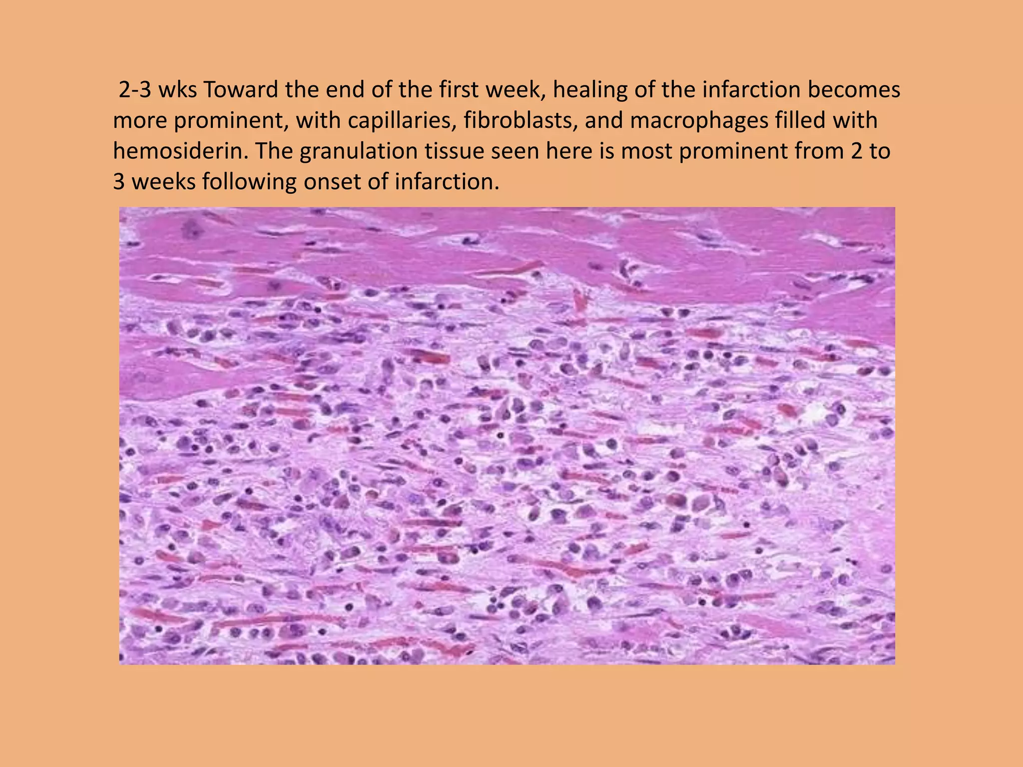
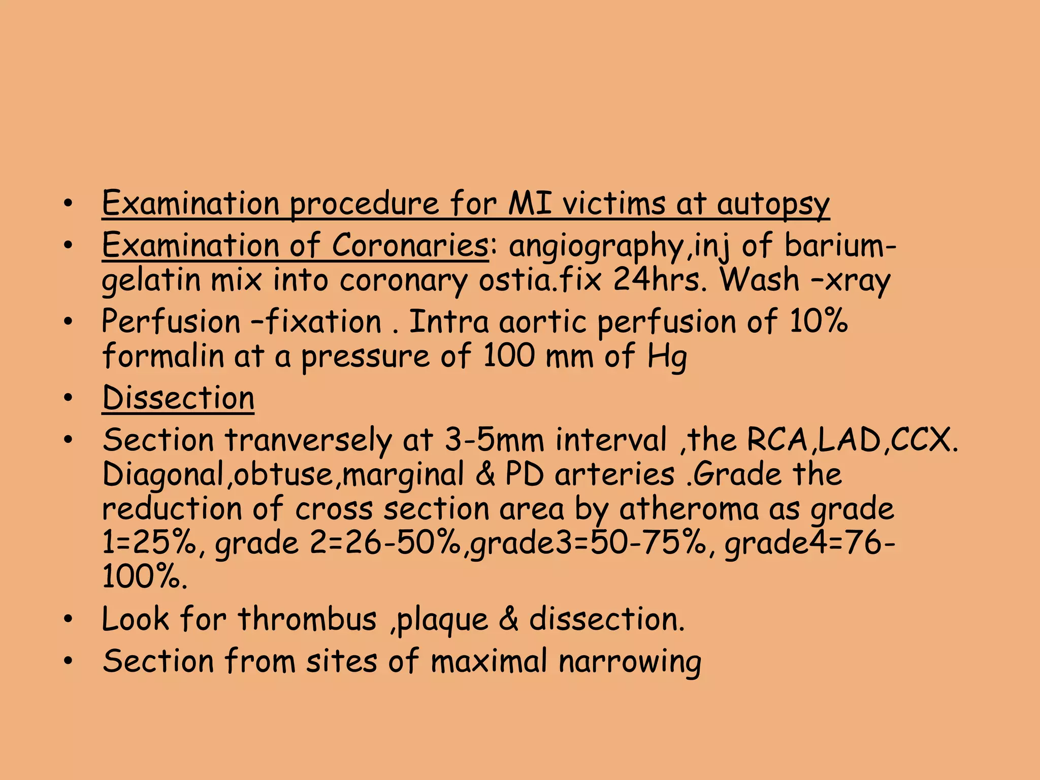
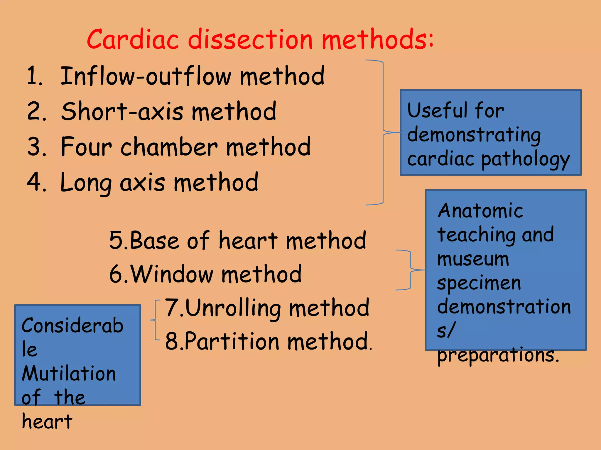
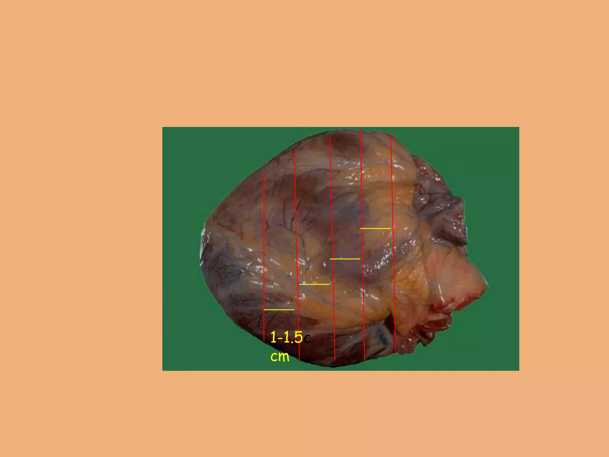
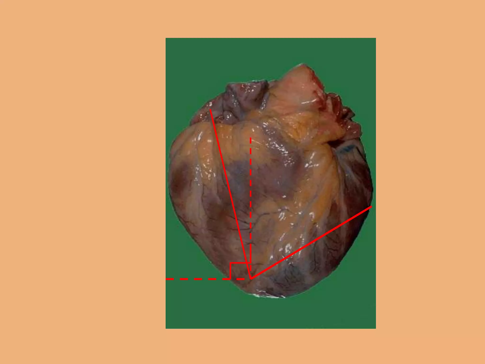
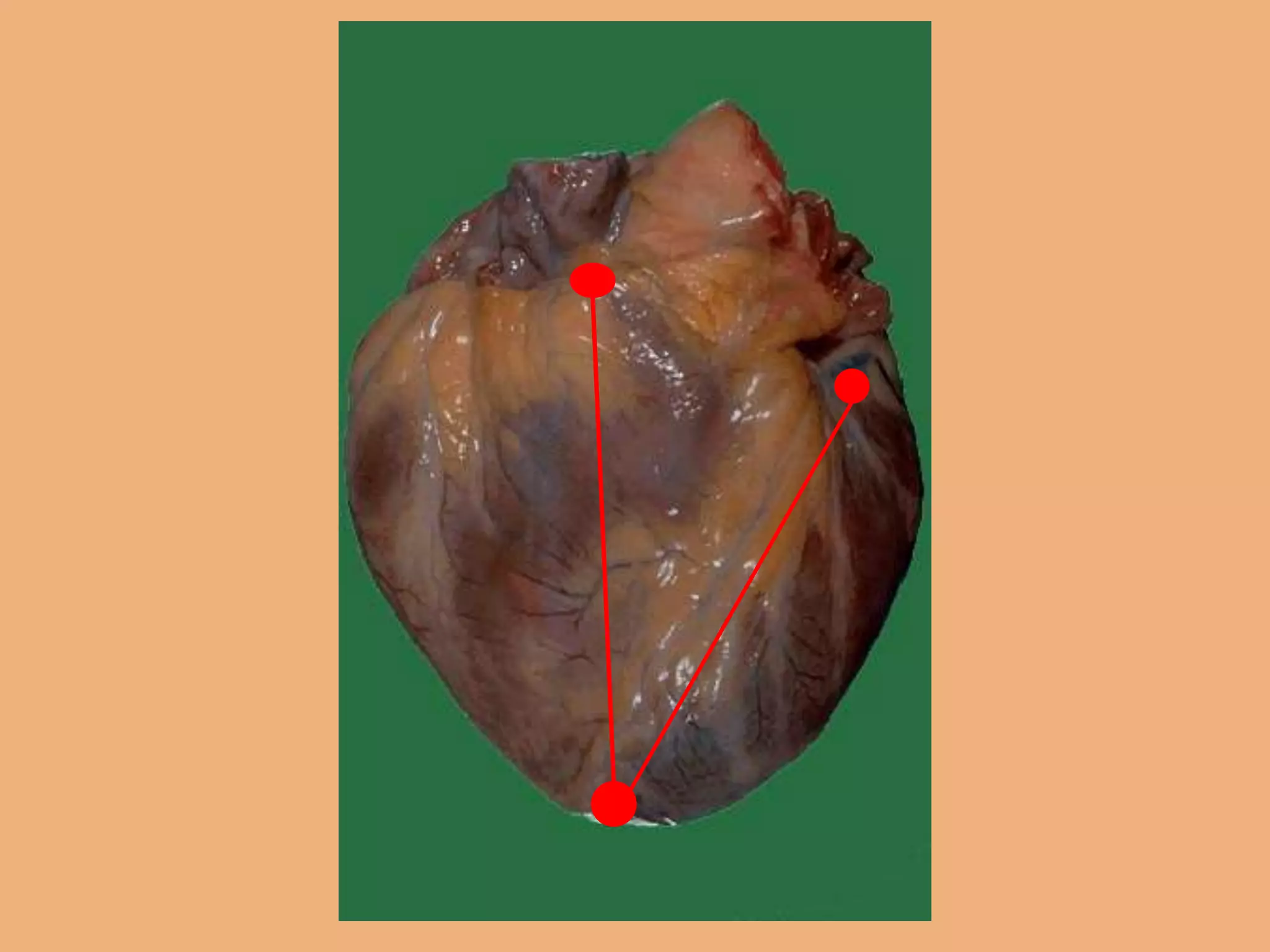
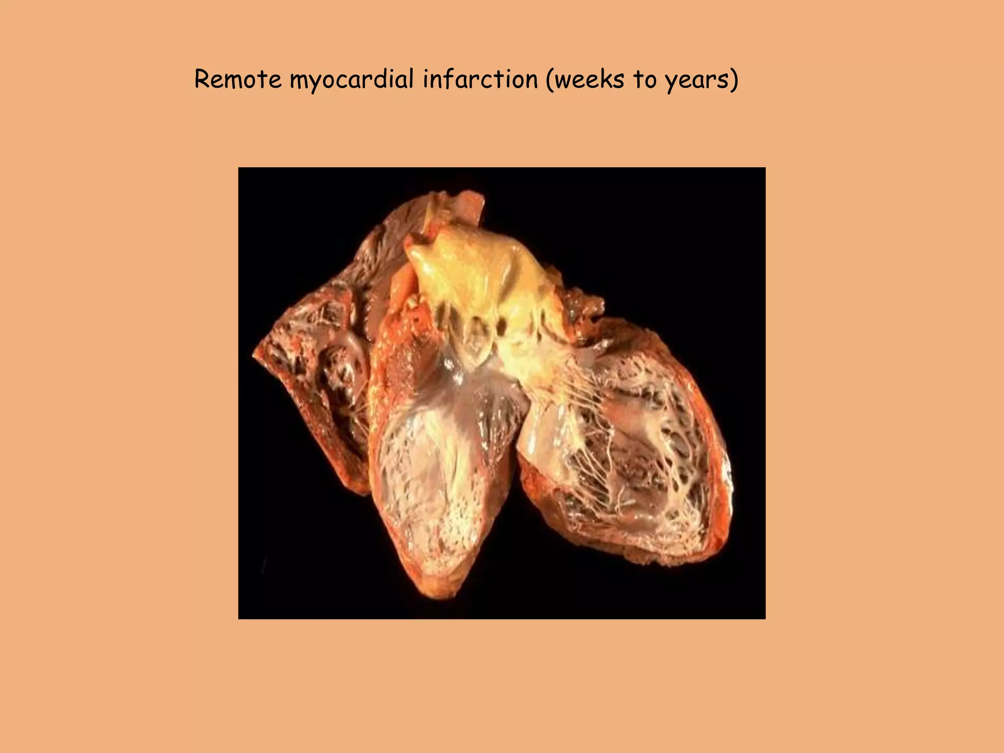
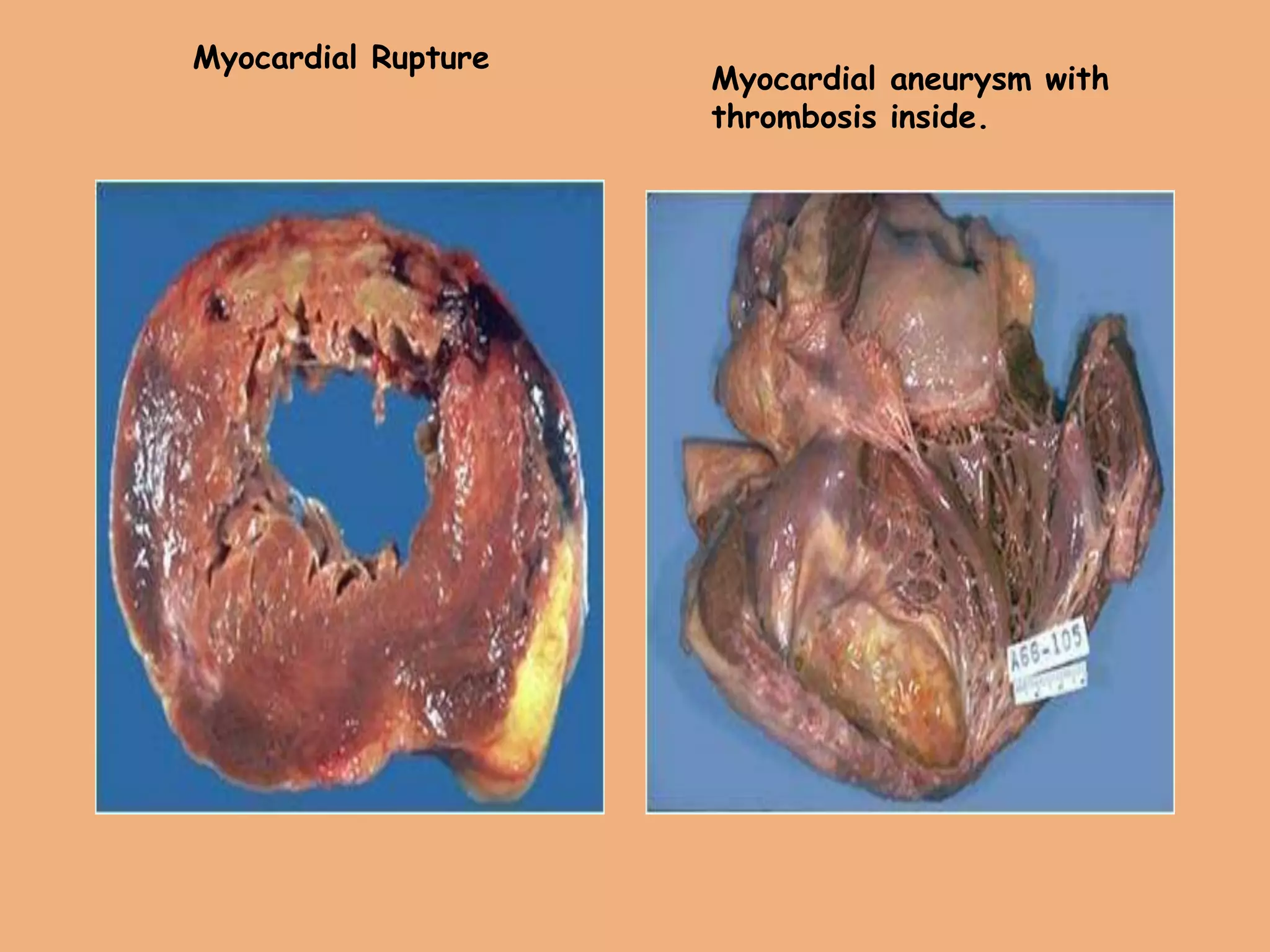
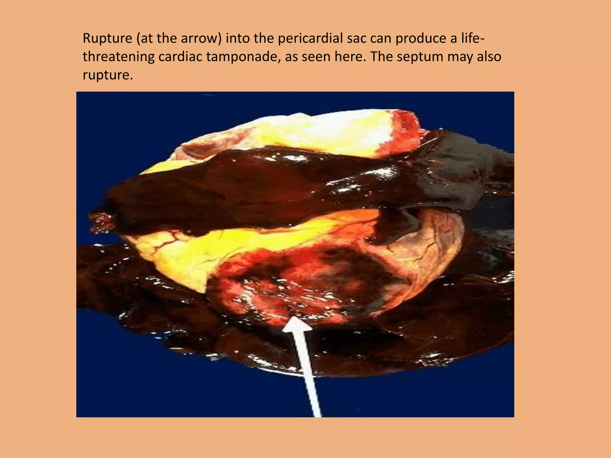
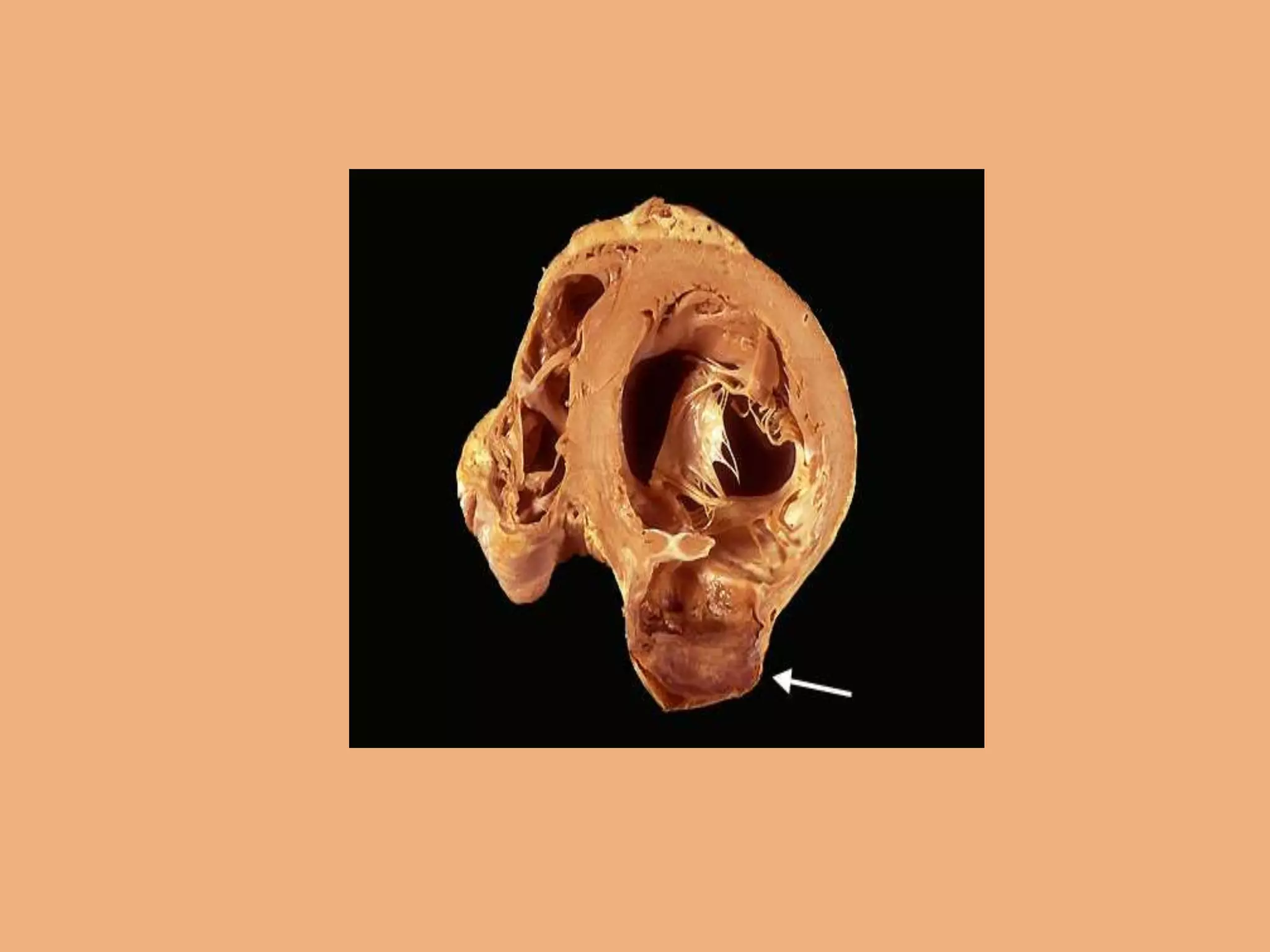
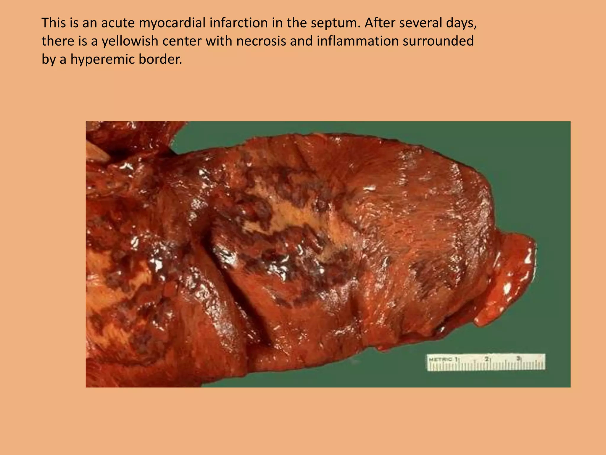
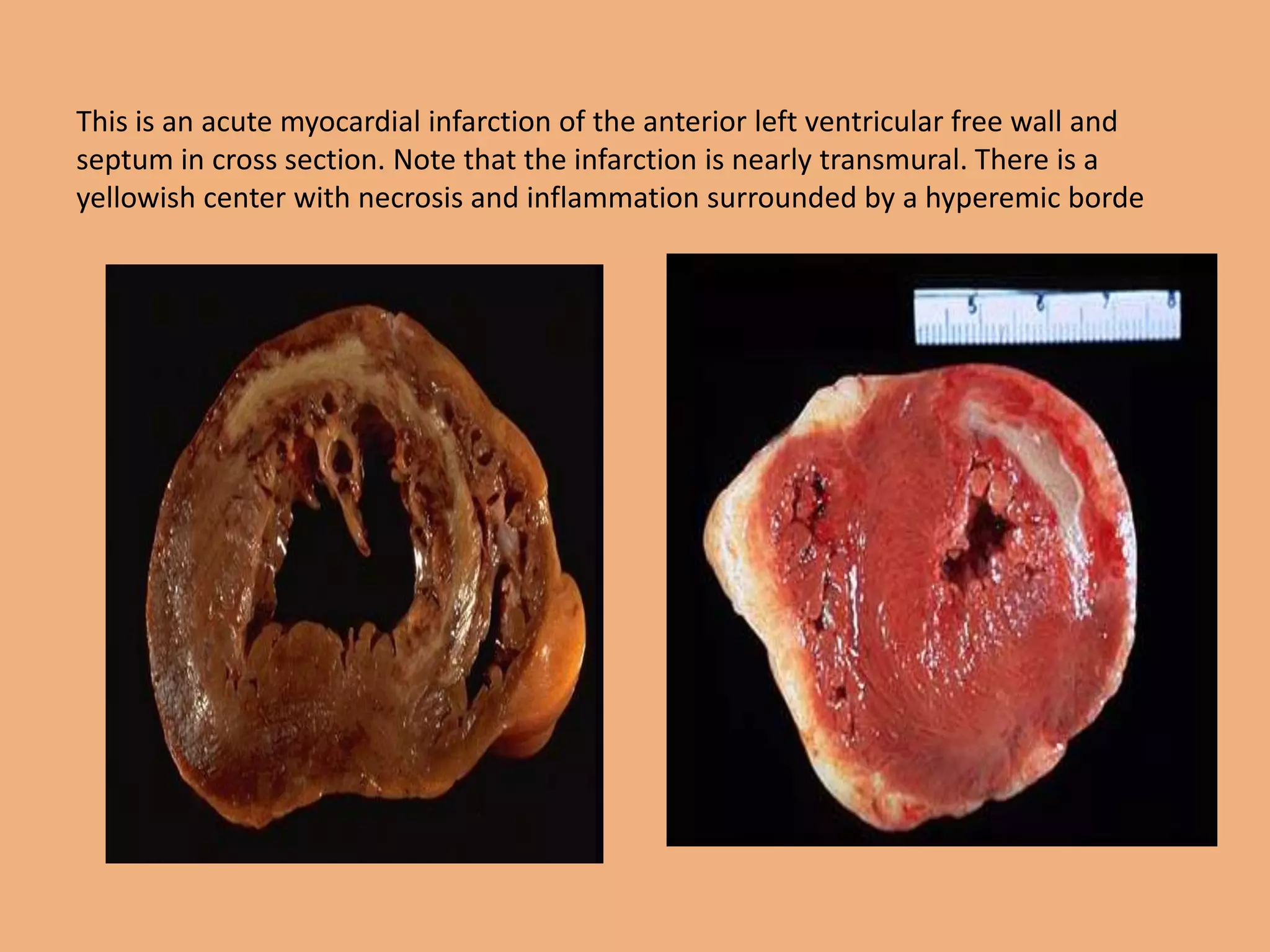
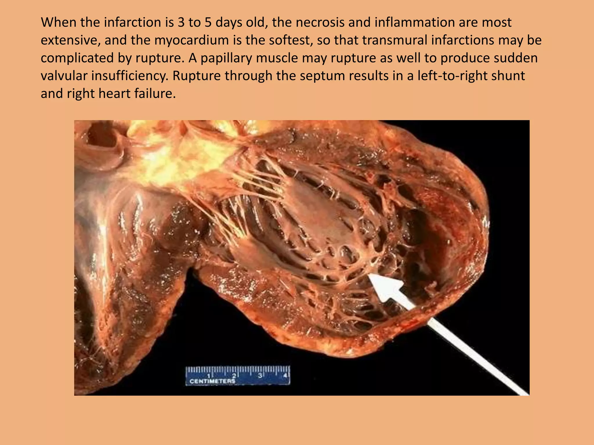
Myocardial Infarction
• Presenter : Dr Gopal G Hargi
» PG in dept of FM & T
• Acute Myocardial infarction is one of
the pathological condition caused by
prolonged ischemia where there is
irreversible necrosis of the myocardial
tissue
• IHD refers to a group of closely related syndromes
caused by imbalance between myocardial oxygen demand &
blood supply.
• Ischemia is the most common type of cell injury, (in
contrast to hypoxia ,the injury is faster)
• IHD - 90% due to atherosclerosis affecting coronary
arteries
• 10% other causes
• Most common cause of ischemia
Narrowing of coronaries by atherosclerosis
Depending on rate & severity of myocardial injury one of 4
syndromes may develop
1)Angina pectoris
2)MI
3)Sudden cardiac death
4)Chr IHD& CCF
Other less common sequele (non cardiac) due to atherosclerosis
• Perepheral vascular disease
• aneurysms
• Ischemic encephalopathy
• mesentric artery occlusion
Pathology of atherosclerosis: affects primarily
the intima of large & medium sized muscular
arteries and is characterized by endothelial
thickenings, fibro fatty plaques or atheromas.
(atheros-porridge –ref to soft lipid rich material in
the centre of atheroma.sclerosis- CT in plaque.
Common sites – Aorta/Coronaries/Cerebral vessels
• Clinical manifestation may develop at any age but
more common in older adults with peak incidence
after age of 60 yrs in men & 70 yrs in women
• Risk factors
• Major : constitutional: age/sex/genetics/familial &
race
acquired : ^ lipid/HT/DM/smoking
• Minor : Environmental/obesity/hormones/physical
inactivity / stress/alcohol/infections(cmv ,
herpes)
• Atherosclerosis formation – no of theories
• 1) insudation theory ( virchows ) :also called
response to injury theory.
cellular proliferation of intimal cells from imbibing
of lipids from blood.
2) Encrustation theory ( Rokitansky) : Atheroma
causes a form of encrustation on the arterial wall
from the components of blood to form thrombi (
platelets ,fibrin, leucocytes)
(currently reaction to injury& monoclonal theory
based on proliferation of SMC is accepted )
• The 4 key significant factors responsible are
• Arterial SMC
• Endothelial cells
• Blood Monocytes
• Hyperlipidemia
Reaction to injury is taken as the endothelial
injury followed by SMC proliferation so that early
lesion consists of SMC mainly .The lipoproteins enter
into the intima ,followed by lipid accumulation in
macrophages( foam cells)
• Earliest pathological lesions in atherosclerosis are
• 1)Intimal thickening-
• 2)Fatty streaks fore-runners in the evolution
• of atherosclerosis.
Fatty streaks : these lie under the endothelium
& composed of closely packed foam cells ,lipid
containing SMC & few lymphoid cells.
3) Atheromatous plaques: also called fibrous plaque ,
fibro fatty plaque & atheromas
• Complication:(fate of atheroma)
• covering endothelium remains intact but danger
to life is caused by luminal reduction from bulge
of enlarging plaque
• Fibroendothelial cap begins to breakdown under
pressure by central necrosing process & erosions
caused by blood flow
• May rupture into lumen leading to acute
obstruction causing death
• Contents of the plaque may get washed down
causing embolism
Changes
• Calcification
• Ulceration
• Thrombosis: plaque - internal necrosis - surface
eroded –fibrofatty content exposed
- nidus for thrombosis
• Hemorrhages : rupture of blood vessels in the
periphery
• aneurysm
• IHD - 90% due to atherosclerosis affecting coronary
arteries
• 10% other causes
Other causes ( non atherosclerotic )
• Vasospasm
• Stenosis of coronary ostia: syphilitic aortitis
• Arteritis: kawasaki disease/TB/polyarteritis
• Embolism: infective endocarditis
• Thrombotic disease: polycythemia vera , SSA , TTP
• Aneurysms: DAA into coronary artery
• Compression: tumour of heart
• Trauma: contusion to muocardium
• MI is the most feared consequence of CAD .Many die within
few hrs ,remaining suffer from effects of impaired cardiac
functions.
• According to WHO statistics ,globally ,MI is responsible for
nearly 32% & 27% of all the mortalities in males & females
respectively
• 13.5 million alive today with history of MI
• 5% of MI in people under 40 years , particularly with HTN
• In 48% of men and 33% of women who died suddenly of MI,
there were no previous symptoms
Acute MI is one of the pathological condition caused
by prolonged ischemia where there is irreversible
necrosis of the myocardial tissue due to
coronary blood flow myocardial demand,
hypertrophy
Risk factors
• HTN
• DM
• Cigarettes
• Dislipidemia with familial hypercholesteremia
• Depending upon site
• LV . LAD- 40-50% . LCCA- 15-20% . RCA- 30-40 %
• MI caused by ischemic injury .
• Types are
• 1)Full thickness or transmural(regional/focal) : chr
atherosclerosis
• 2)Subendocardial or laminar: thrombi absent (diffuse
stenosing coronary atherosclerosis & therefore no
plaque disruption or thrombosis.- hypoperfusion.
MI typically begins in subendocardial region
1) last area to be perfused
2) intra mural pressure- inflow of blood
• Age of infarct is difficult to establish because onset
of symptoms are often much later ( 1- 1.5hrs)than the
onset of the pathological lesions precipitated by the
occlusion
• Various methods of detection
• Histochemical method
• Flouroscent method
• Enzyme chemistry
• Immunohistochemical method
• Diagnosis
• Clinical features: sudden onset /pain /shock /
indigestion / oliguria /acute pul oedema
• ECG changes/ serum cardiac markers (troponin
• c,troponin t, CCK-MB ) / SGOT,PT
• 10%formol soln- non infarcted area-stain ,infarcted
• Haematoxylin –eosin stain – eosinophilia & oedema
• PAS- peripheral pink stain ,infarcted. Grey blue –
healthy
• Phosphotungstic acid haematoxylin- change in
striation pattern of MI fibres
• Histochemical –LDH activity of succin
dehydrogenase,malic dehydrogenase
• Floroscene test-normal-brown.infarcted-green
• 1cc thick slices ,across ventricles ,washed .The slices
dipped into 1% soln of 2:3:5 triphenyl tetrazolium
chloride .After this the slices are treated with 10%
formalin which fixes the stain .The brown stained
area indicates increased activity of dehydrogenase
enzyme and viable tissue .The necrosed tissue
appears pale. These detected tissues can be collected
for histological examination
Myocardial infarction
A heart attack or acute myocardial infarction (MI) occurs when one of the
arteries that supplies the heart muscle becomes blocked. Blockage may be
caused by spasm of the artery or by atherosclerosis with acute clot formation.
The blockage results in damaged tissue and a permanent loss of contraction of
this portion of the heart muscle.
Ohr 2hr
24hr
Time from Onset Gross Morphologic Finding
18 - 24 Hours Pallor of myocardium
24 - 72 Hours
Pallor with some
hyperemia
3 - 7 Days
Hyperemic border with
central yellowing
10 - 21 Days
Maximally yellow and soft
with vascular margins
7 weeks White fibrosis
Gross morphologic changes evolve over time as follows:
Microscopic features:
• Within 1 hour of ischemic injury, there is
intercellular edema and “wavy fibers” may be
present at the periphery of the infarct. These
are noncontrctile dead fibers, stretched by the
adjacent viable contracting myocytes
• Electron microscopy shows reversible changes
(swelling of mitochondria & endoplasmic
reticulum and relaxation of myofibrils).
• Histochemically, there is loss of oxidative
enzyme & fall of glycogen.
• In 12 to 72 hours, there is infiltration of
neutrophils with progressive coagulative
necrosis of myocytes. Dead myocytes become
hypereosinophilic with loss of nuclei.
•
This is normal myocardium. There are cross striations and central nuclei
This is an early acute myocardial infarction. (<iday) Note the
prominent pink contraction bands.
1-2 daysThis is an early acute myocardial infarction. There is increasing loss of
cross striations, and some contraction bands are also seen, and the nuclei are
undergoing karyolysis. Some neutrophils are beginning to infiltrate the
myocardium.
3-4 days This is an acute myocardial infarction of several days'
duration. There is a more extensive neutrophilic infiltrate along
with the prominent necrosis and hemorrhage.
• Between 3 and 7 days after onset, dead myocytes begin to
disintegrate and are removed by macrophages and enzyme
proteolysis. There is proliferation of fibroblasts with formation of
granulation tissue, which progressively replaces necrotic tissue.
• After 6 weeks, healing is complete by fibrosis.
Contraction band necrosis: Contraction band necrosis, contracted
myofibrils characterized by hypereosinophilic transverse bands of
precipitated myofibrils in dead myocytes is usually seen at the
edge of an infarct or with reperfusion (e.g. with thrombolytic
therapy).
• Reperfusion of an infarct: Reperfusion of an infarct is also
associated with more hemorrhage, acute inflammation, less
limitation of the acute inflammation to the periphery in the first
few days, reactive stromal cells, more macrophage infiltration
earlier and a more patchy distribution of necrosis, especially
around the periphery.
2-3 wks Toward the end of the first week, healing of the infarction becomes
more prominent, with capillaries, fibroblasts, and macrophages filled with
hemosiderin. The granulation tissue seen here is most prominent from 2 to
3 weeks following onset of infarction.
• Examination procedure for MI victims at autopsy
• Examination of Coronaries: angiography,inj of barium-
gelatin mix into coronary ostia.fix 24hrs. Wash –xray
• Perfusion –fixation . Intra aortic perfusion of 10%
formalin at a pressure of 100 mm of Hg
• Dissection
• Section tranversely at 3-5mm interval ,the RCA,LAD,CCX.
Diagonal,obtuse,marginal & PD arteries .Grade the
reduction of cross section area by atheroma as grade
1=25%, grade 2=26-50%,grade3=50-75%, grade4=76-
100%.
• Look for thrombus ,plaque & dissection.
• Section from sites of maximal narrowing
Cardiac dissection methods:
1. Inflow-outflow method
2. Short-axis method
3. Four chamber method
4. Long axis method
5.Base of heart method
6.Window method
7.Unrolling method
8.Partition method.
Useful for
demonstrating
cardiac pathology
Anatomic
teaching and
museum
specimen
demonstration
s/
preparations.
Considerab
le
Mutilation
of the
heart
1-1.5c
cm
• Examination of myocardium
• Location/extent/distribution
• Inflow-outflow method
• Complications: It depends on the size , location
duration of the lesion.
With in minutes to 3 days of onset:
1. Arrythmias :75-95% i) ventricular fibrillation ; ii)
block of A-V bundles and its branches causing acute
heart failure.
2. Cardiogenic shock 10-15%(usually in large infarct)
causing acute heart failure.
3. Thrombotic complication- 15-40% mural thrombus
over infarct area or Atrial thrombus, causing
embolism to brain, kidney etc.
4. Rupture of heart.
• 3-14 days:
Large infarct: There is softening of dead muscle
(myomalacia cordis) leading to rupture & death.
Site of rupture is ventricular wall, papillary muscle &
interventricular septum.
5. Acute fibrinous or hemorrhagic pericarditis - over
infarct area.
After weeks or months:
6. Chronic heart failure
7. Cardiac aneurysm, which may rupture producing
hemopericardium and death.
Complications
• Arrythmias
• CCF
• Mural thrombi & thromboemnbolism
• Rupture
• Cardiac aneurysms
• Pericarditis
• Cardiac Tamponade
• Post MI syndrome
Remote myocardial infarction (weeks to years)
Myocardial Rupture
Myocardial aneurysm with
thrombosis inside.
Rupture (at the arrow) into the pericardial sac can produce a life-
threatening cardiac tamponade, as seen here. The septum may also
rupture.
This is an acute myocardial infarction in the septum. After several days,
there is a yellowish center with necrosis and inflammation surrounded
by a hyperemic border.
This is an acute myocardial infarction of the anterior left ventricular free wall and
septum in cross section. Note that the infarction is nearly transmural. There is a
yellowish center with necrosis and inflammation surrounded by a hyperemic borde
When the infarction is 3 to 5 days old, the necrosis and inflammation are most
extensive, and the myocardium is the softest, so that transmural infarctions may be
complicated by rupture. A papillary muscle may rupture as well to produce sudden
valvular insufficiency. Rupture through the septum results in a left-to-right shunt
and right heart failure.
Thank You

Presentation of mi & mli

  • 1.
    Myocardial Infarction • Presenter: Dr Gopal G Hargi » PG in dept of FM & T
  • 2.
    • Acute Myocardialinfarction is one of the pathological condition caused by prolonged ischemia where there is irreversible necrosis of the myocardial tissue
  • 3.
    • IHD refersto a group of closely related syndromes caused by imbalance between myocardial oxygen demand & blood supply. • Ischemia is the most common type of cell injury, (in contrast to hypoxia ,the injury is faster) • IHD - 90% due to atherosclerosis affecting coronary arteries • 10% other causes • Most common cause of ischemia Narrowing of coronaries by atherosclerosis
  • 4.
    Depending on rate& severity of myocardial injury one of 4 syndromes may develop 1)Angina pectoris 2)MI 3)Sudden cardiac death 4)Chr IHD& CCF Other less common sequele (non cardiac) due to atherosclerosis • Perepheral vascular disease • aneurysms • Ischemic encephalopathy • mesentric artery occlusion
  • 5.
    Pathology of atherosclerosis:affects primarily the intima of large & medium sized muscular arteries and is characterized by endothelial thickenings, fibro fatty plaques or atheromas. (atheros-porridge –ref to soft lipid rich material in the centre of atheroma.sclerosis- CT in plaque. Common sites – Aorta/Coronaries/Cerebral vessels
  • 6.
    • Clinical manifestationmay develop at any age but more common in older adults with peak incidence after age of 60 yrs in men & 70 yrs in women • Risk factors • Major : constitutional: age/sex/genetics/familial & race acquired : ^ lipid/HT/DM/smoking • Minor : Environmental/obesity/hormones/physical inactivity / stress/alcohol/infections(cmv , herpes)
  • 7.
    • Atherosclerosis formation– no of theories • 1) insudation theory ( virchows ) :also called response to injury theory. cellular proliferation of intimal cells from imbibing of lipids from blood. 2) Encrustation theory ( Rokitansky) : Atheroma causes a form of encrustation on the arterial wall from the components of blood to form thrombi ( platelets ,fibrin, leucocytes) (currently reaction to injury& monoclonal theory based on proliferation of SMC is accepted )
  • 9.
    • The 4key significant factors responsible are • Arterial SMC • Endothelial cells • Blood Monocytes • Hyperlipidemia Reaction to injury is taken as the endothelial injury followed by SMC proliferation so that early lesion consists of SMC mainly .The lipoproteins enter into the intima ,followed by lipid accumulation in macrophages( foam cells)
  • 10.
    • Earliest pathologicallesions in atherosclerosis are • 1)Intimal thickening- • 2)Fatty streaks fore-runners in the evolution • of atherosclerosis. Fatty streaks : these lie under the endothelium & composed of closely packed foam cells ,lipid containing SMC & few lymphoid cells. 3) Atheromatous plaques: also called fibrous plaque , fibro fatty plaque & atheromas
  • 13.
    • Complication:(fate ofatheroma) • covering endothelium remains intact but danger to life is caused by luminal reduction from bulge of enlarging plaque • Fibroendothelial cap begins to breakdown under pressure by central necrosing process & erosions caused by blood flow • May rupture into lumen leading to acute obstruction causing death • Contents of the plaque may get washed down causing embolism
  • 15.
    Changes • Calcification • Ulceration •Thrombosis: plaque - internal necrosis - surface eroded –fibrofatty content exposed - nidus for thrombosis • Hemorrhages : rupture of blood vessels in the periphery • aneurysm
  • 16.
    • IHD -90% due to atherosclerosis affecting coronary arteries • 10% other causes Other causes ( non atherosclerotic ) • Vasospasm • Stenosis of coronary ostia: syphilitic aortitis • Arteritis: kawasaki disease/TB/polyarteritis • Embolism: infective endocarditis • Thrombotic disease: polycythemia vera , SSA , TTP • Aneurysms: DAA into coronary artery • Compression: tumour of heart • Trauma: contusion to muocardium
  • 17.
    • MI isthe most feared consequence of CAD .Many die within few hrs ,remaining suffer from effects of impaired cardiac functions. • According to WHO statistics ,globally ,MI is responsible for nearly 32% & 27% of all the mortalities in males & females respectively • 13.5 million alive today with history of MI • 5% of MI in people under 40 years , particularly with HTN • In 48% of men and 33% of women who died suddenly of MI, there were no previous symptoms
  • 18.
    Acute MI isone of the pathological condition caused by prolonged ischemia where there is irreversible necrosis of the myocardial tissue due to coronary blood flow myocardial demand, hypertrophy Risk factors • HTN • DM • Cigarettes • Dislipidemia with familial hypercholesteremia
  • 19.
    • Depending uponsite • LV . LAD- 40-50% . LCCA- 15-20% . RCA- 30-40 %
  • 22.
    • MI causedby ischemic injury . • Types are • 1)Full thickness or transmural(regional/focal) : chr atherosclerosis • 2)Subendocardial or laminar: thrombi absent (diffuse stenosing coronary atherosclerosis & therefore no plaque disruption or thrombosis.- hypoperfusion. MI typically begins in subendocardial region 1) last area to be perfused 2) intra mural pressure- inflow of blood
  • 23.
    • Age ofinfarct is difficult to establish because onset of symptoms are often much later ( 1- 1.5hrs)than the onset of the pathological lesions precipitated by the occlusion • Various methods of detection • Histochemical method • Flouroscent method • Enzyme chemistry • Immunohistochemical method
  • 24.
    • Diagnosis • Clinicalfeatures: sudden onset /pain /shock / indigestion / oliguria /acute pul oedema • ECG changes/ serum cardiac markers (troponin • c,troponin t, CCK-MB ) / SGOT,PT
  • 25.
    • 10%formol soln-non infarcted area-stain ,infarcted • Haematoxylin –eosin stain – eosinophilia & oedema • PAS- peripheral pink stain ,infarcted. Grey blue – healthy • Phosphotungstic acid haematoxylin- change in striation pattern of MI fibres • Histochemical –LDH activity of succin dehydrogenase,malic dehydrogenase • Floroscene test-normal-brown.infarcted-green
  • 26.
    • 1cc thickslices ,across ventricles ,washed .The slices dipped into 1% soln of 2:3:5 triphenyl tetrazolium chloride .After this the slices are treated with 10% formalin which fixes the stain .The brown stained area indicates increased activity of dehydrogenase enzyme and viable tissue .The necrosed tissue appears pale. These detected tissues can be collected for histological examination
  • 27.
    Myocardial infarction A heartattack or acute myocardial infarction (MI) occurs when one of the arteries that supplies the heart muscle becomes blocked. Blockage may be caused by spasm of the artery or by atherosclerosis with acute clot formation. The blockage results in damaged tissue and a permanent loss of contraction of this portion of the heart muscle.
  • 28.
  • 29.
    Time from OnsetGross Morphologic Finding 18 - 24 Hours Pallor of myocardium 24 - 72 Hours Pallor with some hyperemia 3 - 7 Days Hyperemic border with central yellowing 10 - 21 Days Maximally yellow and soft with vascular margins 7 weeks White fibrosis Gross morphologic changes evolve over time as follows:
  • 30.
    Microscopic features: • Within1 hour of ischemic injury, there is intercellular edema and “wavy fibers” may be present at the periphery of the infarct. These are noncontrctile dead fibers, stretched by the adjacent viable contracting myocytes • Electron microscopy shows reversible changes (swelling of mitochondria & endoplasmic reticulum and relaxation of myofibrils). • Histochemically, there is loss of oxidative enzyme & fall of glycogen. • In 12 to 72 hours, there is infiltration of neutrophils with progressive coagulative necrosis of myocytes. Dead myocytes become hypereosinophilic with loss of nuclei. •
  • 31.
    This is normalmyocardium. There are cross striations and central nuclei
  • 32.
    This is anearly acute myocardial infarction. (<iday) Note the prominent pink contraction bands.
  • 33.
    1-2 daysThis isan early acute myocardial infarction. There is increasing loss of cross striations, and some contraction bands are also seen, and the nuclei are undergoing karyolysis. Some neutrophils are beginning to infiltrate the myocardium.
  • 34.
    3-4 days Thisis an acute myocardial infarction of several days' duration. There is a more extensive neutrophilic infiltrate along with the prominent necrosis and hemorrhage.
  • 35.
    • Between 3and 7 days after onset, dead myocytes begin to disintegrate and are removed by macrophages and enzyme proteolysis. There is proliferation of fibroblasts with formation of granulation tissue, which progressively replaces necrotic tissue. • After 6 weeks, healing is complete by fibrosis. Contraction band necrosis: Contraction band necrosis, contracted myofibrils characterized by hypereosinophilic transverse bands of precipitated myofibrils in dead myocytes is usually seen at the edge of an infarct or with reperfusion (e.g. with thrombolytic therapy). • Reperfusion of an infarct: Reperfusion of an infarct is also associated with more hemorrhage, acute inflammation, less limitation of the acute inflammation to the periphery in the first few days, reactive stromal cells, more macrophage infiltration earlier and a more patchy distribution of necrosis, especially around the periphery.
  • 36.
    2-3 wks Towardthe end of the first week, healing of the infarction becomes more prominent, with capillaries, fibroblasts, and macrophages filled with hemosiderin. The granulation tissue seen here is most prominent from 2 to 3 weeks following onset of infarction.
  • 37.
    • Examination procedurefor MI victims at autopsy • Examination of Coronaries: angiography,inj of barium- gelatin mix into coronary ostia.fix 24hrs. Wash –xray • Perfusion –fixation . Intra aortic perfusion of 10% formalin at a pressure of 100 mm of Hg • Dissection • Section tranversely at 3-5mm interval ,the RCA,LAD,CCX. Diagonal,obtuse,marginal & PD arteries .Grade the reduction of cross section area by atheroma as grade 1=25%, grade 2=26-50%,grade3=50-75%, grade4=76- 100%. • Look for thrombus ,plaque & dissection. • Section from sites of maximal narrowing
  • 38.
    Cardiac dissection methods: 1.Inflow-outflow method 2. Short-axis method 3. Four chamber method 4. Long axis method 5.Base of heart method 6.Window method 7.Unrolling method 8.Partition method. Useful for demonstrating cardiac pathology Anatomic teaching and museum specimen demonstration s/ preparations. Considerab le Mutilation of the heart
  • 39.
  • 42.
    • Examination ofmyocardium • Location/extent/distribution • Inflow-outflow method
  • 43.
    • Complications: Itdepends on the size , location duration of the lesion. With in minutes to 3 days of onset: 1. Arrythmias :75-95% i) ventricular fibrillation ; ii) block of A-V bundles and its branches causing acute heart failure. 2. Cardiogenic shock 10-15%(usually in large infarct) causing acute heart failure. 3. Thrombotic complication- 15-40% mural thrombus over infarct area or Atrial thrombus, causing embolism to brain, kidney etc. 4. Rupture of heart.
  • 44.
    • 3-14 days: Largeinfarct: There is softening of dead muscle (myomalacia cordis) leading to rupture & death. Site of rupture is ventricular wall, papillary muscle & interventricular septum. 5. Acute fibrinous or hemorrhagic pericarditis - over infarct area. After weeks or months: 6. Chronic heart failure 7. Cardiac aneurysm, which may rupture producing hemopericardium and death.
  • 45.
    Complications • Arrythmias • CCF •Mural thrombi & thromboemnbolism • Rupture • Cardiac aneurysms • Pericarditis • Cardiac Tamponade • Post MI syndrome
  • 46.
  • 47.
  • 48.
    Rupture (at thearrow) into the pericardial sac can produce a life- threatening cardiac tamponade, as seen here. The septum may also rupture.
  • 50.
    This is anacute myocardial infarction in the septum. After several days, there is a yellowish center with necrosis and inflammation surrounded by a hyperemic border.
  • 51.
    This is anacute myocardial infarction of the anterior left ventricular free wall and septum in cross section. Note that the infarction is nearly transmural. There is a yellowish center with necrosis and inflammation surrounded by a hyperemic borde
  • 52.
    When the infarctionis 3 to 5 days old, the necrosis and inflammation are most extensive, and the myocardium is the softest, so that transmural infarctions may be complicated by rupture. A papillary muscle may rupture as well to produce sudden valvular insufficiency. Rupture through the septum results in a left-to-right shunt and right heart failure.
  • 53.