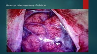
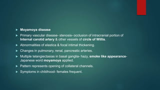
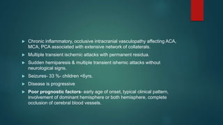
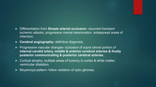
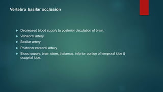
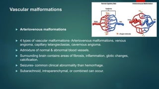
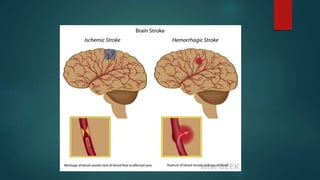
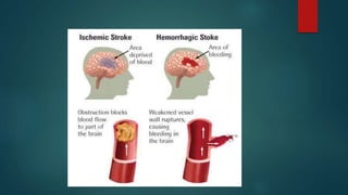
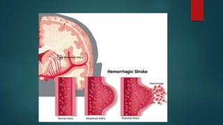
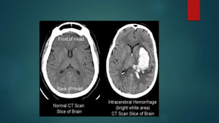
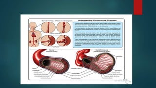
Strokes can occur in children and are usually caused by arterial blockages or venous clots. The incidence of arterial ischemic stroke and cerebral venous thrombosis is approximately 5 per 100,000 children per year. While relatively rare in children, strokes are an important cause of acquired brain injury in newborns and children. The main causes of pediatric strokes include congenital heart defects, infections, vascular abnormalities, genetic conditions, and trauma.
This document discusses various causes and presentations of pediatric stroke. Some key points:
- Arterial ischemic stroke and cerebral venous thrombosis have incidences of 5/100,000/yr and 1 in 2000 newborns respectively. Neonates have a higher risk than older children.
- Common causes of pediatric stroke include cardioembolism from congenital heart defects, arteriopathies like moyamoya disease, hematologic disorders like sickle cell anemia, and various genetic/metabolic conditions.
- Presentations depend on age but can include seizures, motor deficits, headaches, and decreased consciousness. Diagnosis involves imaging like MRI/MRA while treatment depends on the underlying cause but may include
1. Myasthenia gravis is an autoimmune disease causing muscle weakness and fatigability due to antibodies blocking acetylcholine receptors at the neuromuscular junction.
2. Diagnosis involves eliciting a history of fluctuating weakness and physical exam findings of rapidly fatigable weakness, as well as repetitive stimulation tests and tensilon tests.
3. Treatment includes acetylcholinesterase inhibitors, immunosuppressive drugs, thymectomy, and management of myasthenic crisis with IV immunoglobulin or plasma exchange. Long-term management requires balancing medication side effects in children.
The document provides information on pediatric stroke. It defines stroke and describes the different types that can occur in children, including arterial ischemic stroke, cerebral sinovenous thrombosis, and hemorrhagic stroke. Risk factors and potential causes are discussed for each type. Clinical features may include seizures, weakness on one side of the body, difficulty speaking or swallowing. Diagnosis involves neuroimaging like CT or MRI along with other lab tests. Treatment focuses on neuroprotection, recanalization of blocked vessels, and anticoagulation or antiplatelet therapies to prevent further clotting.
This document discusses pediatric stroke. It begins with definitions, types, epidemiology, etiology, and pathophysiology of pediatric stroke. The main types are ischemic and hemorrhagic stroke. Risk factors in children include structural heart disease, vasculopathies, hematological disorders, and prothrombotic states. Clinical features can include focal neurological deficits like hemiparesis. Diagnosis involves neuroimaging such as MRI and distinguishing stroke from other conditions. Management aims to prevent recurrence and support rehabilitation.
The document provides information on approaching patients with potential neurodegenerative disorders, including two patient scenarios. It discusses:
1) Classifying neurodegenerative disorders as either gray matter or white matter diseases based on features like age of onset, head size, seizures, cognition, and exam findings.
2) The key is obtaining a thorough history and physical exam to determine if it is a neurodegenerative process and rule out other treatable conditions.
3) Common inherited and acquired neurodegenerative disorders are described based on features like onset age, neurological exam, investigations and whether they primarily affect gray or white matter.
The document discusses neurodegenerative disorders in children. It describes how these disorders involve the progressive deterioration of neurological function, often with regression of developmental milestones. The disorders can affect gray matter, white matter, or both. Investigations aim to identify the underlying genetic or metabolic cause, while management focuses on treating complications and providing supportive care.
This document discusses childhood stroke, including:
- Childhood stroke differs from adult stroke in its causes, which include cardiac abnormalities, infections, genetic conditions, and hematologic disorders rather than atherosclerosis.
- Diagnosing childhood stroke is challenging due to its rarity and non-specific clinical presentations. Imaging and laboratory tests are used to determine the cause and guide treatment.
- Treatment depends on the underlying cause but may include thrombolysis, anticoagulation, surgery, or lifestyle changes. Recurrence risks vary based on identified risk factors. Outcomes range from full recovery to lasting deficits, though prognosis is generally better than in adult strokes.
This document discusses heart failure in children, including its definition, types, causes, symptoms, diagnosis, complications, and management. Heart failure occurs when the heart cannot pump enough blood to meet the body's needs. In children, common causes include congenital heart disease, rheumatic heart disease, and cardiomyopathy. Symptoms vary by age but may include feeding issues, sweating, poor growth, and edema. Diagnosis involves exams, chest x-rays, electrocardiograms, and echocardiograms. Complications can include arrhythmias, infections, and damage to other organs. Treatment focuses on supportive care, medications to improve heart function, and treating the underlying cause. Prognosis depends on the cause,
1) Pediatric strokes account for less than 5% of all strokes and affect 2-3 in 100,000 newborns and 12 in 100,000 children under 18 years of age. The annual incidence of pediatric strokes is reported to be between 2.5-2.7 per 100,000 children.
2) Risk factors for pediatric strokes include congenital heart defects, sickle cell anemia, coagulation disorders, and other conditions.
3) Prognosis after a pediatric stroke varies depending on the underlying cause, with 80% of children surviving 10 years after an ischemic stroke though most have residual hemiparesis. Hemorrhagic strokes carry a higher mortality risk than ischemic strokes.
This document discusses stroke in children. Key points include:
- Stroke in children differs from adults and can have developmental, genetic, or environmental causes rather than lifestyle factors.
- Presentation is often subtle with a wide differential diagnosis. Risk factors are multiple and poorly understood.
- Neonates are at highest risk. Incidence of ischemic stroke is around 1 in 4000-5600 term births. Cerebral venous thrombosis accounts for 0.67 cases per 100,000 children per year.
- Evaluation involves detailed history and physical exam looking for signs of bleeding disorders, infections, cardiac abnormalities, or genetic syndromes. Imaging and lab tests are needed to make an accurate diagnosis.
Epilepsy syndromes can be categorized based on age of onset from neonates to childhood. Syndromes include self-limited, developmental and epileptic encephalopathies, and genetic generalized epilepsies. Syndromes are defined by seizure type, EEG findings, development, and treatment response/prognosis. Examples provided include benign familial neonatal epilepsy, Dravet syndrome, childhood absence epilepsy, Lennox-Gastaut syndrome, and Landau-Kleffner syndrome.
This document discusses pediatric stroke, including definitions, incidence, causes, investigations, management, and prognosis. Some key points include:
- Pediatric stroke can be ischemic or hemorrhagic and has a variety of potential causes including congenital heart disease, sickle cell anemia, infections, and hypercoagulable states.
- Brain MRI is the preferred imaging modality to diagnose stroke in children. Additional tests may include MRA, CTA, echocardiogram, and lab work to investigate underlying conditions.
- Initial management involves supportive care while determining the cause. Long-term treatment depends on the etiology but may include anticoagulation/antiplatelet therapy and management of underlying conditions to
This document outlines immunology and immunity to infection. It discusses the immune system's role in fighting infection, including humoral immunity from B cells and cellular immunity from T cells. It also addresses intracellular and extracellular pathogens. The document then focuses on primary and secondary immunodeficiency, describing the types of primary immunodeficiencies including B cell, T cell, phagocytic, and complement defects. It provides guidance on clinical evaluation and diagnostic approach for patients with suspected immunodeficiency.
Stroke can occur in children and presents with rapidly developing neurological deficits lasting more than 24 hours. Mortality from pediatric stroke is 6-20% and over 50% of children have residual disabilities. There are several types of stroke in children including hemorrhagic, caused by bleeding, and ischemic, caused by blocked blood vessels. Etiologies of pediatric stroke include various vascular, cardiac, hematologic and other conditions. Aggressive management in the PICU focuses on treating the underlying cause, preventing secondary brain injury, and managing complications.
ATAXIA IN CHILDREN -CAUSES, MANAGEMENT, INVESTIGATIONS, TYPES, COMMONEST ATAXIA IN CHILDREN IN DETAIL, HOW WILL YOU FIND OUT THE CAUSE FOR ATAXIA IN CHILDREN FLOWCHART, DEFINITION, TREATMENT
This document contains a series of multiple choice questions related to pediatric neurology. It covers topics like cerebrospinal fluid findings in different conditions, genetic disorders presenting with floppy babies, developmental milestones, seizures types and their characteristics, inborn errors of metabolism, cerebral palsy subtypes and their causes. The questions assess knowledge on clinical features, investigations, management and genetics of various pediatric neurological disorders.
This document discusses the approach to hypoglycemia in childhood. It begins by defining hypoglycemia and describing the importance of glucose for brain development. It then discusses the pathophysiology of hypoglycemia, focusing on how the body maintains blood glucose levels through glycogenolysis, gluconeogenesis, and lipolysis. The clinical features of hypoglycemia are presented, distinguishing between sympathetic overactivity and neuroglycopenic symptoms. Common etiologies like hyperinsulinism, metabolic disorders, and systemic illnesses are outlined. The document concludes with recommendations for investigating hypoglycemia, managing acute episodes, and treating underlying causes to prevent long-term neurological consequences.
1. The document provides guidance on evaluating and diagnosing anemia in children. It outlines key signs, symptoms, and pointers that suggest a child may have anemia.
2. Laboratory tests that can help determine the severity and type of anemia include complete blood count, hematocrit, reticulocyte count, blood indices, and peripheral smear.
3. A thorough history, physical exam, and lab work are needed to assess if a child is anemic, determine the severity, and identify the potential cause and type, such as blood loss, decreased red blood cell production, or increased red blood cell destruction.
Iron-deficiency anemia is the most common nutritional disorder worldwide. It occurs when iron levels in the body are low and there is not enough iron to produce normal red blood cells. Symptoms can include pallor, fatigue, and irritability. Diagnosis involves blood tests showing low iron levels, smaller and fewer red blood cells. Treatment is oral iron supplementation which leads to improved hemoglobin levels within weeks. Prevention focuses on breastfeeding, iron-fortified formula for infants, and limiting milk intake after age 1.
Childhood demyelinating syndromes
In the past decade, the number of studies related to demyelinating diseases in children has exponentially increased. Demyelinating disease in children may be monophasic or chronic. Typical monophasic disorders in children are acute disseminated encephalomyelitis and clinically isolated syndromes, including optic neuritis and transverse myelitis. However, some cases of acute disseminated encephalomyelitis or clinically isolated syndrome progress to become chronic disorders, including multiple sclerosis and neuromyelitis optica. This review summarizes the current knowledge on monophasic and chronic demyelinating disorders in children, focusing on an approach to diagnosis and management.
Iron deficiency anemia is the most common nutritional deficiency in children worldwide. It occurs most frequently in infants aged 6-24 months, especially those who are artificially fed or from low socioeconomic backgrounds. Iron deficiency anemia develops in stages from iron depletion to iron deficiency to anemia, characterized by low iron stores, serum iron, and high total iron binding capacity. Clinically, it presents with pallor, fatigue, and impact on development in children. Treatment involves oral or parenteral iron replacement therapy, addressing the underlying cause, and ensuring an adequate iron intake.
This document discusses various conditions that can mimic epilepsy in children. It notes that epilepsy is sometimes underdiagnosed or overdiagnosed due to unusual symptom presentations or epilepsy mimics. Several common epilepsy mimics are described in detail for different age groups, including breath holding spells in infants, tics and parasomnias in children, and syncope in adolescents. Tables compare features of epilepsy mimics to epileptic seizures during sleep and wakefulness. In conclusion, the document emphasizes taking an age- and state-based approach to differentiating epilepsy from conditions it may imitate.
Pediatric ARDS is a common cause of respiratory failure in children. It is defined by acute onset hypoxemia that cannot be explained by cardiac failure, with bilateral lung opacities on chest imaging. Management involves controlling the underlying cause, lung protective ventilation with low tidal volumes, permissive hypercapnia, prone positioning, and consideration of recruitment maneuvers, HFOV, surfactant, inhaled nitric oxide, or ECMO in severe cases. Noninvasive ventilation may be tried initially for mild disease but intubation is often required for more severe pediatric ARDS. The goals of management are to maintain adequate oxygenation and ventilation while minimizing ventilator induced lung injury.
aplastic anemia pediatrics
It compromises a group of disorders of the hematopoietic stem cells resulting in the suppression of one or more of erythroid, myeloid and megakaryotic cell lines.
thrombocytopenia
This document provides an overview of leukodystrophies and discusses their clinical presentation and neuroimaging features. It begins with definitions of leukodystrophies and outlines their age of onset. Common clinical features are then described, including neurological, non-neurological, ophthalmological, and radiological findings. A stepwise approach to the neuroimaging of leukodystrophies is presented, focusing on patterns of white matter involvement that can help differentiate genetic from acquired causes.
This document provides an overview of hemolytic anemia in children. It defines hemolytic anemia as anemia resulting from increased red blood cell destruction. The document describes the different types of hemolytic anemia including hereditary, immune, and non-immune causes. It outlines the pathophysiology, clinical features, diagnostic approach and management of common forms of hemolytic anemia in children such as hereditary spherocytosis, thalassemia, sickle cell anemia, and G6PD deficiency. Investigations for diagnosis include blood counts, peripheral smear, reticulocyte count, hemoglobin electrophoresis and enzyme or genetic testing depending on etiology.
Pediatric stroke can be caused by a variety of factors such as cardiac diseases, infections like varicella, sickle cell disease, moyamoy disease, cerebral sinus thrombosis, and genetic conditions like MELAS. The presentation of pediatric stroke depends on the location and size of the lesion in the brain. Diagnosis involves imaging techniques like CT, MRI, MRA and angiography. Early diagnosis and treatment is important to prevent long term neurological deficits in children.
This document discusses ataxia in children. It describes the different types of ataxia including sensory ataxia and cerebellar ataxia. It outlines the characteristic features, locations of lesions, physical exam findings, and hints to differentiate between sensory and cerebellar ataxia. The document also provides guidance on evaluating a child with ataxia including taking a thorough history, performing a full physical and neurological exam, ordering appropriate tests and imaging, and considering possible consultations. Common causes of ataxia in childhood are discussed such as congenital, degenerative/genetic, infectious, metabolic, neoplastic, toxic, traumatic and vascular etiologies.
This document summarizes pathology related to cerebrovascular disease. It discusses different types of strokes including ischemic, hemorrhagic, and transient ischemic attacks. Specific factors that can contribute to tissue damage in ischemic strokes are explained. Locations and appearances of infarcts over time are described. Causes and presentations of hemorrhagic strokes including those from aneurysms and arteriovenous malformations are covered.
This document summarizes information about strokes (cerebrovascular disease). It discusses that strokes are caused by reduced blood flow to the brain and can be ischemic (lack of blood flow) or hemorrhagic (bleeding). The most common causes are atherosclerosis and hypertension. Ischemic strokes are more common and can be thrombotic, embolic, or lacunar. Clinical signs depend on the location and size of the affected brain area. Investigations help determine the type and severity of stroke.
This document summarizes the management of patients with cerebrovascular disorders such as stroke. It discusses the two main types of strokes - ischemic and hemorrhagic. Ischemic strokes are caused by blockage of blood flow to the brain while hemorrhagic strokes involve bleeding into or around the brain. Risk factors, pathophysiology, clinical manifestations, diagnostic assessments, medical and nursing management are described for both types of strokes. Surgical procedures like carotid endarterectomy are mentioned as prevention and treatment options for ischemic strokes.
Stroke is the third leading cause of death and can be caused by thrombosis, embolism, or hemorrhage. The main types of cerebral vascular diseases that cause stroke are thromboembolic infarction, intracerebral hemorrhage, and subarachnoid hemorrhage. Risk factors for stroke include age, gender, hypertension, cardiac diseases, diabetes, smoking, and high alcohol intake. Transient ischemic attacks are focal neurological symptoms lasting less than 24 hours and often precede a major stroke.
A cerebrovascular accident (CVA), also known as a stroke, is caused by the loss of blood flow to an area of the brain resulting in cell death. It is a leading cause of death and disability. The majority of strokes are ischemic and caused by blockage of an artery by a clot or debris. Hemorrhagic strokes occur when a blood vessel ruptures, causing bleeding into or around the brain. Treatment focuses on prevention through risk factor control as well as acute interventions like clot-busting drugs. Rehabilitation aims to maximize recovery of function and independence.
Subarachnoid hemorrhage is caused most commonly by the rupture of a saccular aneurysm. The rupture causes blood to fill the subarachnoid space, which can lead to neurological deficits or death. Treatment involves securing the aneurysm through surgical clipping or endovascular coiling to prevent rebleeding, as well as managing complications like vasospasm, hydrocephalus, and seizures. Outcomes depend on the grade and location of the initial bleed and development of delayed cerebral ischemia.
This document defines stroke as an acute CNS injury resulting from reduced blood flow to the brain. Strokes can be ischemic, caused by blockage of a blood vessel, or hemorrhagic, caused by rupture of a blood vessel. The main types of ischemic stroke are thrombotic, caused by clots within blood vessels, and embolic, caused by clots or debris traveling from elsewhere. Cardioembolic strokes are a type of embolic stroke where clots originate from the heart. Risk factors, signs and symptoms, treatment, and the effects of left versus right brain strokes are described. Transient ischemic attacks are also summarized.
Cerebrovascular accident (CVA), also known as stroke, results from ischemia or hemorrhage in the brain and is a leading cause of death and disability. Approximately 750,000 strokes occur in the US each year. Risk factors include age, gender, race, family history, hypertension, diabetes, smoking, heart disease, and atrial fibrillation. Treatment involves stabilizing the patient, performing diagnostic imaging to determine the type of stroke, administering thrombolytics if appropriate, treating underlying risk factors, and long-term rehabilitation. The goals are to prevent strokes through risk factor control and lifestyle modifications.
This document provides an outline and overview of key topics related to stroke. It begins with definitions and classifications of stroke, including transient ischemic attack (TIA) and different types of stroke. It then covers risk factors, pathophysiology, signs and symptoms, investigations, and management approaches for stroke. Specific sections address hemorrhagic versus ischemic stroke, localization of stroke syndromes, and differentiating features between anterior and posterior circulation strokes. Differential diagnoses are also listed. The document aims to present essential information on stroke for medical education purposes.
This document provides an outline for a presentation on stroke. It begins with an introduction defining stroke and classifying it as either transient ischemic attack (TIA), progressive stroke, or completed stroke. It then covers the types and risk factors of stroke, including modifiable and non-modifiable risk factors. The pathophysiology of both ischemic and hemorrhagic stroke is explained. Signs and symptoms of stroke are outlined, including localization of symptoms based on hemisphere affected. Investigations, prognostic factors, and management of both acute stroke and long-term prevention are summarized.
This document provides an overview of cerebrovascular accidents (CVAs), also known as strokes. It defines CVAs, describes the blood supply to the brain, and classifies the major types of strokes as ischemic (caused by reduced blood flow) or hemorrhagic (caused by bleeding). It discusses the causes, risk factors, clinical presentation, progression, and management of different stroke subtypes, including transient ischemic attacks, infarction, and intracerebral hemorrhage.
750,000 strokes occur in the US annually, making it the third leading cause of death and the leading cause of disability. A stroke is caused by ischemia or hemorrhage in the brain resulting in death of brain cells. Risk factors include age, gender, race, family history, hypertension, diabetes, smoking, heart disease, and atrial fibrillation. Treatment involves stabilizing the patient, administering fibrinolytic drugs if applicable, treating underlying causes, and long-term rehabilitation.
The document discusses stroke, including its types, risk factors, pathophysiology, and clinical manifestations based on the artery affected. Ischemic stroke is more common than hemorrhagic and results from blockage of an artery depriving the brain of blood flow. Clinical features vary depending on the specific artery involved, such as contralateral weakness with middle cerebral artery stroke or visual field defects with posterior cerebral artery stroke. Complications can include altered consciousness, speech/language issues, and emotional or cognitive changes.
Cerebral microbleeds are small brain hemorrhages detected by MRI that are caused by leakage of blood from damaged small vessel walls. They are increasingly recognized in patients with cerebrovascular disease, Alzheimer's disease, vascular cognitive impairment, and normal elderly populations. Microbleeds in lobar regions may indicate cerebral amyloid angiopathy and link vascular and amyloid neuropathologies, while deep or infratentorial microbleeds often reflect hypertensive vasculopathy. Detection of microbleeds provides insight into cerebral small vessel disease and its relationship to cognitive impairment and dementia.
Neurological Conditions and Diseases (At birth)Liew Boon Seng
This document discusses various neurological conditions and diseases that can cause macrocephaly in infants and children. It describes conditions present at birth such as caput succedaneum, subgaleal hemorrhage, cephalohematoma, osteopetrosis, subdural hematomas, benign enlargement of the subarachnoid space, megalencephaly, vein of Galen aneurysm, and hydrocephalus. Hydrocephalus and its causes, clinical presentation, assessment, treatments including shunts, and complications are discussed in detail. Posthemorrhagic hydrocephalus as a consequence of intraventricular hemorrhage is also outlined.
The document defines various terms related to strokes, including stroke, TIA, progressive stroke, completed stroke, and hemorrhagic vs ischemic stroke. It discusses the epidemiology, risk factors, types, clinical features, investigations, and differential diagnosis of strokes. Specifically, it provides details on the clinical presentations and neurological deficits associated with occlusion of different arteries in the anterior and posterior circulations. It also outlines the objectives and modalities used to investigate a potential stroke, including non-invasive tests like CT, MRI, and Doppler ultrasound and invasive tests like angiography.
This document discusses pathology of the nervous system. It covers various disease mechanisms including increased intracranial pressure, vascular and circulatory disorders, trauma, infections, tumors, demyelinating diseases, and developmental abnormalities. It describes the main cell types in the central nervous system and their functions. Key pathologies covered include strokes, herniations, hydrocephalus, and effects of trauma. Specific vascular disorders like ischemia, infarction, hemorrhage, and their causes are explained.
CVA and its causes and sign , symptoms treatmentwajidullah9551
The document discusses cerebrovascular accidents (CVAs), also known as strokes. It covers the definition, risk factors, types, signs and symptoms, diagnostic studies, treatment goals, and management of strokes. The two main types of strokes are ischemic, resulting from blocked blood flow, and hemorrhagic, caused by bleeding in the brain. Management involves stabilizing the patient, performing diagnostic imaging, treating underlying causes, and rehabilitation to recover functions.
This document provides information about stroke, including:
- Strokes are caused by a blockage or rupture of an artery in the brain, depriving brain tissue of oxygen.
- Symptoms vary depending on the area of brain affected but can include weakness, difficulty speaking or swallowing, and visual problems.
- Diagnosis involves medical history, physical exam, and brain imaging tests like CT scans or MRI.
- Treatment depends on the type of stroke but may include clot-busting drugs, surgery to repair blood vessels, and rehabilitation therapies. Prevention focuses on controlling risk factors like high blood pressure and smoking.
1. Childhood stroke is more common than brain tumors and is among the top 10 causes of death in childhood. The incidence is about 8 per 100,000 children and risk factors include congenital heart disease and prematurity.
2. The most common causes of acute ischemic stroke are arteriopathy, cardioembolism from structural heart disease, and hematological conditions like sickle cell anemia. Diagnosis involves CT, MRI, and angiography. Treatment focuses on antithrombotics and rehab.
3. Hemorrhagic stroke risk factors include vascular malformations, blood disorders, and trauma. Subarachnoid hemorrhage is the most common type. Cerebral sinovenous
This document discusses hepatitis, primarily caused by five hepatotropic viruses - hepatitis A, B, C, D, and E viruses. Hepatitis A virus and hepatitis B virus are the most common causes of acute viral hepatitis in children in India. Most cases of acute viral hepatitis improve spontaneously without treatment. Prolongation of prothrombin time is a reliable laboratory marker of worsening liver function or potential liver failure. Chronic infection with hepatitis B or C can potentially lead to chronic hepatitis.
This document discusses persistent pulmonary hypertension of the newborn (PPHN). It defines PPHN as sustained elevation of pulmonary vascular resistance, disrupting normal circulatory transition after birth. PPHN carries risks of chronic lung disease and neurodevelopmental issues. Treatment aims to reverse hypoxemia through respiratory support, pulmonary vasodilation (e.g. inhaled nitric oxide), and hemodynamic support (e.g. medications, ECMO). The prognosis of PPHN depends on the underlying cause and response to treatment.
The document describes the fetal circulation and changes that occur at birth. In the fetus, oxygenated blood from the placenta travels to the heart and body while bypassing the lungs. At birth, the onset of breathing causes pulmonary vascular resistance to decrease and systemic resistance to increase. This results in blood shunting to the lungs, closure of the ductus arteriosus, and closure of the foramen ovale. The umbilical vessels also close, completing the transition to extrauterine life.
The heart develops from cardiac progenitor cells that migrate to the cardiogenic field and form blood islands that fuse to create the endothelial heart tube. The heart tube undergoes cardiac looping and septation to form the four-chambered heart. During septation, the atria, ventricles, and great arteries are partitioned. The aortic arches give rise to major blood vessels of the head and neck. By the end of the 7th week, septation is largely complete and the heart has transformed into a functional four-chambered pump.
Neonatal infectious diseases jornal 2nd topicRobin Thomas
This document summarizes neonatal sepsis, which is divided into early onset sepsis (EOS) occurring in the first week of life, and late onset sepsis (LOS) occurring after the first week. EOS is often caused by maternal transmission of organisms like Group B Streptococcus. LOS is attributed to postnatal pathogens acquired in the hospital and common organisms include coagulase-negative Staphylococci. Preterm and very low birth weight infants are especially at risk due to immunological immaturity. Blood cultures remain the gold standard for diagnosis but biomarkers like CRP and PCT are also used. Prevention strategies focus on identifying at-risk mothers, intrapartum antibiotic prophylaxis, and reducing hospital-acquired
Journal: Approach to Common Bacterial Infections: Community acquired pneumoniaRobin Thomas
1. Community acquired pneumonia (CAP) presents differently in children and adults, with two main challenges being defining CAP in young children who often have viral and bacterial co-infections, and identifying the pathogen to avoid unnecessary antibiotic use.
2. The most prominent bacterial pathogens causing CAP across all age groups are Streptococcus pneumoniae and atypical organisms like Mycoplasma pneumoniae, while viruses account for the majority of CAP in children under 2 years old.
3. Clinical diagnosis and treatment of CAP in children is typically based on age, with guidelines recommending antibiotics for presumed bacterial CAP and observation without antibiotics for presumed viral CAP in preschool aged children.
share - Lions, tigers, AI and health misinformation, oh my!.pptxTina Purnat
• Pitfalls and pivots needed to use AI effectively in public health
• Evidence-based strategies to address health misinformation effectively
• Building trust with communities online and offline
• Equipping health professionals to address questions, concerns and health misinformation
• Assessing risk and mitigating harm from adverse health narratives in communities, health workforce and health system
Adhd Medication Shortage Uk - trinexpharmacy.comreignlana06
The UK is currently facing a Adhd Medication Shortage Uk, which has left many patients and their families grappling with uncertainty and frustration. ADHD, or Attention Deficit Hyperactivity Disorder, is a chronic condition that requires consistent medication to manage effectively. This shortage has highlighted the critical role these medications play in the daily lives of those affected by ADHD. Contact : +1 (747) 209 – 3649 E-mail : sales@trinexpharmacy.com
TEST BANK For An Introduction to Brain and Behavior, 7th Edition by Bryan Kol...rightmanforbloodline
TEST BANK For An Introduction to Brain and Behavior, 7th Edition by Bryan Kolb, Ian Q. Whishaw, Verified Chapters 1 - 16, Complete Newest Versio
TEST BANK For An Introduction to Brain and Behavior, 7th Edition by Bryan Kolb, Ian Q. Whishaw, Verified Chapters 1 - 16, Complete Newest Version
Promoting Wellbeing - Applied Social Psychology - Psychology SuperNotesPsychoTech Services
A proprietary approach developed by bringing together the best of learning theories from Psychology, design principles from the world of visualization, and pedagogical methods from over a decade of training experience, that enables you to: Learn better, faster!
8 Surprising Reasons To Meditate 40 Minutes A Day That Can Change Your Life.pptxHolistified Wellness
We’re talking about Vedic Meditation, a form of meditation that has been around for at least 5,000 years. Back then, the people who lived in the Indus Valley, now known as India and Pakistan, practised meditation as a fundamental part of daily life. This knowledge that has given us yoga and Ayurveda, was known as Veda, hence the name Vedic. And though there are some written records, the practice has been passed down verbally from generation to generation.
Histololgy of Female Reproductive System.pptxAyeshaZaid1
Dive into an in-depth exploration of the histological structure of female reproductive system with this comprehensive lecture. Presented by Dr. Ayesha Irfan, Assistant Professor of Anatomy, this presentation covers the Gross anatomy and functional histology of the female reproductive organs. Ideal for students, educators, and anyone interested in medical science, this lecture provides clear explanations, detailed diagrams, and valuable insights into female reproductive system. Enhance your knowledge and understanding of this essential aspect of human biology.
Basavarajeeyam is a Sreshta Sangraha grantha (Compiled book ), written by Neelkanta kotturu Basavaraja Virachita. It contains 25 Prakaranas, First 24 Chapters related to Rogas& 25th to Rasadravyas.
Integrating Ayurveda into Parkinson’s Management: A Holistic ApproachAyurveda ForAll
Explore the benefits of combining Ayurveda with conventional Parkinson's treatments. Learn how a holistic approach can manage symptoms, enhance well-being, and balance body energies. Discover the steps to safely integrate Ayurvedic practices into your Parkinson’s care plan, including expert guidance on diet, herbal remedies, and lifestyle modifications.
TEST BANK For Basic and Clinical Pharmacology, 14th Edition by Bertram G. Kat...rightmanforbloodline
TEST BANK For Basic and Clinical Pharmacology, 14th Edition by Bertram G. Katzung, Verified Chapters 1 - 66, Complete Newest Version.
2. Stroke-important cause of acquired brain injury in newborns and children.
Relatively rare-children- Arterial or Venous stroke.
Incidence of Arterial ischemic stroke (AIS) and cerebral sinovenous
thrombosis (CSVT)-5/100,000/yr and affects 1 in 2000 newborns.
18. Pathophysiology
Interruption of blood flow to the part of the brain
Rupture of blood vessels with bleeding into cerebral parenchyma.
Blood supply of brain- carotid & vertebro-basilar circulations
Numerous anastomosis at the level of circle of willis & through smaller
vessels in leptomeninges.
Diencephalon- supplied by end arteries- anastomosis not adequate-arterial
occlusions have devastating effect.
Water shed zones- portion of cerebral cortex b/w 2 major arteries- less
affected by arterial occlusions –damage when cerebral perfusion pressure
is reduced.
19. Pathophysiology
Interruption of blood flow of arterial or thrombotic d/s- Ischemic stroke.
Rupture of blood vessels with bleeding into cerebral parenchyma-
Hemorrhagic stroke.
Trauma or intimal tears- ICA injury.
Interruption of function – infections, inflammation of intima- severe meningitis,
post varicella angiopathy, mycoplasma pneumonia, borrelia, chlamydia, HIV,
Helicobactor, Hemolysing streptococci.
Embolisation- Cardioembolism- CCHD, RHD, Prosthetic or Prolapsed
cardiac valves, Cardiomyopathy, Arrhythmias.
20. Decreased cerebral blood flow- high cerebral metabolic rate & paucity of
energy stores in CNS.
Cerebral metabolic rate for oxygen- 3.5 ml/100mg brain/min.
Cerebral hypoxia- oxygen partial pressure < 40 torr.
Glucose storage in brain- survival of cerebral tissue for 90 min.
21. Preterm infants
Sub ependymal germinal matrix- highly cellular well vascularized area
beneath the ependyma of lateral ventricles- major site of neurogenesis.
Sub ependymal hemorrhage & Intraventricular hemorrhage- common form
of CVD in preterm infants.
Bleeding in highly vascular sub ependymal germinal matrix & then rupture
into lateral ventricle.
Hemorrhagic infarction of periventricular white matter-15% of infants with
intraventricular hemorrhage.
22. Several factors- fluctuating cerebral blood flow, increase in cerebral
venous pressure, immature capillaries in germinal matrix, abnormal
platelet & coagulation function, low Apgar scores, low birth weight,
prolonged labour, apnea, bradycardia, large PDA, pneumothorax, group B
streptococcal sepsis, hypoxia, hypercapnia, acidosis.
Bed side Ultrasonography- better understanding of subependymal
hemorrhage & intraventricular hemorrhage.
23. Term infants
Intra cranial hemorrhage- birth trauma- forceps & breech deliveries.
Tears of falx cerebri & tentorium cerebelli.
C/F- apathy, seizures, high pitched cry, irregular respirations.
Tense fontanel, moro reflex abnormalities.
Focal ischemic lesions can occur- DIC, placental infarcts, embolism,
trauma to blood vessels.
Ultrasonography & CT scans- diagnosis.
24. Neuronal injury & death in hypoxic states- release of excitatory
neurotransmitters: L-glutamate & L-aspartate.
Increased post synaptic stimulation of N-methyl-D-aspartate receptors-
entry of sodium & calcium into neurons & cell death.
Delayed cell death- calcium influx- mitochondrial dysfunction with
breakdown of cell components & free radical formation.
Major factor controlling cerebral metabolic rate-degree of neuronal activity.
Seizures- cerebral metabolic rate increases & reduced to low levels in
coma.
25. Cerebral blood flow maintained at high levels- substrate for brain
metabolic activity.
< 3 yrs- 30-60 ml/100 mg brain/min.
3-10 yrs- 105 ml/100 mg brain/min.
Adults- 50 ml/100 mg brain/min.
Mature brain- phenomenon of Auto regulation.
Blood flow increases with increased neuronal activity.
Perfusion pressure, intracranial pressure & vascular resistance.
26. Increasing conc. of CO2- increase of blood flow- dilatation of intracranial
blood vessels.
High conc. of O2- reducing blood flow- causing vasoconstriction.
Most cerebral vascular accidents in children- impairment of arterial blood
flow- result of thrombosis or embolism.
Localized region of metabolic acidosis- dilatation of surrounding blood
vessels- increased vascularity- luxury perfusion.
Damage to neurons & glia- destruction of blood brain barrier- localized
cerebral edema- compress capillaries.
27. Occlusion of venous structures- increased venous pressure- tendency for
blood vessel to rupture- bleeding & raised intra cranial pressure.
Hemorrhage- intra parenchymal or extra cerebral- acts as mass lesion-
rise in intra cranial pressure.
Damage to blood-brain barrier- promotes cerebral edema.
Blood & blood products of erythrocytes- produce vascular spasm-add to
preexisting damage- cause meningeal irritation- can cause hydrocephalus.
28. Cerebral infarction pathologic changes- neuronal death & perivascular
hemorrhage- influx of polymorpho-nuclear leukocytes- mononuclear cells
& macrophages.
Astrocytosis results in formation of glial scar.
Preterm infant with sub ependymal germinal matrix hemorrhage-
destruction of white matter- periventricular leukomalacia.
Ventricular dilatation – destruction & disappearance of periventricular white
matter, post hemorrhagic hydrocephalus.
41. Acute infantile hemiplegia
Sudden onset of pediatric stroke-no specific cause delineated.
Thrombotic occlusions of carotid artery or branches of middle cerebral
artery- frequently documented causes of strokes in children.
3 dimensional MRA- demonstrate significant vascular abnormalities in 75%
of children with strokes. (Wiznitzer & Masaryk, 1991)
Infants- seizures, motor signs few, abnormal hand preference.
Older children- sudden onset of hemiparesis, seizures.
42. CT or MRI scan- mass lesion, intracranial hemorrhage or arteriovenous
malformation.
Angiography or MRA- moyamoya disease & fibro-muscular dysplasia-
visualization of major blood vessels.
Lumbar puncture- infectious etiological condn.
49. Moyamoya disease
Primary vascular disease- stenosis- occlusion of intracranial portion of
Internal carotid artery & other vessels of circle of Willis.
Abnormalities of elastica & focal intimal thickening.
Changes in pulmonary, renal, pancreatic arteries.
Multiple telangiectasias in basal ganglia- hazy, smoke like appearance-
Japanese word moyamoya applied.
Pattern represents opening of collateral channels.
Symptoms in childhood- females frequent.
50. Chronic inflammatory, occlusive intracranial vasculopathy affecting ACA,
MCA, PCA associated with extensive network of collaterals.
Multiple transient ischemic attacks with permanent residua.
Sudden hemiparesis & multiple transient ishemic attacks without
neurological signs.
Seizures- 33 %- children <6yrs.
Disease is progressive
Poor prognostic factors- early age of onset, typical clinical pattern,
involvement of dominant hemisphere or both hemisphere, complete
occlusion of cerebral blood vessels.
51. Differentiation from Simple arterial occlusion- recurrent transient
ischemic attacks, progressive mental deterioration, widespread areas of
infarction.
Cerebral angiography- definitive diagnosis.
Progressive vascular changes- occlusion of supra clinoid portion of
internal carotid artery, middle & anterior cerebral arteries & finally
posterior communicating & posterior cerebral arteries.
Cortical atrophy, multiple areas of lucency in cortex & white matter,
ventricular dilatation.
Moyamoya pattern- follow radiation of optic gliomas.
59. Fibro muscular dysplasia
Non atherosclerotic, non inflammatory vascular disease that causes
abnormal growth within wall of artery.
Common arteries- carotid & renal arteries.
Cause for childhood stroke & secondary hypertension.
Fibromuscular dysplasia of intra cranial vessels in children rare.
Angiographic finding of string of beads appearance of artery.
60.
61. Migraine
Hemiplegic migraine- transient hemiparesis with severe headache.
Familial forms & sporadic forms.
Transient loss of vision in one eye- amaurosis fugax- adolescent
migraineurs.
64. Vertebro basilar occlusion
Sudden onset or stuttering progression
Brain stem localization- corticospinal & cerebellar signs with oculomotor
abnormalities.
Locked in syndrome- infarction at the level of Basilar artery. (Golden etal
1987)
Child alert, quadriplegia, facial diplegia, absent horizontal eye movements.
Child cannot speak, preserved vertical eye movements.
Diagnosis considered in a child in coma after a vascular accident but has
spontaneous eye opening.
65. Subclavian steal syndrome
Retrograde flow of blood in vertebral artery, due to proximal stenosis of
subclavian artery.
Follows correction of coarctation of aorta
Headache, dizziness, visual field defects after exercise, seizures.
Angiographic findings characteristic.
Treatment- ligating left vertebral artery or placing a subclavian artery graft.
66.
67. Cerebrovascular disease secondary to medical conditions
Cardiac disease
Clinical scenario
9/12 old infant k/c/o TOF came with h/o Left sided hemiparesis, seizures,
depressed state of consciousness.
MRI brain showed Right MCA infarct with cerebral edema.
68. CVA- complication of cyanotic congenital heart disease.
TOF, TGA- common.
Mech : Arterial thrombosis, venous sinus thrombosis, or embolism.
“Any child < 2 yrs with CHD , who has acute onset of neurological
signs- CVA should be considered as primary diagnosis ‘’
After 2 yrs , Brain abscess most common.
Embolic strokes- children with cyanotic congenital heart disease- R-L
shunt bypasses lungs, which normally filter small emboli.
69. Bacterial endocarditis- congenital heart disease & Rheumatic heart
disease- potential sources of emboli.
Thrombi can form on prosthetic cardiac valves- imp. cause of cerebral
emboli.
70. Hematologic & Neoplastic diseases
Sickle cell anemia (SCA)
most common Hemoglobinopathy assoc. with CVA.
Stroke in children <19 yrs of age with SCA- 8 %.
Incidence- 700 per 1 lakh children with SCA.
Stroke in SCA- large vessel disease, venous occlusion, subarachnoid or
intracerebral hemorrhage.
Neuroimaging- occlusion of large cerebral vessels or watershed infarction
secondary to disease of large vessels.
Fewer shows isolated subcortical or small cortical branch occlusion.
71. Neuro pathological examn. confirm infarctions in area supplied by anterior-
middle cerebral artery & thrombi in distal cervical & proximal intracranial
carotid arteries.
Strokes in children with SCA- highest incidence in 5-10 yrs.
Hemiparesis- most common symptom.
Aphasia- 20 %, Seizures- 15 %, TIA- 10 %
Persistent neurologic deficits & neuro psychologic abnorm.
Children with HBSS or SB thalassemia- highest incidence of stroke-
monitor with trans cranial USG.
72. Blood transfusion & exchange transfusion- standard mode of treatment of
acute stroke in SCA.
Periodic blood transfusions- decrease productions of sickle cells- reduce
recurrence of strokes by 90 %.
Intracranial hemorrhage- serious complication of any bleeding disorder.
Intracerebral, subarachnoid, subdural, intra spinal hemorrhage.
Symptoms- headache, seizures, depressed state of consciousness.
Intra spinal hemorrhages- weakness, back pain.
73. Hemophilia
Bleeding occurs in 25 %.
Bleeding more common in factor IX deficiency.
Intra cranial Hemorrhage common in children <18 yrs, esp. <3yrs.
Serious permanent deficit- 50 %, mortality- 35 %.
Complications of labour or delivery produce intracranial hemorrhage in
newborns with hemophilia.
Treatment- replace deficient clotting factors- performed prophylactically
after h/o head trauma.
74. Homozygous deficiency of Protein C in newborns- purpura fulminans &
venous thrombosis- thrombosis of cerebral veins.
Strokes –significantly reduced levels of protein C.
ITP- major intracranial hemorrhage can occur.
Subdural, intra parenchymal, intra ventricular hemorrhage- infants b/w 2
weeks & 6/12- Vitamin K deficiency.
Hemolytic uremic syndrome- seizures, depressed consciousness,
subarachnoid hemorrhage, hemiparesis, thrombotic strokes.
75. Leukemia
Intracranial hemorrhage- 20%.
Intracranial bleeding occurs in acute stage of leukemia with extremely
elevated high leukocyte count.
Intracerebral or extra cerebral hemorrhage- results from increased blood
viscosity.
Multiple small thrombi, damage to blood vessels.
Children with high leukocyte count- develop dural sinus occlusion with
increased intracranial pressure & headache.
CVA also occur after bone marrow transplantation.
76. Infectious diseases
Acute bacterial meningitis- treatment delayed- infectious arteritis-
multiple areas of arterial narrowing & occlusion.
Occlusion of veins or dural sinuses- complication of meningitis & follow
otitis media, mastoiditis, sinusitis & infection of scalp & face.
Clinical picture- convulsions, coma, changing neurologic signs, nuchal
rigidity, signs of infection.
Otitis media & mastoiditis- cause lateral sinus thrombosis associated
with abducens palsy & increased intracranial pressure.
77. Facial skin & para nasal sinus infections- produce cavernous sinus
thrombosis with proptosis, conjunctival reddening, retinal hemorrhages &
extra ocular palsies.
Retropharyngeal abscess- produces compression or thrombosis of
carotid artery.
Post varicella angiopathy, mycoplasma pneumonia, borrelia-burgdorferi,
chlamydia pneumonia, HIV, helicobactor pylori, hemolysing streptococci-
predisposition for stroke.
78. Cranial infections
Stroke common sequel of Severe meningitis- H. Influenza, Pneumococcal,
Tuberculous meningitis.
Purulent material around basal cisterns & orbito frontal area, circle of
Willis- envelops small arteries & veins- vasculitis & thrombus.
80. Metabolic disorders
Infants with fever & dehydration- primary venous or sinus thrombosis.
C/F- multiple seizures, changing neurological signs, convulsions, coma,
increased intracranial pressure.
Hypernatremic dehydration- seizures, depressed state of
consciousness.
Pathology- multiple hemorrhagic lesion in white matter.
Juvenile onset Insulin dependent Diabetes mellitus- acute
hemiparesis.
81. MELAS SYNDROME- epilepsia partialis continua or status epilepticus,
repeated strokes.
MRI- multiple areas of hyper intense signal in cortex & subcortical white
matter, sparing deep white matter.
82. Trauma & physical agents
Trauma to carotid artery- delayed onset of neurological signs-
thrombosis in vessel & extension into cerebral vessels.
Severe cerebral edema- death.
Permanent neurological residua- seizures & neuropsychological deficits.
Children- falling on stick held in mouth, lolli pop injury .
83. External trauma to carotid artery- hematoma on lateral portion of neck,
Horners syndrome, TIA followed by lucid interval –then sudden onset of
hemiplegia or hemiparesis.
Bone abnormalities of upper cervical spine & odontoid, trauma to cervical
spine- sudden twisting or jerking of head- injure carotid or vertebral
arteries.
Basilar skull fracture can cause laceration of carotid artery at foramen
magnum- severe bleeding from mouth & ipsilateral ear.
Radiation & chemotherapy.
84. Vascular malformations
Arteriovenous malformations
4 types of vascular malformations- Arteriovenous malformations, venous
angioma, capillary telangiectasias, cavernous angioma.
Admixture of normal & abnormal blood vessels.
Surrounding brain contains areas of fibrosis, inflammation, glotic changes,
calcification.
Seizures- common clinical abnormality than hemorrhage.
Subarachnoid, intraparenchymal, or combined can occur.
85. Intracerebral hematoma- focal neurological signs & increased intracranial
pressure.
Subarachnoid hemorrhage- sudden onset of headache, meningeal signs.
Vascular malformations may be located in cerebellum & brainstem.
50 % of children with intracranial arteriovenous malformations have bruits
heard over head.
“A cranial bruit heard in an infant younger than 4 months of age ,
even in the presence of loud cardiac murmur , is always assoc. with
intracranial arteriovenous malformation” (Cohen and Levin 1978)
86. CT with contrast, MRI, Arteriography.
RX- surgical accessibility of lesion.
Total surgical excision is curative, but best approach to treatment ??
Embolization of lesion may be effective.
Stereotactic radiosurgery with linear accelerator – effective modality for
some patients.
87. Vein of Galen malformations
Arteriovenous malformation of Vein of Galen- direct connection between
branches of carotid or vertebral circulation & Vein of Galen.
Vein undergoes aneurysmal dilatation because of high pressure & arteries
divide forming a network of vessels adjacent to the vein.
Development of malformation in infancy forms a hemodynamically
significant arteriovenous shunt.
88. Neonatal period- signs & symptoms of high output congestive heart failure.
Children- systolic heart murmur, cranial bruit, cardiomegaly, hepatomegaly,
tachycardia, respiratory distress, & pulmonary edema.
Death – cardiac failure.
Presentation in later infancy- Hydrocephalus, Subarachnoid hemorrhage.
Dilated veins over scalp, intracranial bruits.
Poor prognosis- death from hemorrhage, increased intracranial pressure or
cardiac failure.
89. Presentation in later life- Headache, signs of intracranial hemorrhage-
convulsions & focal neurological signs.
Signs of brain stem dysfunction & raised intracranial pressure.
Calcification within malformation on CT scan.
Arteriography- diagnostic.
RX- difficult- location, surrounding network of blood vessels, poor
cardiovascular status of pt.
Microsurgical techniques & staged surgical procedures.
Embolization effective in some.
90. Aneurysms
Uncommon in children less than 10 yrs.
Located in either anterior or posterior circulation.
Sudden onset of massive subarachnoid hemorrhage & depressed state of
consciousness.
CN II, III or both.
Commonly occur on anterior cerebral artery or internal carotid artery ,
distal portions of cerebral vasculature.
Usually are >1cm – intracranial hemorrhage, seizures.
92. Surgery, microsurgical techniques- definitive RX for aneurysms.
Aneurysm not removed- 50 % will bleed- serious neurological deficits.
Mycotic aneurysms- bleeding.
93. Neuro cutaneous syndromes
Sturge weber syndrome
Port wine stain on face & scalp , capillary venous angioma of meninges,
vascular abnormality within cortex & white matter of ipsilateral hemisphere.
Highest risk for brain involve.- bilateral port-wine stain, unilateral with
involv. of all three divisions of trigeminal nerve, involves eyelid.
Brain- gliosis, calcification & neuronal loss.
Seizures, hemiparesis, mental retardation.
Eye- glaucoma, angioma of retina & choroid.
94. Progressive abnormalities with areas of calcification, intractable seizures,
intellectual & behavioural deterioration.
Abnormalities of regional cerebral blood flow & progressive cerebral
atrophy.
Early excision of abnormal areas of cortex.
Affected area is large- hemispherectomy.
Lasers- reduce or eliminate port-wine stain.
95. Dural venous sinus thrombosis
Venous sinuses- major pathway for drainage of intracranial circulation.
Significant proportion of CSF drains into sagittal sinus through pacchionian
granulations.
Thrombosis of major venous sinuses causes increased intracranial
pressure by impeding venous outflow & interfering with resorption of CSF.
96. Sagital sinus thrombosis
Sagital sinus drains vast majority of cortical veins over brain convexity.
Partial or complete.
Occlusion of sagittal sinus causes stasis & thrombosis in connecting
cortical veins- assoc. hemorrhage over brain surface.
Common in children under 3 yrs, often during first year of life.
Thrombosis extends into cortical veins- rapidly increasing intracranial
pressure, changes in level of consciousness, seizures, focal motor
impairment.
97. Septic venous sinus thrombosis- most common in neonates.
Spinal fluid resorption impaired-compromise of spinal flow between
arachnoidal granulations & blood in sinus-communicating hydrocephalus.
Venous stasis promotes vascular congestion of brain parenchyma-
increase in intracranial pressure.
Sagital sinus thrombosis-distension of veins over scalp & superior
forehead.
98. Occlusion of sagittal sinus in older children- syndrome of pseudo tumor
cerebri , headache, CN 6 palsy- false localizing sign, papilledema, visual
loss.
Good prognosis.
Predisposing factors leading to sagittal sinus thrombosis- dehydration,
malnutrition, debilitating d/s, febrile illness, congenital heart disease,
hypercoagulable states.
Spinal fluid studies-normal in early course, later xanthochromic or flankly
blood with increased protein concn.
99. CT scan- increased density within sinuses-diagnosis of venous sinus
thrombosis. Superficial hemorrhage & cerebral edema.
Delta sign- CT scan with contrast shows enhancement around thrombotic
sinus.
MRA- flow void- thrombosis in the area of sinus.
Conventional angiogram- venous phase of study well documented.
No role for anticoagulant therapy.
Interventional thrombolytic therapy- useful in some.
RX for raised intracranial tension.
100. Lateral sinus thrombosis
Clinical- seizures, increased intracranial tension, decreased level of
consciousness.
Predisposing factors- otitis media, mastoiditis.
Otitic hydrocephalus- when otitis media & mastoiditis led to lateral sinus
thrombosis & increased intracranial pressure.
Vigorus RX for otitis media & mastoiditis, surgical intervention?
101. Cavernous sinus thrombosis
Cavernous sinus- CN 3, 4, 6, ophthalmic division of CN5, internal carotid
artery.
Rupture of artery- massive arteriovenous shunt with proptosis, bruit,
involve. of CNs.
Internal carotid artery becomes thrombosed in segment- cavernous sinus-
massive hemispherical infarction.
Predisposing factors- infection of orbit, paranasal sinus, skin of periorbital
& malar areas.
102. Clinical- conjunctival suffusion- peripheral conjunctival capillaries,
conjunctival edema, retinal edema.
Ptosis- CN 3.
External opthalmoplegia- CN3, 4, 6.
Septic cavernous sinus thrombosis- medical emergency- vigorous
antibiotic therapy.
No role for anticoagulant therapy.
103. Spinal cord vascular abnormalities
Thrombotic & embolic d/s of spinal cord- rare.
Arteriovenous malformation- back pain, gait abnormalities, bladder &
bowel dysfunction.
Neurological examn.- long tract signs, asymmetric tendon reflexes.
Angioma of skin of back- 20 % accentuated by Valsalva maneuver.
Bruits over spine- rare.
Subarachnoid hemorrhage, no localizing neurological abnorm.
Spinal cord arteriovenous malformation- multiple episodes of subarachnoid
hemorrhage?
104. High resolution CT or MRI scans- diagnosis.
Selective spinal angiography- outline feeding vessels & extend of
malformation.
Surgical excision possible in many.
106. Clinical presentation
Older children : Hemiplegia, Hemi sensory loss, Aphasia &
other neurological deficits.
Younger children : subtle, variable findings, seizure,
early hand preference, limp during walking.
107. Conditions which mimic stroke
Todds paralysis ( Transient post ictal hemiparesis )
Hemiplegic migraine
Syndrome of alternating hemiplegia
ICSOL ( Intracranial space occupying lesion )
Acute disseminated encephalomyelitis
108. History & Physical examination
H/O ear, throat, mastoid infection.
H/O intra oral or neck trauma.
H/O cardiac d/s.
H/O Hematological disorders.
H/O multifocal seizures, raised intracranial pressure, vomiting- ? Superior
sagittal sinus thrombosis.
H/O Hemiparesis & seizures in first two years of life- ?
Arterial occlusions.
109. Localization of Hemiplegia
1. Hemispheric lesion
a. Cortex - cortical features like seizures, dysplasia.
motor deficits are minimum.
Cortical type of sensory loss - Parietal lobe function-
( loss of tactile localization, 2 point discrimination,
stereognosis, graphaesthesia, sensory inattention ).
b. Corona radiata - absence of cortical features
motor deficits- unequal weakness of limbs on
C/L side. Either UL>LL or LL>UL.
Dulling of Primary modalities of sensation- touch,
superficial pain, temperature.
110. c. Internal capsule : C/L hemiplegia, uniform weakness of limbs on
C/L side, dulling of primary modalities of
sensation-touch, superficial pain, temperature.
Homonymous hemianopia
2. Brain stem : Crossed hemiplegia
I/L LMN cranial nerve palsy
C/L hemiplegia
112. Diagnostic Approach
Confirmation of presence of Cerebrovascular lesion.
Exclude other causes of neurological dysfunction.
Etiology of stroke
113. Evaluation of child with Stroke
Standard Evaluation
CT scan (plain)
If CT scan is normal , MRI scan (plain)
117. Treatment
Arterial Ischemic Stroke
No randomized control trial on children with AIS.
Treatment primary directed towards stabilizing systemic factors &
management of underlying causes.
Supportive care
Manage raised intracranial pressure, blood pressure & fluid balance.
Blood glucose carefully monitored-Hyperglycemia exacerbate infarct size.
Maintain normal body temperature.
118. Aggressive antiepileptic treatment.
Antithrombotic therapies
Use of anticoagulant therapy increasing in pediatric AIS.
Avoid anticoagulation in hemorrhage, hypertension, or bleeding diathesis.
Heparin
Use anticoagulation in children at high risk of recurrence/extension of
thromboembolic stroke & who are at minimum risk of secondary
hemorrhage.
119. Loading dose of Heparin 75-100 U/kg iv over 10 min followed by initial
maintenance dose of 28 U/kg/hr in children>1yr & 18 U/kg/hr in
older children.
Adjust Heparin dose to maintain APTT in range of 60-85 sec.
Low molecular weight Heparin
Greater safety & efficacy.
Monitoring done-Antifactor Xa assay once weekly or monthly.
SVT in infants- LMW Heparin for 7-14 days followed by Coumadin for 3/12.
120. Antiplatelet agents
Traditional role of aspirin in prevention of recurrence after TIA or ischemic
stroke.
Adults-Aspirin reduces stroke by 25 %.
Clopidogrel-no control trials, but may be good choice.
Dose-3-5 mg/kg/day.
121. Oral anticoagulation
Warfarin used for secondary prevention of stroke if aspirin fails.
Congenital or acquired heart disease
Severe hypercoagulable states
Arterial dissection
Recurrent AIS or TIA while on aspirin.
123. Surgical evacuation of hematomas, insertion of ventricular or
lumboperitoneal shunts & rarely revascularization procedures.
Revascularization procedures like Encephalo duroarterio synangiosis
(EDAS) or Pirl Synangiosis- important in treatment of moya moya
disease.
Rehabilitation therapy
Speech therapy
Occupational therapy
Physical & psychological therapy
124. Summary
Stroke in children relatively rare.
There are fundamental, etiologic & developmental differences in children
compared with adults .
Multiple causes for stroke in children & many risk factors.
Ischemic stroke-interruption of blood flow of arterial or thrombotic d/s.
Hemorrhagic stroke-rupture of blood vessels with bleeding into cerebral
parenchyma.
Hemorrhagic stroke higher mortality than ischemic stroke.
125. Bibliography
1. Adam K, Gabrielle D. Pediatric stroke syndromes. Nelson textbook of
Pediatrics.19th edition;2080-2086.
2. Gerald SG, Keeneth FS. Pediatric Neurology. Edition; 787-803.
3. Veena K. Stroke in the children. 2nd edition;239-246.
4. Gerald MF. Clinical Pediatric Neurology.6th edition; 249-265.
5. Vishram S. Textbook of Clinical Neuro anatomy.3rd edition; 230-249.
6. Amlie LC, Sebire G, Fullerton HJ. Recent developments in childhood
arterial ischemic stroke. Lancet Neurol 2010;7:425-435.
7. Benseler SM, Silverman E, Aviv RI etal. Primary central nervous system
vasculitis in children. Arthritis Rheum 2011; 54:1291-1297.
126. 8. Birnbaum J, Hellmann DB. Primary angitis of the central nervous
system. Arch Neurol.2008;66:704-709.
9. Danchaivijitr CT, Saunders DE etal. Evolution of cerebral arteriopathies
in childhood arterial ischemic stroke.2006;59:620-626.
10. Chollet F, Tardy J etal. Fluoxitine for motor recovery after acute
ischemic stroke (FLAME): a randomized placebo controlled trial. Lancet
Neurol.2011;10:123-130.