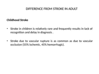
DIFFERENCE FROM STROKEIN ADULT
Childhood Stroke
• Stroke in children is relatively rare and frequently results in lack of
recognition and delay in diagnosis .
• Stroke due to vascular rupture is as common as due to vascular
occlusion (55% ischemic, 45% hemorrhagic).
5.
• Permanent aphasiais rare in children below 4 yrs, even
with involvement of dominant hemisphere.
• Neurodeficit is mild because collateral blood flow is
better than adults.
6.
EPIDEMIOLOGY
•
•
Incidence world overvaries between 1.2 per lakh to 2.7 per lakh.
In India hospital based studies indicate incidence of stroke in less
than 1 percent of all pediatric admission (According to
N.I.M.H.A.N.S. the incidence of stroke in children is 0.70% of
pediatric hospitalization).
• Slightly male preponderance.
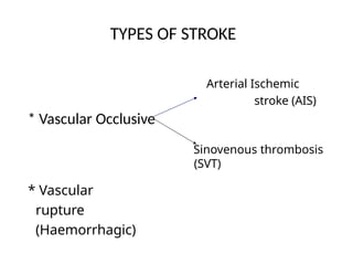
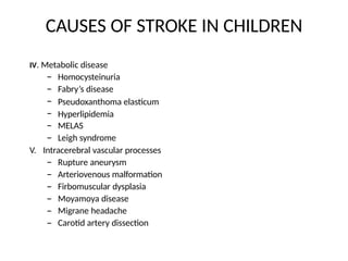
CAUSES OF STROKEIN CHILDREN
II. Hematologic Abnormalities
•
•
•
•
•
Sickle cell anemia.
Polycythemia
Thrombocytopenia / Thrombocytosis
Leukemia, lymphoma
Disorders of coagulation
– Protein C and S deficiency
– Antithrombin III deficiency
– Lupus Anticoagulant
– DIC
– Paroxysmal nocturnal
hemoglobinuria.
9.
CAUSES OF STROKEIN CHILDREN
III. Inflammatory Disorder
•
•
•
•
Meningitis : Viral, bacterial or tuberculosis
Systemic : Viremia, Bacteremia
Autoimmune diseases
Drug induced inflammation
– Amphetamine
– Cocaine
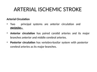
ARTERIAL ISCHEMIC STROKE
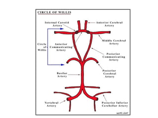
ArterialCirculation
• Two principal systems are anterior circulation and
posterior
circulation.
Anterior circulation has paired carotid arteries and its major
branches anterior and middle cerebral arteries.
Posterior circulation has vertebro-basilar system with posterior
cerebral arteries as its major branches.
•
•
14.
• Anterior andposterior circulation are linked by ACA and PCA to form the
circle of Willis.
Small perforator or lenticulostriate branches arise from the stems of
anterior, middle and posterior cerebral arteries.
•
16.
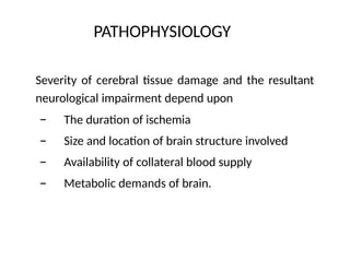
PATHOPHYSIOLOGY
Severity of cerebraltissue damage and the resultant
neurological impairment depend upon
–
–
–
–
The duration of ischemia
Size and location of brain structure involved
Availability of collateral blood supply
Metabolic demands of brain.
17.
PATTERN OF ARTERIALISCHEMIC STROKE
Transient ischemic attacks (TIA)
• Clinical deficit is usually extremely brief < 24 hours usually < 1
hour.
• Also called mini, warning or transient stroke.
18.
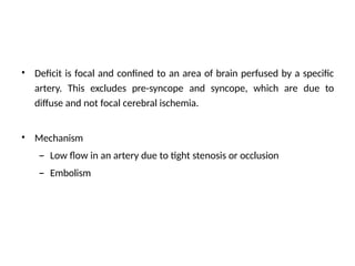
• Deficit isfocal and confined to an area of brain perfused by a specific
artery. This excludes pre-syncope and syncope, which are due to
diffuse and not focal cerebral ischemia.
• Mechanism
– Low flow in an artery due to tight stenosis or occlusion
– Embolism
19.
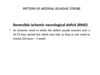
PATTERN OF ARTERIALISCHEMIC STROKE
Reversible ischemic neurological deficit (RIND)
An ischemic event in which the deficit usually recovers over a
24-72 hour period but which may take as long as one week to
resolve (24 hours – 1 week)
•
20.
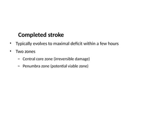
Completed stroke
•
•
Typically evolvesto maximal deficit within a few hours
Two zones
–
–
Central core zone (irreversible damage)
Penumbra zone (potential viable zone)
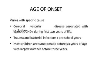
AGE OF ONSET
Varieswith specific cause
• Cerebral vascular
occlusive
disease associated with
cyanotic CHD : during first two years of life.
Trauma and bacterial infections : pre-school years
Most children are symptomatic before six years of age
with largest number before three years.
•
•
23.
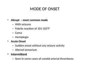
MODE OF ONSET
•Abrupt – most common mode
– With seizures
– Febrile reaction of 101-103ºF
– Coma
– Hemiplegia
Acute Onset
– Sudden onset without any seizure activity
– Altered sensorium
Intermittent
– Seen in some cases of carotid arterial thrombosis.
•
•
24.
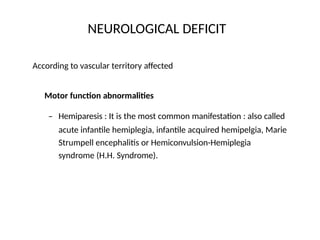
NEUROLOGICAL DEFICIT
According tovascular territory affected
Motor function abnormalities
– Hemiparesis : It is the most common manifestation : also called
acute infantile hemiplegia, infantile acquired hemipelgia, Marie
Strumpell encephalitis or Hemiconvulsion-Hemiplegia
syndrome (H.H. Syndrome).
25.
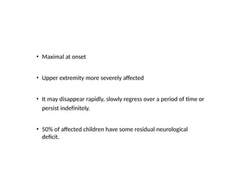
• Maximal atonset
• Upper extremity more severely affected
• It may disappear rapidly, slowly regress over a period of time or
persist indefinitely.
• 50% of affected children have some residual neurological
deficit.
26.
• Spasticity appearsabout 2-3 weeks after the onset & contractures
1-3 months later.
Persistent weakness 2-3 weeks after the onset indicates residual
motor deficit in later life.
•
Involuntary movements
•
•
•
Athetosis, dystonia and choreiform movements
Implies basal ganglia involvement
Imitative or mirror movements.
27.
Deep Tendon Reflexes
•Absent or hypotonic during flaccid phase
• Exaggerated in spastic phase
• Babinski response : soon after onset and persists for indefinite time
28.
Cranial Nerve Function
•Eye deficit – homonymous
hemianopia,
strabismus,
amblyopia, severe myopia.
Facial paralysis
Bulbar – Psuedobulbar
paralysis
•
•
Sensory loss
•
•
Superficial modalities (touch and pain) may be intact
Stereognosis, position sense
discrimination are often impaired
Hemi-atrophy
and two point
•
29.
Speech deficit :Dependent upon the age of child at onset and
hemisphere involved
* >4 years dysphasia common if dominant hemisphere is involved
* <4 year : relatively rare .
Never seen in <2 years of age.
* Normal speech pattern returns after 2-21 days.
30.
Vasomotor function
During acutestage, involved extremity may be warmer, edematous
and skin may be erythematous.
Development of spasticity causes affected extremity to to be cold.
31.
Epilepsy
• 50% ofchildren with acute hemiplegia subsequently develop
recurrent seizures.
• More common if onset <2 years of age or multiple seizures at
the time of onset.
.
32.
Defects in learning,behavior and intellect
• Usually mild intellectual deficit is present. Approximately 25% are
mentally retarded.
• Severe intellectual impairment is most common observed when the
hemiplegia is complicated by seizures refractory to anticonvulsants
33.
• Learning defectsare due to motor deficits, perceptual
disturbances, visiospatial problems and spatial orientation.
• Behavioral problems are typically a child with hyperkinetic behavior
with short attention span, impulsivity and distractibility.
• Child may be passive or underactive, particularly for the initial 1-5
months.
34.
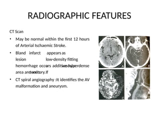
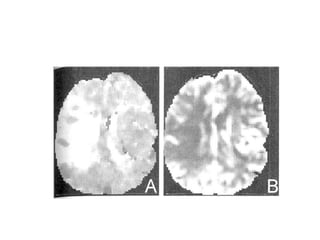
RADIOGRAPHIC FEATURES
CT Scan
•May be normal within the first 12 hours
of Arterial Ischaemic Stroke.
• Bland
lesion
infarct appearsas
low-density fitting
a vascular
territory.If
hemorrhage occurs additive hyperdense
area are seen.
CT spiral angiography :It identifies the AV
malformation and aneurysm.
•
35.
RADIOGRAPHIC FEATURES
MRI andMRA : MRI is better than CT scan as
• It is more sensitive than CT in detecting small and multiple
infarcts.
In posterior fossa, it is more sensitive than CT scan
•
• More sensitive at detecting hemorrhagic conversion of
infarct.
MRA can be performed at the same time. It is able to visualize
the flow in major cerebral, vertebral and external carotid
arteries.
•
37.
RADIOGRAPHIC FEATURES
Angiography
• Itis the definite method of visualizing the extra and
intracranial vasculature including the medium and
small sized arteries, which are not seen in MRA.
Other neuroimaging techniques
• Doppler imaging of carotid arteries and transcranial
Doppler for detecting large vessel vasculopathy.
38.
RADIOGRAPHIC FEATURES
X.ray skull
•Early X-ray are normal but after a period of years
they may show Dyke-Davidoff-Mason (DDM)
syndrome – thickening of cranial vault,
overdevelopment of frontal and ethmoid sinuses
and elevation of the
petrous pyramid
contralateral side
of temporal bone on
the due to
cerebral
hemiatrophy,
specially if onset is at <3 years of age.
39.
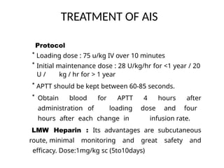
TREATMENT OF AIS
•Non anti-thrombotic
• Anti thrombotic
Anti thrombotic :
Heparin – considered if there is high risk of recurrence and low
risk of secondary hemorrhage. Nowadays It has been replaced
by low molecular weight heparin
40.
TREATMENT OF AIS
Protocol
*Loading dose : 75 u/kg IV over 10 minutes
* Initial maintenance dose : 28 U/kg/hr for <1 year / 20
U / kg / hr for > 1 year
* APTT should be kept between 60-85 seconds.
* Obtain blood for APTT 4 hours after
administration of loading dose and four
hours after each change in infusion rate.
LMW Heparin : Its advantages are subcutaneous
route, minimal monitoring and great safety and
efficacy. Dose:1mg/kg sc (5to10days)
41.
TREATMENT OF AIS
Indicationof heparin and LMWH
•
•
•
•
Arterial dissection
Hypercoagulable states
High risk of embolism
Progressive neurological
hemorrhage .
deficits not caused by cerebral
• Antifactor Xa is used for monitoring of LMWH(.5 to 1 unit/ml)
42.
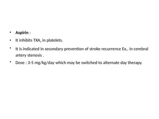
•
•
•
Aspirin :
It inhibitsTXA2 in platelets.
It is indicated in secondary prevention of stroke recurrence Ex,. in cerebral
artery stenosis .
Dose : 3-5 mg/kg/day which may be switched to alternate day therapy.
•
43.
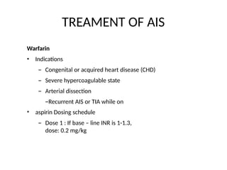
TREAMENT OF AIS
Warfarin
•
•Indications
– Congenital or acquired heart disease (CHD)
– Severe hypercoagulable state
– Arterial dissection
–Recurrent AIS or TIA while on
aspirin Dosing schedule
– Dose 1 : If base – line INR is 1-1.3,
dose: 0.2 mg/kg
44.
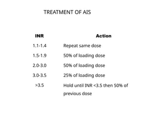
INR Action
1.1-1.4 Repeatsame dose
1.5-1.9 50% of loading dose
2.0-3.0 50% of loading dose
3.0-3.5 25% of loading dose
>3.5 Hold until INR <3.5 then 50% of
previous dose
TREATMENT OF AIS
45.
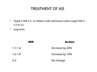
TREATMENT OF AIS
•Target is INR 2-3 . In children with mechanical valves target INR is –
2.5 to 3.5
Long term
•
INR Action
1.1-1.4 Increase by 20%
1.5-1.9 Increase by 10%
2-3 No change
46.
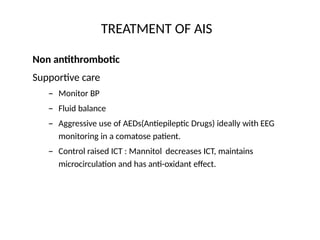
TREATMENT OF AIS
Nonantithrombotic
Supportive care
– Monitor BP
– Fluid balance
– Aggressive use of AEDs(Antiepileptic Drugs) ideally with EEG
monitoring in a comatose patient.
– Control raised ICT : Mannitol decreases ICT, maintains
microcirculation and has anti-oxidant effect.
47.
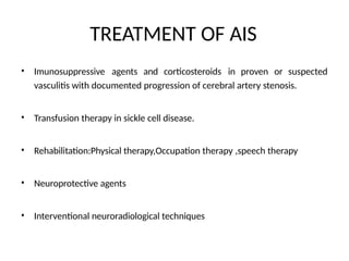
TREATMENT OF AIS
•Imunosuppressive agents and corticosteroids in proven or suspected
vasculitis with documented progression of cerebral artery stenosis.
• Transfusion therapy in sickle cell disease.
• Rehabilitation:Physical therapy,Occupation therapy ,speech therapy
• Neuroprotective agents
• Interventional neuroradiological techniques
PATHOPHYSIOLOGY
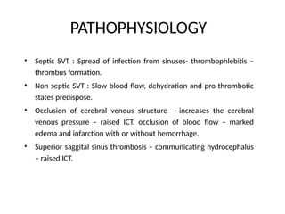
• Septic SVT: Spread of infection from sinuses- thrombophlebitis –
thrombus formation.
Non septic SVT : Slow blood flow, dehydration and pro-thrombotic
states predispose.
Occlusion of cerebral venous structure – increases the cerebral
venous pressure – raised ICT. occlusion of blood flow – marked
edema and infarction with or without hemorrhage.
Superior saggital sinus thrombosis – communicating hydrocephalus
– raised ICT.
•
•
•
50.
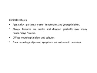
Clinical Features
•
•
Age atrisk : particularly seen in neonates and young children.
Clinical features are subtle and develop gradually over many
hours / days / weeks.
Diffuse neurological signs and seizures
Focal neurologic signs and symptoms are not seen in neonates.
•
•
51.
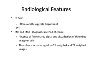
Radiological Features
• CTScan
– Occasionally suggests diagnosis of
SVT
• MRI and MRA : Diagnostic method of choice
– Absence of flow related signal and visualization of thrombus
in a given vein
– Thrombus – increase signal on T1 weighted and T2 weighted
images.
52.
• Ultrasonogram
– Canscreen SVT in neonate with open AF.
– Regular cranial USG can monitor the presence and severity of
ICH associated with SVT.
Angiography
– Gold standard
– Diagnostic : Partial or complete lack of flow
– Suggestive
• Delayed venous emptying
• Reversal of flow
• Abnormal cortical veins
•
53.
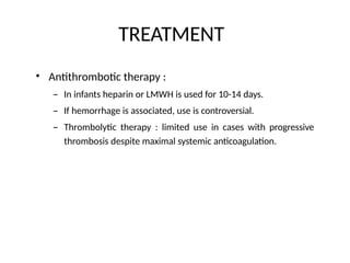
TREATMENT
• Antithrombotic therapy:
– In infants heparin or LMWH is used for 10-14 days.
– If hemorrhage is associated, use is controversial.
– Thrombolytic therapy : limited use in cases with progressive
thrombosis despite maximal systemic anticoagulation.
54.
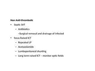
Non Anti-thrombotic
•
• SepticSVT
– Antibiotics
–Surgical removal and drainage of infected
focus Raised ICT
– Repeated LP
– Acetazolamide
– Lumboperitoneal shunting
– Long term raised ICT – monitor optic fields
55.
HEMORRHAGIC STROKE
• Hemorrhagicstroke : Rupture of normal cerebral blood vessels as
in bleeding diathesis, or abnormal blood vessels as with aneurysms
or vascular malformations.
• Hemorrhagic stroke is as common in children as ischemic stroke.
• Frequently requires neurosurgical intervention.
56.
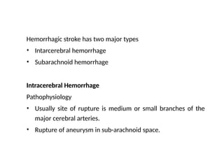
Hemorrhagic stroke hastwo major types
•
•
Intarcerebral hemorrhage
Subarachnoid hemorrhage
Intracerebral Hemorrhage
Pathophysiology
•
• Usually site of rupture is medium or small branches of the
major cerebral arteries.
Rupture of aneurysm in sub-arachnoid space.
57.
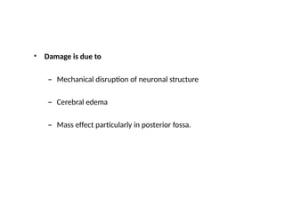
• Damage isdue to
– Mechanical disruption of neuronal structure
– Cerebral edema
– Mass effect particularly in posterior fossa.
58.
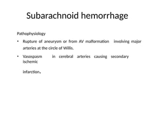
Subarachnoid hemorrhage
Pathophysiology
• Ruptureof aneurysm or from AV malformation
arteries at the circle of Willis.
involving major
• Vasospasm in cerebral arteries causing secondary
ischemic
infarction.
59.
Diagnosis
Radiological features
•
•
•
CT scan: Hyperdense areas
MRI and MRA:More sensitive for posterior fossa lesions
Conventional angiography
CSF
• In case of SAH, CSF is blood stained and xanthochromic
after six hours, but LP should never be performed
on child presenting with suggestive of
haemorrhage without prior CT scan.
60.
MANAGEMENT
•
•
Hypertension should beavoided and if present, treated
Straining at stool and anything producing Valsalva-like maneuver should
be avoided.
Bed rest
Anti-emetics and sedation
Analgesia for headache
Control of seizure
Monitor ICP
Do not use antifibrinolytic
agents
•
•
•
•
•
•
61.
• Neurosurgical intervention: In children who present with SAH or
intracerebral bleed, due to an aneurysm or AV malformation
surgery is management of choice.
• If child is in coma, if angiography shows aneurysm is fusiform or if
there is any question of a mycotic aneurysm then conservative
medical management is indicated.
• Overall prognosis for cerebral hemorrhage in childhood is poor with
50% mortality.
62.
PROGNOSIS
• Mortality afterstroke in children from 20-30% depending upon the
location and underlying cause.
• Hemorrhagic stroke has higher mortality
• Residual neurological deficit is present in more than 50% of
survivors and is more common after ischemic stroke.
63.
• References:
• PiyushGupta PG text book of
Pediatrics
• Pediatric Neurology by: Veena Kalra


![ACUTE_AND_CHRONIC_RENAL_FAILxxxxx1].pptx](https://cdn.slidesharecdn.com/ss_thumbnails/acuteandchronicrenalfail1-250803044151-1ee4884c-thumbnail.jpg?width=640&height=640&fit=bounds)


![CTEV [ clubfoot] DR ARUN LAL ,DR MOHAMED ASHRAF travancore medical college k...](https://cdn.slidesharecdn.com/ss_thumbnails/ctevclubfootdrarunlaldrmohamedashraftravancoremedicalcollegekollamkeralaindia-260208063247-18fc466c-thumbnail.jpg?width=640&height=640&fit=bounds)
![PERI-PROSTHETIC FRACTURE NAIL-PLATE CONSTRUCT [NPC].pptx](https://cdn.slidesharecdn.com/ss_thumbnails/drarunkumardrmohamedashrafperiprostheticfrasturenail-plateconstructnpc-260209164459-7e9d15a1-thumbnail.jpg?width=640&height=640&fit=bounds)


