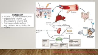
This document discusses hypovitaminosis D (vitamin D deficiency). It covers the roles, metabolism, effects, risk groups, clinical presentation, screening, treatment, and guidelines regarding vitamin D levels. Some key points include:
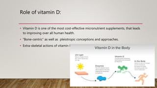
- Vitamin D has important roles in bone health and many other body systems. It is obtained mostly from sun exposure and dietary sources.
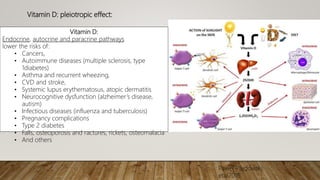
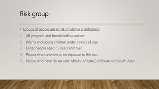
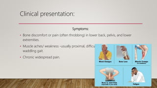
- Deficiency can cause rickets in children and osteomalacia in adults, and is also linked to increased risk of various diseases. Risk groups include pregnant/breastfeeding women and those with limited sun exposure.
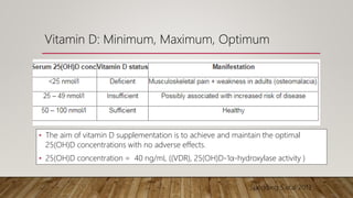
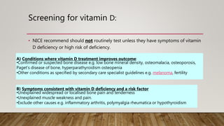
- Screening may be done in symptomatic patients or those at high risk. Treatment involves supplementation to reach/maintain sufficient vitamin D levels,
This document discusses megaloblastic anemia, its causes, symptoms, and treatments. It is characterized by abnormally large red blood cells due to a deficiency in vitamin B12 or folic acid, which is needed for DNA synthesis. The deficiencies can result from inadequate intake, malabsorption, or increased demand. Treatments include injections or supplements of vitamin B12, folic acid, or erythropoietin to stimulate red blood cell production. Adverse reactions are also discussed.
This document discusses hypervitaminosis, or vitamin toxicity, for several vitamins:
- Vitamin A toxicity can cause headaches, abdominal pain, and impaired consciousness from acute high doses, or bone pain and loss of appetite from chronic excessive intake.
- High vitamin D levels from supplements or medications can lead to polyuria, polydipsia, hypercalcemia, and in severe cases, death.
- Excessive vitamin E intake above 1 g/kg can cause bruising and bleeding due to reduced platelet function and impaired immunity.
- High vitamin B6 doses over 200 micrograms daily taken long-term may damage nerves and cause numbness, poor coordination, and tiredness.
Iron Deficiency Anemia occurs when iron levels are too low to support normal red blood cell production. It has various causes like bleeding, low dietary iron intake, or loss of iron in the urine. Symptoms include fatigue and pale skin. Investigations show low hemoglobin, ferritin and iron levels with microcytic red blood cells. Treatment involves oral or intravenous iron supplementation depending on severity. Complications can include impaired development in children or increased risk of falls in the elderly. Prevention focuses on consuming iron-rich foods and supplements. Differential diagnoses include thalassemia and anemia of chronic disease.
Megaloblastic anaemia is a red blood cell disorder due to the inhibition of DNA synthesis during erythropioesis.
Mitotically, the inhibition of the DNA synthesis impaires the progression of the cell cycle development from G2 to (M) stage.
Vitamin B12 deficiency can result from decreased absorption in the ileum, decreased intrinsic factor production in the stomach, inadequate dietary intake, or prolonged medication use. Clinical manifestations include neurological, psychiatric, hematological and cutaneous issues. Screening is recommended for those with risk factors or suspected symptoms. Treatment involves intramuscular B12 injections or high dose oral replacement, indefinitely for irreversible causes or until deficiency is corrected for reversible causes. Prevention focuses on supplementation in high risk groups.
Hemolytic anemia is characterized by accelerated red blood cell destruction and vigorous blood regeneration. It can be classified as intrinsic or extrinsic, congenital or acquired. The site of red blood cell destruction can be intravascular or extravascular. Common causes of hemolytic anemia include hereditary spherocytosis, thalassemias, sickle cell anemia, glucose-6-phosphate dehydrogenase deficiency, paroxysmal nocturnal hemoglobinuria, and immune-mediated hemolytic anemia. Evaluation of hemolytic anemia involves determining whether the anemia is hemolytic, the site of red blood cell destruction, the etiology, and severity through blood smears, reticulocyte counts, LDH and
Megaloblastic anemias are caused by impaired DNA synthesis due to vitamin B12 or folate deficiency. The summary examines megaloblastic anemias, including causes such as vitamin B12 or folate metabolism defects, clinical features like pallor and neurological symptoms, investigation findings in peripheral blood and bone marrow showing megaloblasts and macroovalocytes, and treatment involving vitamin B12 or folate supplementation.
Iron deficiency anemia is the most common type of anemia globally. It results from inadequate iron intake or absorption to meet physiological needs. Common symptoms include pallor, weakness, and fatigue. Diagnosis involves blood tests showing microcytic hypochromic anemia, low serum iron and ferritin levels, and high total iron binding capacity. Treatment consists of oral iron supplementation in the form of ferrous salts to replenish iron stores.
Diagnosis and management of folic acid deficiency anaemiasubhrajit das
Folic acid deficiency anemia is a problem in 3-4% of pregnancies in India. It is caused by poor diet, malabsorption issues, increased demand during pregnancy, and certain drugs. Symptoms include loss of appetite, vomiting, and diarrhea. Signs include pallor, enlarged organs, and neuropathy. Complications for the mother include preeclampsia and placental abruption, while complications for the fetus include neural tube defects, abortion, growth issues, and prematurity. Diagnosis is based on low hemoglobin, MCV, and serum folate levels as well as megaloblastic changes in blood cells. Treatment involves folic acid supplementation before and during pregnancy to prevent deficiencies.
Haemolytic anaemias are a group of anemias caused by the premature breakdown of red blood cells in the bloodstream or spleen. There are two main types - intrinsic defects that cause red blood cell damage from within, such as hereditary spherocytosis, and extrinsic defects that cause damage from outside factors like immune mediated hemolysis. Symptoms include anemia, jaundice, splenomegaly and gallstones. Laboratory tests show signs of increased red blood cell breakdown like elevated bilirubin and LDH, as well as signs of the bone marrow attempting to compensate with reticulocytosis and nucleated red blood cells. Intravascular hemolysis specifically causes hemoglobinemia,
Vitamin B12 deficiency is a common cause of megaloblastic anemia and neuropsychiatric symptoms, especially in older persons. There are several risk factors for vitamin B12 deficiency including prolonged metformin use. While major organizations do not recommend routine screening, high-risk patients such as those with malabsorptive disorders may warrant screening through CBC, serum B12, methylmalonic acid, and homocysteine levels. Treatment involves intramuscular B12 injections or high dose oral B12 supplementation.
Megaloblastic anemia is caused by a deficiency of vitamin B12 or folate, which are essential for DNA synthesis and the production of red blood cells. This leads to large, abnormally developed red blood cells seen in the bone marrow. Symptoms include anemia, fatigue, and neurological issues. Diagnosis involves blood tests to check vitamin B12 and folate levels. Treatment consists of lifelong vitamin B12 injections or high dose oral supplements to replenish stores, along with folate supplements for a period of time.
Vitamin D Deficiency, by Dr. Mihir Adhikari Mihir Adhikari
Vitamin D deficiency can cause rickets in children and osteomalacia in adults. It is caused by lack of vitamin D from diet and sun exposure. The body synthesizes the inactive form cholecalciferol from cholesterol in skin upon sun exposure and the active form calcitriol is produced in the kidneys. Vitamin D plays a key role in calcium absorption and bone mineralization. Deficiency is highly prevalent in India due to low dietary intake and sun exposure. It is associated with many systemic disorders including musculoskeletal, autoimmune, cardiovascular and infectious diseases.
Vitamin K is essential for blood clotting and plays an important role in bone health. Insufficient vitamin K can lead to excessive bleeding and easy bruising. Low vitamin K levels have been associated with low bone density and increased risk of fractures. Vitamin K deficiency is most common in newborns and those taking anticoagulant medications or antibiotics, which can interfere with vitamin K levels. Treatment involves oral or injectable vitamin K supplementation.
This document discusses the causes and types of edema. It categorizes the pathophysiologic causes of edema into increased hydrostatic pressure, reduced oncotic pressure, lymphatic obstruction, sodium retention, and increased capillary permeability. Specific conditions are provided as examples for each category. The document also describes characteristics of generalized versus localized edema and pitting versus non-pitting edema.
Drug treatment of iron deficiency anaemiaNaser Tadvi
This document discusses iron deficiency anemia and its treatment. It defines anemia and identifies iron deficiency as a common cause. It describes how iron is used to form hemoglobin and the signs and symptoms of iron deficiency anemia. The document outlines dietary iron requirements and food sources of iron. It provides details on oral and parenteral iron therapy, including dosages, formulations, and potential adverse effects. It also discusses intravenous iron preparations and the use of desferrioxamine for acute iron poisoning.
Calcium is the most abundant mineral in the body and is primarily stored in bones and teeth. It performs many important biochemical functions including bone and teeth formation, muscle contraction, blood coagulation, and nerve transmission. Calcium levels are regulated by parathyroid hormone, vitamin D, and calcitonin. These hormones work to maintain calcium homeostasis by impacting absorption in the intestine and kidneys and mobilization from bones.
Vitamin C plays important roles in the body including in collagen formation, wound healing, and immune function. A deficiency in vitamin C can lead to scurvy, which causes symptoms like bleeding gums, bruising, and poor wound healing. Scurvy develops when vitamin C levels in the body drop below a critical point, which can occur within 1-3 months if dietary intake is lacking. Several groups are at higher risk for vitamin C deficiency, including smokers, pregnant women, and those with digestive disorders that impact absorption. Treatment involves taking supplemental vitamin C orally to restore levels.
This document discusses severe acute malnutrition (SAM) in children. It defines SAM and describes its etiology as primarily being due to inadequate dietary intake, though secondary causes like infection can also play a role. The pathophysiology involves a vicious cycle between undernutrition and infection that impacts multiple organ systems. Complications of SAM include hypoglycemia, hypothermia, and electrolyte imbalances. The principles of management outline a 10 step process including stabilization, infection treatment, rehydration, micronutrient supplementation, refeeding, and follow up care. Refeeding syndrome is also discussed as a potential complication.
Rickets is a disease characterized by softening and weakening of bones in children, caused by a vitamin D deficiency or impaired metabolism of calcium or phosphorus. It most commonly affects children between 6-24 months old and can lead to fractures and bone deformities. While historically more prevalent, rickets decreased in developed countries in the 20th century but remains common in developing nations and populations with dark skin where access to vitamin D-fortified foods is limited. Treatment involves increasing dietary intake of calcium, phosphorus and vitamin D through supplements, exposure to sunlight, or vitamin D-fortified foods and formula. Physical therapy can help reduce bone and muscle pain through stretching and strengthening exercises.
Hemolytic anemias are caused by increased red blood cell destruction. They are characterized by normochromic, normocytic anemia with reticulocytosis, increased indirect bilirubin and LDH, and absent haptoglobin. Causes include membrane defects, metabolic abnormalities, hemoglobinopathies, and immune or non-immune mechanisms. Specific conditions discussed include hereditary spherocytosis, glucose-6-phosphate dehydrogenase deficiency, paroxysmal nocturnal hemoglobinuria, drug-induced hemolysis, alloimmune hemolytic anemia, and warm or cold autoimmune hemolytic anemia. Management depends on the underlying cause and may involve transfusions, medications, or splenectomy.
This document discusses vitamin D, including its functions, sources, daily requirements, deficiency, toxicity, and management. Some key points:
- Vitamin D promotes calcium absorption and is important for bone health. Deficiency can cause rickets in children and osteomalacia in adults.
- Good dietary sources include fatty fish, fish liver oils, eggs, and dairy. Sun exposure also enables vitamin D production.
- Recommended daily intake is 10-20 micrograms for adults. Deficiency is managed with high dose vitamin D supplementation. Toxicity risks include excessive intake.
Iron deficiency anemia is a common type of microcytic anemia caused by low levels of iron available to produce hemoglobin. It affects people with high iron demands like pregnant women, young children, and menstruating women. Symptoms include fatigue, weakness, and shortness of breath. Treatment involves oral iron supplementation and dietary changes to restore iron stores. Prevention focuses on adequate iron intake through diet or supplements in at-risk groups.
This document summarizes scurvy, a disease caused by vitamin C deficiency. It discusses that vitamin C is required in the diet and its functions include collagen synthesis and acting as an antioxidant. Scurvy's clinical features include bleeding gums, joint pain, and weakness. In infants, signs include irritability and pseudoparalysis. Diagnosis is usually clinical but may include low vitamin C blood levels. Treatment involves vitamin C supplementation with a loading dose followed by daily doses. Prevention recommendations include breastfeeding, vitamin C supplementation in artificial feeding, and including citrus fruits and other vitamin C sources in the diet.
Blood system in children of different age groupsEneutron
This document discusses anatomical and physiological features of the blood system in children of different ages. It covers the development of hematopoiesis and blood forming organs from embryogenesis through toddlerhood. Key stages include embryonic hematopoiesis in the yolk sac, liver, spleen, thymus, lymph nodes and bone marrow. The document also outlines the major functions of blood and features of a full-term newborn's blood.
This document summarizes an academic presentation on approaches to patients with anemia. It discusses definitions and classifications of anemia, as well as common types including iron deficiency anemia, anemia of chronic disease, megaloblastic anemias from B12 or folate deficiency, and sickle cell anemia. For each type, it covers etiologies, symptoms, diagnostic findings, bone marrow characteristics, and treatment approaches. Iron deficiency anemia is the most common worldwide and is discussed in depth, including stages of deficiency and management strategies depending on severity and underlying cause of iron loss.
This document discusses vitamin D, its natural sources, deficiency, and importance. It notes that vitamin D is a fat-soluble vitamin essential for bone and calcium health. While sunshine is the main natural source, few foods contain significant amounts. Vitamin D deficiency is very common worldwide and increases risk of bone abnormalities and other diseases. Deficiency can result from inadequate sun exposure, skin pigmentation, obesity, lack of nutritional intake, and other factors. At-risk groups include breastfed infants, older adults, those with limited sun exposure or dark skin, and the obese. Blood tests can identify deficiency which is treated through dietary sources and supplements.
Vitamin D deficiency is very common globally, including in Saudi Arabia where prevalence is around 90-95%. Vitamin D is important for bone and skeletal health and development during childhood, as it regulates calcium and phosphorus balance and bone mineralization. Beyond skeletal benefits, vitamin D has roles in reducing infection risk, autoimmune disease, asthma, COPD and cancer. Deficiency is diagnosed through blood tests measuring vitamin D levels, with normal being 30-100 ng/ml. Deficiency can cause rickets or osteomalacia and is often due to lack of sun exposure or intake of vitamin D sources like fortified foods. Prevention involves sun exposure, intake of vitamin D foods or supplements. Treatment requires high dose vitamin D
Vitamin D deficiency is common, affecting approximately 66% of the global population. It is defined as a serum 25-hydroxyvitamin D level below 20 ng/mL. Symptoms include bone and muscle pain. At-risk groups include those with dark skin, obesity, or malabsorption issues. Screening is recommended for high-risk individuals. Treatment involves oral vitamin D supplements of 50,000 IU weekly or 6000 IU daily to raise levels above 30 ng/mL. Maintenance doses of 800-1000 IU daily are used thereafter to prevent recurrence.
Vitamin D deficiency can lead to rickets in children and osteomalacia in adults. It is caused by insufficient vitamin D intake from food or sun exposure, impaired vitamin D absorption or metabolism. Symptoms include weakened, soft bones and muscle weakness. Diagnosis involves measuring blood levels of vitamin D and calcium. Treatment consists of high dose vitamin D supplementation.
This document summarizes guidelines from the Indian Academy of Pediatrics (IAP) on preventing and treating vitamin D and calcium deficiencies in children and adolescents. It reports that deficiencies of these nutrients are widespread in India, affecting 30-90% of people. The guidelines define sufficient, insufficient, and deficient vitamin D levels and recommend routine supplementation of 400IU of vitamin D for all infants up to 1 year old. Children over 1 year and adolescents should take 600IU daily. At-risk groups may require higher doses. Treatment of deficiencies generally involves 2000-6000IU of vitamin D and 500-800mg of calcium daily for 3 months. Maintaining adequate vitamin D and calcium intake is important for bone health.
Vitamin D plays an important role in bone and mineral metabolism and is also involved in preventing many chronic diseases. It is synthesized in the skin upon exposure to sunlight and is also obtained through dietary sources. Most cells in the body have receptors for vitamin D. Deficiency is linked to increased risk of diseases such as cancer, cardiovascular disease, diabetes and multiple sclerosis. Adequate vitamin D levels can be maintained through sensible sun exposure, dietary sources like fatty fish, and supplementation.
VITAMIN D, very important drug in most of the situation. content-introduction,brand and pharmacological name,available forms,dosage and routes, classification , mechanism of action,pharmacokinetics, indications,precautions, side effects,nursing responsibilities and patient education.
This document discusses the treatment and prevention of osteoarthritis (OA). It begins by outlining the goals of OA treatment, which include educating patients, maintaining joint mobility, limiting impairment, and relieving pain and stiffness to improve quality of life. Treatment approaches include both pharmacological (e.g. analgesics, NSAIDs, supplements) and non-pharmacological (e.g. physical therapy, diet, exercise) options. The document also provides steps to prevent OA, such as maintaining a healthy weight, exercising regularly, avoiding injuries, and eating a nutritious diet high in omega-3s, vitamin C and vitamin D.
Vitamin D is important for bone and immune system health. It is obtained through sun exposure and dietary sources. Vitamin D aids calcium absorption to strengthen bones and helps T cells activate to fight pathogens. Studies show vitamin D may reduce risks of multiple sclerosis, asthma symptoms, diabetes, heart disease, depression, and some cancers. Ensuring adequate vitamin D intake through diet and sun exposure can support overall health and wellness.
Vitamin d insufficiency and deficiency in children and adolescentsAzad Haleem
Vitamin D insufficiency and deficiency can occur in children and adolescents. The document discusses the forms and pathways of vitamin D in the body. Risk factors for deficiency include dark skin, limited sun exposure, exclusive breastfeeding, obesity, and genetic disorders. Deficiency can lead to rickets in children or osteomalacia. Diagnosis is made by measuring 25-hydroxyvitamin D levels in the blood. Treatment involves vitamin D supplementation, with dosage depending on age and severity of deficiency. Monitoring of vitamin D levels is important during and after treatment.
Vitamin d in health and disease august 2020Khaled Saad
Vitamin D plays an important role in skeletal health and the immune system. The document discusses vitamin D deficiency as an underrecognized problem, and summarizes evidence that vitamin D supplementation can reduce infections in children and may help prevent autoimmune disorders and childhood asthma. Vitamin D is involved in calcium regulation, bone growth, cell growth, and immune function. Deficiency has been linked to increased risk of various diseases while supplementation may decrease risk of some chronic conditions.
Vitamin A is essential for vision, immune function, and cell growth. Deficiency can cause night blindness and susceptibility to infection. Treatment involves oral vitamin A supplements. Toxicity from excessive intake is rare but can cause headaches and bone pain.
Vitamin D is important for calcium absorption and bone health. Deficiency causes rickets in children, characterized by bowed legs. Treatment involves vitamin D supplements and exposure to sunlight. Toxicity from high intake can raise calcium levels and cause nausea.
Thiamine (B1) deficiency, known as beriberi, affects nerve function and heart health. It is treated with thiamine supplements administered orally or intravenously.
Vitamin D deficiency is common in critically ill neonates. A study found serum 25-OH vitamin D levels were significantly lower in critically ill neonates compared to healthy newborns, with no correlation to disease severity except in pneumonia cases. The study recommended measuring 25-OH vitamin D levels in critically ill neonates and ensuring adequate maternal vitamin D intake during pregnancy and lactation, as well as vitamin D supplementation for breastfed infants. Guidelines for treating vitamin D deficiency in children include daily or weekly high dose vitamin D supplementation for 4-8 weeks, followed by maintenance doses, while insufficiency is managed with biweekly or monthly lower dose supplementation.
This document discusses vitamin D, including its functions, sources, daily requirements, deficiency, and management. Some key points:
- Vitamin D promotes calcium absorption in the intestine and supports bone mineralization. It is produced in the body after sun exposure or obtained through dietary sources like fish, liver, and eggs.
- Deficiency can cause rickets in children, characterized by bone deformities and growth issues. Adults may experience bone pain. Risk factors include limited sun exposure, obesity, and certain medications.
- Diagnosis involves assessing clinical features, radiological findings showing bone abnormalities, and biochemical testing showing low vitamin D and high alkaline phosphatase levels.
- Treatment for deficiency involves high dose vitamin
Vitamins are organic substances that are essential for carrying out normal biochemical processes and physiological functions. They are divided into fat-soluble vitamins (A, D, E, K) and water-soluble vitamins. Fat-soluble vitamins are stored in the liver and excessive intake can lead to toxicity, while water-soluble vitamins are excreted and toxicity does not occur. Vitamins must be activated in the body and deficiencies can cause specific syndromes, while adequate intake through a balanced diet meets daily requirements.
This document provides information on vitamins, including their classification as either water-soluble or fat-soluble, roles and deficiencies. Water-soluble vitamins like C and B vitamins must be replenished daily as they are not stored in the body, while fat-soluble vitamins A, D, E and K can accumulate and potentially cause toxicity. The document outlines the names, roles and recommended daily amounts of each vitamin, along with absorption and storage processes. Sources of vitamins and deficiency symptoms are also summarized.
This market for vitamin D supplements is projected to reach $2.5 billion by 2020, growing at 11% annually. Currently, many vitamin D supplements have poor absorption in the body. A new product called SMART-D3 contains solubilized vitamin D3 in a highly bioavailable formulation as a dry powder capsule. It is the first product of its kind to provide fully bioavailable vitamin D3 in a convenient, once-daily capsule. Vitamin D plays an important role in bone and heart health, immunity, and reducing the risk of various diseases.
Nutritional rickets is caused by defective bone growth and mineralization due to vitamin D deficiency or low calcium intake. It is diagnosed based on history, physical exam, biochemical testing and confirmed by radiographs. Serum 25-hydroxyvitamin D levels below 30 nmol/L indicate deficiency and levels below 50 nmol/L increase risk of rickets. Treatment involves high dose vitamin D supplementation of 2000 IU/day for at least 3 months along with calcium supplementation. Prevention involves adequate dietary vitamin D and calcium intake.
Vitamin D is a fat-soluble vitamin that is important for bone and muscle health. It is synthesized in the skin upon exposure to sunlight or obtained from dietary sources. The liver and kidneys work to activate vitamin D before it can be used by the body. Vitamin D deficiency can lead to rickets in children and osteomalacia or osteoporosis in adults and is associated with increased risk of various diseases. Treatment of deficiency involves obtaining more vitamin D through diet, supplements, or sunlight exposure.
VEXAS syndromes , a diagnostic PuzzlepptxMarwa Besar
This document discusses VEXAS syndrome, a recently discovered autoinflammatory and hematologic condition caused by somatic mutations in the UBA1 gene. It provides details on the clinical manifestations (fevers, inflammatory symptoms affecting skin, joints, lungs, blood vessels), hematological abnormalities (anemia, cytopenias), and challenges in management due to the combined inflammatory and hematological involvement. Diagnosis is confirmed via genetic testing demonstrating UBA1 mutations. Treatment aims to suppress inflammation and eliminate mutant hematopoietic cells, but outcomes remain poor with high mortality.
1. The document discusses when to call an immunologist in an interstitial lung disease (ILD) clinic. It provides information on clinical signs, autoantibodies, and instrumental exams that can help recognize autoimmune ILD associated with rheumatic diseases.
2. Risk factors for developing ILD in autoimmune rheumatic diseases include certain diseases like systemic sclerosis, as well as specific autoantibodies. Monitoring of pulmonary symptoms depends on the underlying rheumatic disease but may include regular testing and imaging.
3. A multidisciplinary approach is important for diagnosing and managing autoimmune ILD, and guidelines recommend evaluating any newly diagnosed ILD for an underlying autoimmune rheumatic disease.
Management of hereditary angioedema involves treating acute attacks, preventing attacks, and improving quality of life. Treatment strategies include treating acute attacks, preventing attacks short-term before procedures, and long-term prophylaxis. Therapies include C1 inhibitor replacement, bradykinin receptor antagonists, attenuated androgens, and antifibrinolytics. An individualized treatment plan is recommended based on a patient's attack frequency, severity and location, as well as their medication access and preferences.
Hidradenitis suppurativa (HS) is a chronic inflammatory skin disease characterized by recurrent abscesses and scarring in the apocrine gland-bearing areas. It typically presents in young adults and is more common in women and people of African descent. Obesity, smoking, family history and certain drugs are associated risk factors. The pathogenesis involves occlusion of hair follicles leading to rupture, inflammation, abscess formation and scarring. Diagnosis is clinical though imaging and biopsy may help in some cases. Treatment aims to control flare-ups and prevent scarring. The course is usually chronic with intermittent flares and remissions.
- The document analyzes IL-18 and IFN-gamma as biomarkers for diagnosis and prognosis in juvenile idiopathic arthritis (JIA) patients. It finds that IL-18 levels correlate with disease activity and can predict response to therapy, while IFN-gamma levels indicate response to therapy. The study compares biomarker and disease activity measurements in 45 JIA patients at initial presentation and after treatment. It concludes that IL-18 and IFN-gamma may be promising markers for detecting prognosis and treatment response in JIA patients.
This document discusses three case studies of patients with autoimmune diseases and thrombosis. It then provides statistics on the increased risk of thrombosis in various autoimmune diseases compared to the general population. The mechanisms of thrombosis in autoimmune diseases are reviewed. Several pearls and myths regarding thrombosis and treatment in autoimmune diseases are discussed, including that all autoimmune diseases confer a hypercoagulable state; anticoagulants are not always effective or safe for Bechet's syndrome; and immunosuppressive drugs may also cause thrombosis. The take home message is that autoimmune diseases are both inflammatory and hypercoagulable, and risk versus benefit of anticoagulants like DOACs must be considered.
This document discusses systemic lupus erythematosus (SLE) and conditions that can mimic its presentation. It describes how SLE often has a waxing and waning chronic course that can vary in severity. Several infectious, inflammatory, and neoplastic conditions can present similarly to SLE through involvement of multiple organ systems and production of autoantibodies. Correct diagnosis requires a thorough history, physical exam, targeted testing, and biopsies to distinguish SLE from its mimickers. Serological similarities alone do not confirm SLE if clinical features are inconsistent.
Pediatric uveitis can be caused by autoimmune or autoinflammatory disorders. The most common type seen in children is chronic anterior uveitis, unlike adults where it is less common. Juvenile idiopathic arthritis (JIA)-associated anterior uveitis is the most frequent cause of uveitis in children. It is typically bilateral and non-granulomatous with a chronic relapsing course. Idiopathic intermediate uveitis (pars planitis) commonly affects children and adolescents and has a low association with systemic diseases. Behcet's disease onset is most common in late childhood, with bilateral recurrent panuveitis and retinal vasculitis seen clinically.
Adult-onset Still's disease (AOSD) is a rare autoinflammatory disorder characterized by high spiking fever, evanescent rash, arthritis, and multiorgan involvement. It was first described in adults in 1971. The disease most commonly affects young adults and women. Diagnosis is based on clinical criteria and exclusion of other conditions. Treatment involves nonsteroidal anti-inflammatories initially followed by disease-modifying drugs like methotrexate. Biologics that inhibit IL-1 or IL-6 are effective for refractory or complicated cases. Prognosis is generally good but complications can include macrophage activation syndrome, organ failure, or amyloidosis.
Vogt-Koyanagi-Harada (VKH) syndrome is a rare autoimmune disease that affects melanin-containing tissues like the eyes, inner ears, skin, and brain. It is characterized by bilateral uveitis and can cause vision loss. The disease occurs in phases including prodromal, acute uveitic, convalescent, and recurrent. Treatment involves high-dose corticosteroids for at least 6 months to reduce inflammation and recurrence risk. Immunosuppressants may be needed for resistant cases. Prognosis depends on factors like early treatment, treatment duration, and presence of complications. Strict monitoring is needed due to risk of vision threatening complications.
Familial Mediterranean Fever is an autosomal recessive disease characterized by recurrent fevers and inflammation localized to the peritoneum, pleura, joints, or skin. It is most common in people of Mediterranean descent and is caused by mutations in the MEFV gene. Symptoms include abdominal pain, fever, arthritis, and erysipelas-like lesions. If left untreated, it can lead to amyloidosis. Treatment involves lifelong colchicine to prevent inflammatory episodes and reduce amyloidosis risk. Some patients may be resistant to colchicine and require alternative treatments like interleukin-1 or tumor necrosis factor inhibitors.
Giant cell arteritis (GCA) is a large vessel vasculitis that commonly affects the branches of the carotid artery and causes headaches, jaw claudication, and vision loss. The pathology involves granulomatous inflammation in the vessel walls. Diagnosis is based on temporal artery biopsy showing giant cells, but imaging such as ultrasound, CT, and PET can also provide supportive evidence of vessel inflammation. Treatment involves high-dose corticosteroids to reduce inflammation and prevent relapses and vision loss, with tapering over 2-5 years. Monitoring for complications like aortic aneurysms is also important given the vessel involvement.
Polyarteritis nodasa and microscopic polyangitisMarwa Besar
This document discusses Polyarteritis Nodosa (PAN) and Microscopic Polyangiitis (MPA). PAN is a necrotizing vasculitis predominantly affecting medium-sized arteries that spares small vessels. It is typically ANCA-negative. MPA is a pauci-immune necrotizing vasculitis involving small vessels and sometimes medium arteries, associated with ANCA positivity. Both diseases can affect multiple organ systems and have variable clinical manifestations. Differentiation is based on vessel size involvement and ANCA status according to the Chapel Hill consensus criteria.
Here is the updated list of Top Best Ayurvedic medicine for Gas and Indigestion and those are Gas-O-Go Syp for Dyspepsia | Lavizyme Syrup for Acidity | Yumzyme Hepatoprotective Capsules etc
These lecture slides, by Dr Sidra Arshad, offer a quick overview of the physiological basis of a normal electrocardiogram.
Learning objectives:
1. Define an electrocardiogram (ECG) and electrocardiography
2. Describe how dipoles generated by the heart produce the waveforms of the ECG
3. Describe the components of a normal electrocardiogram of a typical bipolar lead (limb II)
4. Differentiate between intervals and segments
5. Enlist some common indications for obtaining an ECG
6. Describe the flow of current around the heart during the cardiac cycle
7. Discuss the placement and polarity of the leads of electrocardiograph
8. Describe the normal electrocardiograms recorded from the limb leads and explain the physiological basis of the different records that are obtained
9. Define mean electrical vector (axis) of the heart and give the normal range
10. Define the mean QRS vector
11. Describe the axes of leads (hexagonal reference system)
12. Comprehend the vectorial analysis of the normal ECG
13. Determine the mean electrical axis of the ventricular QRS and appreciate the mean axis deviation
14. Explain the concepts of current of injury, J point, and their significance
Study Resources:
1. Chapter 11, Guyton and Hall Textbook of Medical Physiology, 14th edition
2. Chapter 9, Human Physiology - From Cells to Systems, Lauralee Sherwood, 9th edition
3. Chapter 29, Ganong’s Review of Medical Physiology, 26th edition
4. Electrocardiogram, StatPearls - https://www.ncbi.nlm.nih.gov/books/NBK549803/
5. ECG in Medical Practice by ABM Abdullah, 4th edition
6. Chapter 3, Cardiology Explained, https://www.ncbi.nlm.nih.gov/books/NBK2214/
7. ECG Basics, http://www.nataliescasebook.com/tag/e-c-g-basics
ABDOMINAL TRAUMA in pediatrics part one.drhasanrajab
Abdominal trauma in pediatrics refers to injuries or damage to the abdominal organs in children. It can occur due to various causes such as falls, motor vehicle accidents, sports-related injuries, and physical abuse. Children are more vulnerable to abdominal trauma due to their unique anatomical and physiological characteristics. Signs and symptoms include abdominal pain, tenderness, distension, vomiting, and signs of shock. Diagnosis involves physical examination, imaging studies, and laboratory tests. Management depends on the severity and may involve conservative treatment or surgical intervention. Prevention is crucial in reducing the incidence of abdominal trauma in children.
8 Surprising Reasons To Meditate 40 Minutes A Day That Can Change Your Life.pptxHolistified Wellness
We’re talking about Vedic Meditation, a form of meditation that has been around for at least 5,000 years. Back then, the people who lived in the Indus Valley, now known as India and Pakistan, practised meditation as a fundamental part of daily life. This knowledge that has given us yoga and Ayurveda, was known as Veda, hence the name Vedic. And though there are some written records, the practice has been passed down verbally from generation to generation.
Adhd Medication Shortage Uk - trinexpharmacy.comreignlana06
The UK is currently facing a Adhd Medication Shortage Uk, which has left many patients and their families grappling with uncertainty and frustration. ADHD, or Attention Deficit Hyperactivity Disorder, is a chronic condition that requires consistent medication to manage effectively. This shortage has highlighted the critical role these medications play in the daily lives of those affected by ADHD. Contact : +1 (747) 209 – 3649 E-mail : sales@trinexpharmacy.com
Rasamanikya is a excellent preparation in the field of Rasashastra, it is used in various Kushtha Roga, Shwasa, Vicharchika, Bhagandara, Vatarakta, and Phiranga Roga. In this article Preparation& Comparative analytical profile for both Formulationon i.e Rasamanikya prepared by Kushmanda swarasa & Churnodhaka Shodita Haratala. The study aims to provide insights into the comparative efficacy and analytical aspects of these formulations for enhanced therapeutic outcomes.
Histololgy of Female Reproductive System.pptxAyeshaZaid1
Dive into an in-depth exploration of the histological structure of female reproductive system with this comprehensive lecture. Presented by Dr. Ayesha Irfan, Assistant Professor of Anatomy, this presentation covers the Gross anatomy and functional histology of the female reproductive organs. Ideal for students, educators, and anyone interested in medical science, this lecture provides clear explanations, detailed diagrams, and valuable insights into female reproductive system. Enhance your knowledge and understanding of this essential aspect of human biology.
Recomendações da OMS sobre cuidados maternos e neonatais para uma experiência pós-natal positiva.
Em consonância com os ODS – Objetivos do Desenvolvimento Sustentável e a Estratégia Global para a Saúde das Mulheres, Crianças e Adolescentes, e aplicando uma abordagem baseada nos direitos humanos, os esforços de cuidados pós-natais devem expandir-se para além da cobertura e da simples sobrevivência, de modo a incluir cuidados de qualidade.
Estas diretrizes visam melhorar a qualidade dos cuidados pós-natais essenciais e de rotina prestados às mulheres e aos recém-nascidos, com o objetivo final de melhorar a saúde e o bem-estar materno e neonatal.
Uma “experiência pós-natal positiva” é um resultado importante para todas as mulheres que dão à luz e para os seus recém-nascidos, estabelecendo as bases para a melhoria da saúde e do bem-estar a curto e longo prazo. Uma experiência pós-natal positiva é definida como aquela em que as mulheres, pessoas que gestam, os recém-nascidos, os casais, os pais, os cuidadores e as famílias recebem informação consistente, garantia e apoio de profissionais de saúde motivados; e onde um sistema de saúde flexível e com recursos reconheça as necessidades das mulheres e dos bebês e respeite o seu contexto cultural.
Estas diretrizes consolidadas apresentam algumas recomendações novas e já bem fundamentadas sobre cuidados pós-natais de rotina para mulheres e neonatos que recebem cuidados no pós-parto em unidades de saúde ou na comunidade, independentemente dos recursos disponíveis.
É fornecido um conjunto abrangente de recomendações para cuidados durante o período puerperal, com ênfase nos cuidados essenciais que todas as mulheres e recém-nascidos devem receber, e com a devida atenção à qualidade dos cuidados; isto é, a entrega e a experiência do cuidado recebido. Estas diretrizes atualizam e ampliam as recomendações da OMS de 2014 sobre cuidados pós-natais da mãe e do recém-nascido e complementam as atuais diretrizes da OMS sobre a gestão de complicações pós-natais.
O estabelecimento da amamentação e o manejo das principais intercorrências é contemplada.
Recomendamos muito.
Vamos discutir essas recomendações no nosso curso de pós-graduação em Aleitamento no Instituto Ciclos.
Esta publicação só está disponível em inglês até o momento.
Prof. Marcus Renato de Carvalho
www.agostodourado.com
TEST BANK For Community Health Nursing A Canadian Perspective, 5th Edition by...Donc Test
TEST BANK For Community Health Nursing A Canadian Perspective, 5th Edition by Stamler, Verified Chapters 1 - 33, Complete Newest Version Community Health Nursing A Canadian Perspective, 5th Edition by Stamler, Verified Chapters 1 - 33, Complete Newest Version Community Health Nursing A Canadian Perspective, 5th Edition by Stamler Community Health Nursing A Canadian Perspective, 5th Edition TEST BANK by Stamler Test Bank For Community Health Nursing A Canadian Perspective, 5th Edition Pdf Chapters Download Test Bank For Community Health Nursing A Canadian Perspective, 5th Edition Pdf Download Stuvia Test Bank For Community Health Nursing A Canadian Perspective, 5th Edition Study Guide Test Bank For Community Health Nursing A Canadian Perspective, 5th Edition Ebook Download Stuvia Test Bank For Community Health Nursing A Canadian Perspective, 5th Edition Questions and Answers Quizlet Test Bank For Community Health Nursing A Canadian Perspective, 5th Edition Studocu Test Bank For Community Health Nursing A Canadian Perspective, 5th Edition Quizlet Test Bank For Community Health Nursing A Canadian Perspective, 5th Edition Stuvia Community Health Nursing A Canadian Perspective, 5th Edition Pdf Chapters Download Community Health Nursing A Canadian Perspective, 5th Edition Pdf Download Course Hero Community Health Nursing A Canadian Perspective, 5th Edition Answers Quizlet Community Health Nursing A Canadian Perspective, 5th Edition Ebook Download Course hero Community Health Nursing A Canadian Perspective, 5th Edition Questions and Answers Community Health Nursing A Canadian Perspective, 5th Edition Studocu Community Health Nursing A Canadian Perspective, 5th Edition Quizlet Community Health Nursing A Canadian Perspective, 5th Edition Stuvia Community Health Nursing A Canadian Perspective, 5th Edition Test Bank Pdf Chapters Download Community Health Nursing A Canadian Perspective, 5th Edition Test Bank Pdf Download Stuvia Community Health Nursing A Canadian Perspective, 5th Edition Test Bank Study Guide Questions and Answers Community Health Nursing A Canadian Perspective, 5th Edition Test Bank Ebook Download Stuvia Community Health Nursing A Canadian Perspective, 5th Edition Test Bank Questions Quizlet Community Health Nursing A Canadian Perspective, 5th Edition Test Bank Studocu Community Health Nursing A Canadian Perspective, 5th Edition Test Bank Quizlet Community Health Nursing A Canadian Perspective, 5th Edition Test Bank Stuvia
Local Advanced Lung Cancer: Artificial Intelligence, Synergetics, Complex Sys...Oleg Kshivets
Overall life span (LS) was 1671.7±1721.6 days and cumulative 5YS reached 62.4%, 10 years – 50.4%, 20 years – 44.6%. 94 LCP lived more than 5 years without cancer (LS=2958.6±1723.6 days), 22 – more than 10 years (LS=5571±1841.8 days). 67 LCP died because of LC (LS=471.9±344 days). AT significantly improved 5YS (68% vs. 53.7%) (P=0.028 by log-rank test). Cox modeling displayed that 5YS of LCP significantly depended on: N0-N12, T3-4, blood cell circuit, cell ratio factors (ratio between cancer cells-CC and blood cells subpopulations), LC cell dynamics, recalcification time, heparin tolerance, prothrombin index, protein, AT, procedure type (P=0.000-0.031). Neural networks, genetic algorithm selection and bootstrap simulation revealed relationships between 5YS and N0-12 (rank=1), thrombocytes/CC (rank=2), segmented neutrophils/CC (3), eosinophils/CC (4), erythrocytes/CC (5), healthy cells/CC (6), lymphocytes/CC (7), stick neutrophils/CC (8), leucocytes/CC (9), monocytes/CC (10). Correct prediction of 5YS was 100% by neural networks computing (error=0.000; area under ROC curve=1.0).
Muktapishti is a traditional Ayurvedic preparation made from Shoditha Mukta (Purified Pearl), is believed to help regulate thyroid function and reduce symptoms of hyperthyroidism due to its cooling and balancing properties. Clinical evidence on its efficacy remains limited, necessitating further research to validate its therapeutic benefits.
#cALL# #gIRLS# In Dehradun ꧁❤8107221448❤꧂#cALL# #gIRLS# Service In Dehradun W...
Hypovitaminosis D
1. Hypovitaminosis D
Marwa Abo Elmaaty Besar
Lecturer Of Internal Medicine
(Rheumatology Immunology Unit)
(Pediatric Rheumatology)
2. Role of vitamin D:
• Vitamin D is one of the most cost-effective micronutrient supplements, that leads
to improving over all human health.
• “Bone-centric” as well as pleiotropic conceptions and approaches.
• Extra-skeletal actions of vitamin D.
3. Metabolism
• Vitamin D is a fat-soluble vitamin
• Ergocalciferol (vitamin D2)
• Cholecalciferol (vitamin D3)
• 1microgram cholecalciferol /
ergocalciferol are equivalent to
40units
https://www.researchgate.net/profile/Nicola-Giordano,
2017
4. Vitamin D: pleiotropic effect:
Vitamin D:
Endocrine, autocrine and paracrine pathways
lower the risks of:
• Cancers,
• Autoimmune diseases (multiple sclerosis, type
1diabetes)
• Asthma and recurrent wheezing,
• CVD and stroke,
• Systemic lupus erythematosus, atopic dermatitis
• Neurocognitive dysfunction (alzheimer’s disease,
autism)
• Infectious diseases (influenza and tuberculosis)
• Pregnancy complications
• Type 2 diabetes
• Falls, osteoporosis and ractures, rickets, osteomalacia
• And others
Pawel Pludowski,
etal2016
5. • Vitamin D supplementation had a protective
effect against respiratory tract infections,
leading to a decrease in the number of
antibiotic-prescriptions.
• 25(OH)D concentrations in the lowest quartile
(<40 nmol/L) during the childhood, had
significantly increased risk IMT later in life and
an important risk factor in adult for CVD.
A.C. Norlin,etal2016
Juonala M etal2015
6. https://doi.org/10.3390/cancers3010213
• Women with 25(OH)D
concentrations ≥ 40 ng/ml had a
67% lower risk of any invasive
cancer.
• Vitamin d status is an important
factor in the reduction of risk of
breast , colorectal cancer and
colorectal adenomas
Lappe JM
etal2007
• Very severe vitamin D deficiency
below 10ng/ml (< 25 nmol/L)
had an accelerated risk of
cognitive decline, (alzheimer’s
disease, dementia).
• ??? A casual effect of vitamin d
deficiency on multiple sclerosis
(MS)
Slinin et al 2010
7. Wimalawansa SJ etal2016
Endocrinal action of vitamin
D:
Decrease risk of NAFLD.
Improve insulin resistant.
Decrease inflammatory mediators.
8.
9. Risk group:
• Groups of people are at risk of vitamin D deficiency:
1. All pregnant and breastfeeding women.
2. Infants and young children under 5 years of age.
3. Older people aged 65 years and over.
4. People who have low or no exposure to the sun.
5. People who have darker skin; African, African-Caribbean and South Asian.
10. Clinical presentation:
Symptoms:
• Bone discomfort or pain (often throbbing) in lower back, pelvis, and lower
extremities.
• Muscle aches/ weakness -usually proximal, difficulty rising from sitting and
waddling gait.
• Chronic widespread pain.
11. Vitamin D: Minimum, Maximum, Optimum
• The aim of vitamin D supplementation is to achieve and maintain the optimal
25(OH)D concentrations with no adverse effects.
• 25(OH)D concentration = 40 ng/mL ((VDR), 25(OH)D-1α-hydroxylase activity )
Spedding S,etal 2013
13. Screening for vitamin D:
• NICE recommend should not routinely test unless they have symptoms of vitamin
D deficiency or high risk of deficiency.
A) Conditions where vitamin D treatment improves outcome:
•Confirmed or suspected bone disease e.g. low bone mineral density, osteomalacia, osteoporosis,
Paget’s disease of bone, hyperparathyroidism osteopenia
•Other conditions as specified by secondary care specialist guidelines e.g. melanoma, fertility
B) Symptoms consistent with vitamin D deficiency and a risk factor
•Unexplained widespread or localised bone pain and tenderness
•Unexplained muscle weakness and pain.
•Exclude other causes e.g. inflammatory arthritis, polymyalgia rheumatica or hypothyroidism
14. RISK group:
• Reduced exposure to sunlight e.g. due to being housebound, having skin and dark skin.
• Over 65s, (particularly with a history of falls or in care home not already prescribed Ca + Vit D)
• Pregnant + breastfeeding women, especially teenagers and younger women
• Obese people i.e. BMI>30.
• Those who may have fat malabsorption e.g. CF, Crohns or bariatric patients.
• Those taking medication that may increase vitamin D catabolism e.g. anti-epilepsy drugs,
glucocorticoids, systemic anti-fungal drugs such as ketoconazole, cholestryramine, rifampicin, HIV
drugs.
C) use of anti-resoptive therapy
Particularly IV zoledronic acid or SC denosumab
Required test:
• Vitamin D (25OHD)
• Adjusted Calcium (to exclude hypercalcaemia and provide a baseline for monitoring)
• U+Es
• Parathyroid hormone (PTH) may be elevated in deficiency states, but routine testing is not required.
15. Lifestyle Advice And Prevention
• Ultraviolet B sunlight exposure 80-90%.
During the summer two or three exposures of 20 minutes each week.
• Nutritional supplements on the NHS to these groups is not recommended.
Food sources which can contribute to vitamin D status are (NICE guidelines):
1.Oily fish such as herring, sardines, mackerel, salmon, tuna, pilchards, trout, kippers and eel.
2.Egg yolk, meat, offal, milk and meat contain small amounts but this varies during seasons.
Liver is also a rich source of vitamin A and consumption should be limited to once a week to avoid toxicity
and avoided in pregnancy.
3. Vitamin D fortified foods such as margarines and cereals & powdered milk (check product labels).
16.
17. Vitamin D guidelines:
• The US Institutes of Medicine (IOM), 2010;
• 400 IU/d (10 µg) for infants,
• 600- 1000 IU/d (15 µg) for children, adolescents and adults,
• 800 -1000 IU/d (20 µg) for adults aged over 70 years
• 1,500-2,000 IU/day for obese people
• To maintain a desirable 25(OH)D concentration > 30-50 ng/ml (75 -125nmol/L),
preferred 40-60 ng/m.
• Colecalciferol (vitamin D3) is considered the preferred form of vitamin D for
treatment as raises vitamin D levels more effectively and has a longer duration
of action.
21. Maintenance treatment
Should follow correction of deficiency or insufficiency combined with food and lifestyle
advice.
NHSE recommends maintenance therapy for vitamin D deficiency should NOT routinely be
prescribed in primary care 12.
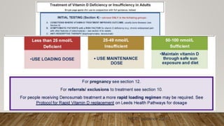
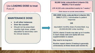
•Colecalciferol tablets 800-2000 unit tablets ONCE daily individualised to patient circumstances.
• Start at lower doses within this range.
• Higher doses (within this range) may be required in Winter months compared to Summer
months.
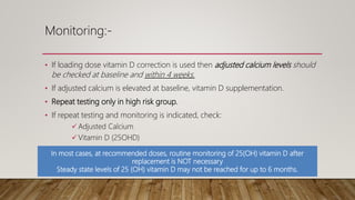
• 25(OH) vitamin D levels may take up to 6 months to reach steady state and rapidly fall once
supplementation is ceased. Therefore maintenance therapy may need to be long-term.
• If the patient continues to be genuinely symptomatic despite 6 months of maintenance
treatment, then evaluate adherence issues.
• If no adherence issues after 6 months of treatment, then check Vitamin D levels and if still
insufficient consider increase in maintenance dose within parameters described or loading
dose followed by maintenance if appropriate.
•Advise to purchase a supplement suitable to supply 20 – 50micrograms (800 to 2000 units) daily.
•Available in supermarkets and pharmacies: ask community pharmacist for latest advice.
OR
•Calcium + vitamin D (calcium as maximum calcium carbonate 2.5g [equivalent to 1.5g calcium]) and
1000 units daily vitamin D3 when given as combined preparation) for frail, institutionalised people to
prevent falls and for those with osteoporosis/osteopenia if daily recommended intake of calcium not
met
22. Monitoring:-
• If loading dose vitamin D correction is used then adjusted calcium levels should
be checked at baseline and within 4 weeks.
• If adjusted calcium is elevated at baseline, vitamin D supplementation.
• Repeat testing only in high risk group.
• If repeat testing and monitoring is indicated, check:
Adjusted Calcium
Vitamin D (25OHD)
U+Es
In most cases, at recommended doses, routine monitoring of 25(OH) vitamin D after
replacement is NOT necessary
Steady state levels of 25 (OH) vitamin D may not be reached for up to 6 months.
23. Safety about supplementation:-
• UK Scientific Advisory Committee on Nutrition (SACN) reviewed that an upper limit
of 4,000 units (100μg) per day is safe for adults, children >11 years of age, pregnant
and lactating women
• Research suggests up to 10,000 units can be taken daily by healthy people for up to
16 weeks without toxicity.
• Excessive intake can rarely lead to hypercalcaemia; symptoms include muscle
weakness, apathy, headache, anorexia, nausea and vomiting.
• High dose, intermittent vitamin D supplementation has been associated with falls
and increased fracture risk and should be avoided, unless there is an urgency to
replace vitamin D
24. Treatment Regimes In Pregnancy:
• Vitamin D use in human pregnancy is not associated with an increased risk of
congenital malformation.
• An upper physiological limit of 10,000units of vitamin D/day has been
suggested.
• Above the upper limit, 10.000 should not used in pregnancy.
• Safety data relate to use in the second or third trimesters, avoided in the first
trimester.
http://www.healthystart.nhs.uk/.
25. Lifestyle advice during pregnancy and breastfeeding:
Safe in all trimesters
For all pregnant women: Vitamin D3 (colecalciferol) tablets 400 units once a day
For at risk pregnant women: Vitamin D3 (colecalciferol) tablets 1000 units a day
Higher Risk pregnant women include;
•Increased skin pigmentation,
•Reduced exposure to sunlight,
•Socially excluded
•Obese BMI ≥ 30kg/m2
For pregnant women at risk of pre-eclampsia a Calcium + vitamin D combination is
recommended.
26. Treatment Of INSUFFICIENCY In PREGNANCY: 25(OH) vitamin D 25-49 nmol/L
Only use this regime in SECOND or THIRD trimester
Vitamin D3 (colecalciferol) capsules: 2000 units DAILY for 10 weeks
Or if weekly dosing required for adherence.
Vitamin D3 (colecalciferol) capsules: 20,000 units ONCE WEEKLY for 8 weeks.
•Monitor adjusted calcium at baseline and 4 weeks after commencing treatment.
•Follow loading dose with MAINTENANCE vitamin D3 (800-2000 units daily)
•Repeat testing of 25(OH) vitamin D levels is NOT usually necessary unless patient remains
symptomatic, or has other risk factors for remaining vitamin D deficient
Treatment of DEFICIENCY / INSUFFICIENCY in PREGNANCY ;
Rapid Correction
Only use this regime in SECOND or THIRD trimester
Can use RAPID CORRECTION regime in the following scenarios:
25(OH) vitamin D levels <25 nmol/L
Third trimester of pregnancy and rapid correction required
Vitamin D3 (colecalciferol) capsules: 4000 units DAILY for 8 weeks
(If more rapid correction than this is required seek advice from Endocrinology)
OR If weekly dosing required for adherence
Vitamin D3 (colecalciferol) capsules: 20,000 units ONCE WEEKLY for 14 weeks**
I if liquid required prescribe:
Colecalciferol 25,000units/1ml oral solution unit dose ampoules sugar free, 1 ampoule once a week for 6 weeks
• Monitor adjusted calcium 4 weeks at baseline and after commencing treatment.
• Follow loading dose with MAINTENANCE vitamin D3 (800-2000 units daily)
• Repeat testing of 25(OH) vitamin D levels is NOT usually necessary unless patient remains symptomatic, or has other
risk factors for remaining vitamin D deficient
27. Reference:
• http://www.evidence.nhs.uk/search?q=%22What+dose+of+vitamin+D+should+be+prescribed+for+the+treatment+of+vitamin+D+defi
ciency%22.
• A.C. Norlin, S. Hansen, E. Wahren-Borgström, C. Granert, L. Björkhem-Bergman , P
. Bergman, Vitamin D3 Supplementation and Antibiotic
Consumption – Results from a Prospective, Observational Study at an Immune-Deficiency Unit in Sweden. PLoS One, 11(9)
(2016):e0163451.
• Juonala M, Voipio A, Pahkala K, Viikari JSA, Mikkila V, Kahonen M, et al. Childhood 25-OH Vitamin D Levels and Carotid Intima-Media
Thickness in Adulthood: The Cardiovascular Risk in Young Finns Study. Journal of Clinical Endocrinology & Metabolism. 2015;100(4):1469-
76.
• Lappe JM, Travers-Gustafson D, Davies KM, Recker RR, Heaney RP
. Vitamin D and calcium supplementation reduces cancer risk: results of
a randomized trial. American Journal of Clinical Nutrition. 2007;85(6):1586-91.
• Slinin Y, Paudel ML, Taylor BC, Fink HA, Ishani A, Canales MT, et al. 25-Hydroxyvitamin D levels and cognitive performance and decline in
elderly men. Neurology. 2010;74(1):33-41.
• Wimalawansa SJ. Associations of vitamin D with insulin resistance, obesity, type 2 diabetes, and metabolic syndrome. J Steroid Biochem
Mol Biol. 2016.
• http://www.cancerresearchuk.org/cancer-
info/prod_consump/groups/cr_common/@nre/@sun/documents/generalcontent/cr_052628.pdf.
• Pearce SHS, Cheetham TD. Diagnosis and management of vitamin D. BMJ 2010; 340: 142‐147.