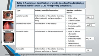
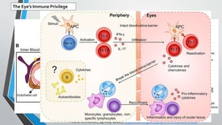
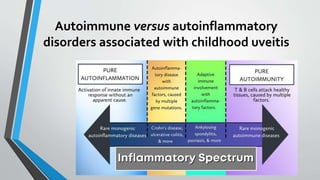
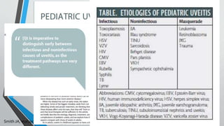
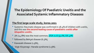
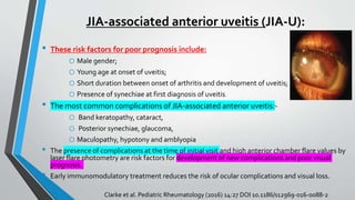
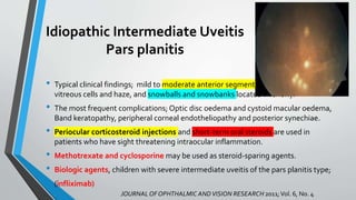
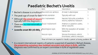
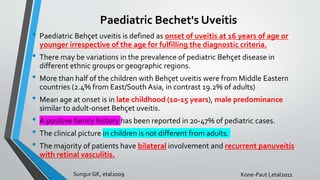
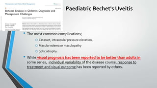
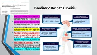
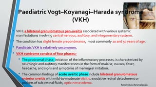
Pediatric uveitis can be caused by autoimmune or autoinflammatory disorders. The most common type seen in children is chronic anterior uveitis, unlike adults where it is less common. Juvenile idiopathic arthritis (JIA)-associated anterior uveitis is the most frequent cause of uveitis in children. It is typically bilateral and non-granulomatous with a chronic relapsing course. Idiopathic intermediate uveitis (pars planitis) commonly affects children and adolescents and has a low association with systemic diseases. Behcet's disease onset is most common in late childhood, with bilateral recurrent panuveitis and retinal vasculitis seen clinically.

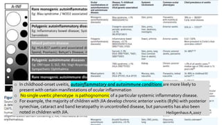


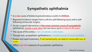
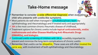
![Uveitis in the paediatric vasculitides
• Vasculitis can be a coexisting disease seen with, or as part of, an
autoinflammatory disorder.
• Retinal vasculitis may be a common end result of a number of inflammatory
events.
• Uveitis is a recognized feature of antineutrophil cytoplasmic antibody (ANCA)-
associated vasculitis (e.g. granulomatosis with polyangiitis),
• Vasculitis within connective tissue disease (e.g. systemic lupus erythematosus or
scleroderma)
• Small or medium vessel vasculitis [e.g. IgA vasculitis/ Henoch–Schönlein purpura
and Kawasaki disease (KD)].
• In KD, a mild, bilateral uveitis in a child with red eyes and a nonspecific fever can be
one of the earliest signs of disease, and early diagnosis of KD is of particular value
as late diagnosis can result in secondary chronic coronary artery disease.](https://image.slidesharecdn.com/pediatricautoimmuneuveitis-221017172710-5d0605eb/85/Pediatric-autoimmune-uveitis-pptx-35-320.jpg)


![jCA-uveitis 2 [Autosaved].pptx](https://cdn.slidesharecdn.com/ss_thumbnails/jca-uveitis2autosaved-230303163346-40ddc821-thumbnail.jpg?width=640&height=640&fit=bounds)


![PERI-PROSTHETIC FRACTURE NAIL-PLATE CONSTRUCT [NPC].pptx](https://cdn.slidesharecdn.com/ss_thumbnails/drarunkumardrmohamedashrafperiprostheticfrasturenail-plateconstructnpc-260209164459-7e9d15a1-thumbnail.jpg?width=640&height=640&fit=bounds)

