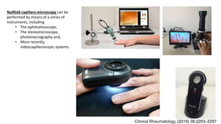
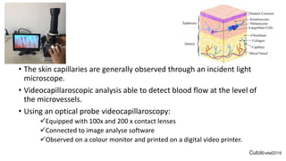
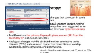
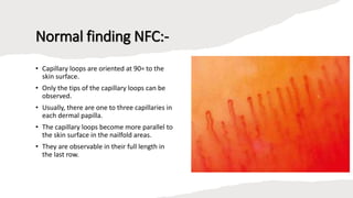
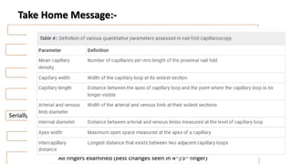
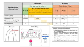
Nailfold capillaroscopy is a valuable, non-invasive diagnostic tool used in rheumatic diseases to detect microvascular changes and differentiate between primary and secondary Raynaud's phenomenon. The procedure involves careful examination under an incident light microscope and the use of videocapillaroscopic systems to assess capillary characteristics such as distribution, shape, density, and dimensions. Proper techniques and patient preparation are critical to ensure accurate measurements and interpretation of results.

![Reference:
• O. Sander, C. Sunderk¨otter, I. K¨otte et al., “Kapillarmikroskopie,” Zeitschrift f¨ur Rheumatologie, vol. 69,no.
3, pp. 253–262, 2010.
• M. Cutolo and V. Smith, “State of the art on nailfold capillaroscopy: a reliable diagnostic tool and putative
biomarker in rheumatology?” Rheumatology, vol. 52, no. 11, pp. 1933–1940, 2013.
• K.-M. Lin, T.-T. Cheng, and C.-J. Chen, “Clinical applications of nailfold capillaroscopy in different rheumatic
diseases,” Journal of InternalMedicine of Taiwan, vol. 20, no. 3, pp. 238–247, 2009.
• M. Berks, P. Tresadern, G. Dinsdale et al., “An automated systemfor detecting andmeasuring nailfold
capillaries,” in Medical Image Computing and Computer-Assisted Intervention—MICCAI 2014, P. Golland, N.
Hata,C.Barillot, J. Hornegger, and R. Howe, Eds., vol. 8673 of Lecture Notes in Computer Science,pp. 658–665,
Springer, Berlin, Germany, 2014.
• C. Hoerth, M. Kundi, R. Katzenschlager, and M. Hirschl, “Qualitative and quantitative assessment of nailfold
capillaries by capillaroscopy in healthy volunteers,” Vasa, vol. 41, no. 1, pp. 19–26, 2012.
• H. M. A. Hofstee, E. H. Sern´e, C. Roberts et al., “A multicentre study on the reliability of qualitative and
quantitative nail-fold videocapillaroscopy assessment,” Rheumatology, vol. 51, no. 4, pp. 749–755, 2012.
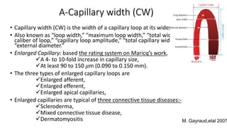
• M. Gayraud, “Raynaud’s phenomenon,” Joint Bone Spine, vol. 74, no. 1, pp. e1–e8, 2007.
• H. R. Maricq, “Comparison of quantitative and semiquantitative estimates of nailfold capillary abnormalities
in scleroderma spectrum disorders,” Microvascular Research, vol. 32, no. 2, pp. 271–276, 1986.
• M. D. P. Kamboj, A study of nail fold capillaroscopy in psoriasis [Ph.D. thesis], Rajiv Gandhi University of
Health Sciences, Bengaluru, India, 2014.](https://image.slidesharecdn.com/capillaroscope-230714201156-3624efbb/85/Capillaroscope-pptx-43-320.jpg)