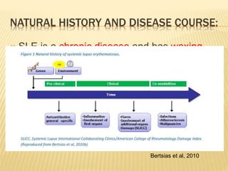
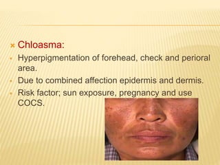
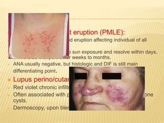
This document discusses systemic lupus erythematosus (SLE) and conditions that can mimic its presentation. It describes how SLE often has a waxing and waning chronic course that can vary in severity. Several infectious, inflammatory, and neoplastic conditions can present similarly to SLE through involvement of multiple organ systems and production of autoantibodies. Correct diagnosis requires a thorough history, physical exam, targeted testing, and biopsies to distinguish SLE from its mimickers. Serological similarities alone do not confirm SLE if clinical features are inconsistent.
Systemic lupus erythematosus (SLE) is a chronic autoimmune disease that can affect multiple organ systems. SLE most commonly affects women in their 20s and 30s. The course of SLE is unpredictable, with periods of illness alternating with remission. Common initial symptoms include fatigue, fever, joint pain, and weight changes. SLE can cause skin rashes, oral ulcers, hair loss, serositis, lung involvement including pleurisy, heart involvement such as pericarditis, and kidney disease. The prognosis for SLE has improved in recent decades due to medical advances, with survival rates of approximately 95% at 5 years and 90% at 10 years. SLE is treated symptomatically mainly with
Connective tissue disorders involve inflammation of connective tissue leading to skin and organ problems. They range from mild skin involvement to severe multi-system diseases. Lupus erythematosus can involve only the skin or be systemic. Discoid lupus erythematosus causes scarring plaques on sun exposed skin that may spread but rarely progresses to systemic lupus. Systemic lupus erythematosus is an autoimmune disease affecting multiple organs with a characteristic rash and internal organ involvement. Dermatomyositis causes skin signs like a lilac rash around the eyes and on fingers along with muscle weakness. Systemic sclerosis causes hardening of the skin that can progress
Systemic lupus erythematosus (SLE) is a rare but severe autoimmune disease in children and adolescents. It can affect any organ system and commonly involves the kidneys and central nervous system. SLE is difficult to diagnose due to its varied manifestations. It often presents with nonspecific symptoms like fever and fatigue as well as skin rashes and oral ulcers. Renal involvement occurs in 50-75% of childhood SLE patients, ranging from mild proteinuria to nephrotic syndrome or kidney failure. Early diagnosis and treatment are important to prevent organ damage and complications. Childhood SLE tends to be a more severe form of the disease compared to adult SLE.
This document summarizes key information about leprosy (Hansen's disease), including:
- It is caused by Mycobacterium leprae bacteria and primarily affects the skin, nerves, respiratory tract and testes.
- There is a spectrum of clinical forms from tuberculoid (high resistance) to lepromatous (low resistance).
- Complications include reactions, nerve damage leading to deformities, and secondary infections.
- Treatment involves multidrug therapy but nerve damage may be permanent without rehabilitation. Advocacy groups continue working to eliminate stigma and provide care.
- Lupus erythematosus is a chronic inflammatory disease associated with abnormalities of the immune system that results from genetic, hormonal, and environmental factors interacting. It can involve the skin and multiple organ systems.
- The document discusses the history, epidemiology, etiology, pathogenesis, clinical findings and classification of the different types of cutaneous lupus erythematosus including acute, subacute, and neonatal lupus. It provides details on the clinical presentations, pathogenic mechanisms, and classification systems for the skin involvement in lupus.
Acute rheumatic fever-definition,pathophysiology,clinical presentation and ma...onlinefreelancer1
A detailed approach to ACUTE RHEUMATIC FEVER,based on Harrison Principles of internal medicine and Braunwald Textbook of Cardiology.Useful for post graduate seminars.
This document provides an overview of sarcoidosis, including its definition, epidemiology, etiology, pathology, clinical features, diagnosis and natural course. Sarcoidosis is a multisystem disorder characterized by noncaseating granulomas and can affect many organs, most commonly the lungs (90%). It has the highest prevalence in Scandinavian countries. While the cause is unknown, it is thought to involve an abnormal immune response to environmental factors in genetically predisposed individuals. Tissue biopsy showing noncaseating granulomas is required for diagnosis, along with supportive tests such as BAL and serum ACE levels. The natural course is variable, with 70% experiencing spontaneous remission and 16% having progressive disease.
The document discusses several autoimmune diseases including systemic lupus erythematosus, rheumatoid arthritis, Sjögren's syndrome, systemic sclerosis, and mixed connective tissue disease. It provides details on the pathogenesis, clinical features, immunological characteristics, and morphology of these conditions. Autoimmune diseases result from a loss of tolerance to self-antigens and can involve deregulated immune responses against tissues and organs, leading to inflammation and damage.
Systemic lupus erythematosus (SLE) is a chronic autoimmune disease that affects multiple organ systems through chronic inflammation. Common symptoms include skin rashes, joint pain, fatigue and oral lesions. SLE particularly impacts women in their 30s and 40s. Kidney disease is a serious complication that can lead to chronic renal failure. Dentists play an important role in managing oral symptoms of SLE and preventing infections by providing antibiotic prophylaxis for procedures in patients with heart valve damage.
RHEUMATIC DISEASE OF CHILDHOOD(BINGHAM UNIVERSITY)_074613.pptxEmmanuelElijah8
This document discusses rheumatic diseases that can affect children, including juvenile rheumatoid arthritis (JRA), systemic lupus erythematosus, and Kawasaki disease. It provides details on the characteristics, symptoms, diagnostic criteria, and treatment approaches for each condition. JRA is regarded as not a single disease but a category with three types of onset. Systemic lupus erythematosus is characterized by autoantibodies against self antigens that can damage multiple organs. Kawasaki disease causes severe vasculitis that predominantly affects medium-sized arteries like the coronary arteries.
The document contains medical images and descriptions of various conditions:
1) Images show lesions and rashes characteristic of conditions like pyoderma gangrenosum, dermatitis herpetiformis, and livedo reticularis.
2) Radiographic images depict abnormalities related to osteonecrosis, DISH syndrome, and Gardner's syndrome.
3) Microscopic slides show cellular features of diseases such as malaria, CLL, and Goodpasture's syndrome.
an overview of Lupus for journalist
Lupus has a wide spectrum of manifestation. Some mild but in most cases it has a high impact of life and quality of life
This document discusses cutaneous pseudolymphoma (CPL). It was first described in 1891 and has been called by various names over time. Pseudolymphoma clinically and sometimes histologically mimics lymphoma, but has benign behavior and does not meet criteria for malignant lymphoma. It is characterized by lymphocytic infiltration in the skin in response to stimuli. CPL is classified into types depending on the predominant cell type (B cell vs T cell) and location of infiltration (nodular vs stripe-like). While CPL has no associated mortality, localized variants can cause minor symptoms. Treatment involves excision, corticosteroids, radiation therapy, and immunosuppressants depending on the subtype.
The skin can provide clues to underlying systemic diseases. Some examples include:
1. Seborrheic dermatitis may be associated with Parkinson's disease or HIV infection. Vitiligo is often associated with autoimmune thyroid disease or diabetes.
2. Blistering diseases like pemphigus vulgaris and bullous pemphigoid are associated with malignancy in elderly patients. Epidermolysis bullosa acquisita may be associated with inflammatory bowel disease.
3. Cutaneous metastases, Paget's disease, acanthosis nigricans, and amyloidosis can indicate underlying internal cancers.
Connective tissue diseases share features of immune dysregulation and autoantibody production directed at nuclear components, causing widespread tissue damage. Systemic lupus erythematosus is characterized by arthritis, rashes, kidney involvement and positive ANA and anti-dsDNA antibodies. Systemic sclerosis involves skin thickening from fibrosis, Raynaud's phenomenon, and autoantibodies like anti-Scl-70. Polymyositis and dermatomyositis cause proximal muscle weakness and inflammation with skin lesions in dermatomyositis.
This document provides an overview of psoriasis and other papulosequamous skin diseases. It describes psoriasis as a chronic inflammatory skin disorder characterized by red papules and plaques covered with silvery scales, most commonly occurring on the elbows and scalp. The cause is believed to be an immune system dysfunction interacting with genetic and environmental factors. Symptoms include itchy lesions that may worsen with stress or injury to the skin. Treatment involves long-term, individualized regimens due to the chronic nature of the disease.
1. Lupus erythematosus (LE) is an autoimmune disease that can cause heterogeneous cutaneous and systemic manifestations. Cutaneous lupus erythematosus (CLE) can occur as a manifestation of systemic lupus erythematosus (SLE) or independently.
2. CLE is classified into LE-specific lesions and LE-nonspecific lesions. Common LE-specific lesions include acute cutaneous lupus erythematosus (ACLE), subacute cutaneous lupus erythematosus (SCLE), and chronic cutaneous lupus erythematosus (CCLE).
3. Diagnosis of CLE
All ACUTE LYMPHOBLASTIC LEUKEMIA BY DR MAGDI SASIcardilogy
1. The document provides information about acute lymphocytic leukemia (ALL), the most common type of cancer in children. It describes the symptoms, signs, classification, and risk factors of ALL.
2. Common symptoms of ALL include fever, fatigue, easy bruising, frequent infections, and bone/joint pain. Physical signs may include pallor, organ enlargement, and lymphadenopathy.
3. ALL is classified according to the type of lymphocytes involved (B-cell vs T-cell lineage) and cellular features. The vast majority of ALL cases involve B-cell lymphocytes. Risk factors for ALL are generally unknown for most people.
Know more about Psoriasis ,Types and TreatmentsiCliniq
Psoriasis is a prototypic papulosquamous skin
diseases characterised by erythematous papules. It is a chronic inflammatory skin disease with increased epidermal proliferation related to dysregulation of the immune system.
It needs long time medication to get it control, the permanent is not found yet.
To Get guidance to treat Psoriasis from a doctor --> https://www.icliniq.com/ask-a-doctor-online/dermatologist/psoriasis
Systemic lupus erythematosus (SLE) is a chronic inflammatory autoimmune disease that can affect multiple organs. It is characterized by the presence of autoantibodies that target nuclear and cytoplasmic components of cells. SLE most commonly affects women between the ages of 20-30. Clinical manifestations vary but often involve the skin, joints, kidneys, lungs, nervous system, and other organs. The course of SLE is variable with periods of remission and relapse.
Autoimmune diseases result from immune reactions against self-antigens and loss of self-tolerance. They can be organ-specific like type 1 diabetes which targets pancreatic beta cells, or systemic like systemic lupus erythematosus which affects multiple organs. Central and peripheral tolerance mechanisms normally prevent autoimmunity, but their failure allows self-reactive T and B cells to escape and cause tissue damage. Common autoimmune diseases include rheumatoid arthritis, Sjögren's syndrome, systemic sclerosis, and inflammatory myopathies.
Pityriasis rosea is a common skin condition that mainly affects children and young adults. It is characterized by the appearance of multiple pink, oval lesions on the trunk that have a distinctive scale. The rash begins with a single larger 'herald' plaque, followed by many smaller plaques over the next few weeks. While the cause is unknown, it is generally not contagious and most cases resolve spontaneously within 2-10 weeks without treatment. Topical steroids or calamine lotion can help relieve itching.
1) Lepra reactions are immunologically inflammatory states that can occur in leprosy patients. There are two main types - Type 1 reactions (downgrading and reversal reactions) and Type 2 reactions (Erythema Nodosum Leprosum or ENL).
2) Type 1 reactions involve the skin and nerves and cause swelling and tenderness. They occur after treatment begins. Type 2 or ENL reactions cause painful swollen skin lesions and other symptoms like fever. They typically occur after treatment in lepromatous patients.
3) Histologically, Type 1 reactions show edema while Type 2 reactions show deep inflammation, necrosis and few bacilli. Both can cause significant morbidity if not properly treated.
Lupus erythematosus (LE) is an autoimmune connective tissue disorder that can affect one or several organs. Circulating autoantibodies and immune complexes are due to loss of normal immune tolerance and are pathogenic. Clinical features of LE are highly variable. LE nearly always affects the skin to some degree.
VEXAS syndromes , a diagnostic PuzzlepptxMarwa Besar
This document discusses VEXAS syndrome, a recently discovered autoinflammatory and hematologic condition caused by somatic mutations in the UBA1 gene. It provides details on the clinical manifestations (fevers, inflammatory symptoms affecting skin, joints, lungs, blood vessels), hematological abnormalities (anemia, cytopenias), and challenges in management due to the combined inflammatory and hematological involvement. Diagnosis is confirmed via genetic testing demonstrating UBA1 mutations. Treatment aims to suppress inflammation and eliminate mutant hematopoietic cells, but outcomes remain poor with high mortality.
1. The document discusses when to call an immunologist in an interstitial lung disease (ILD) clinic. It provides information on clinical signs, autoantibodies, and instrumental exams that can help recognize autoimmune ILD associated with rheumatic diseases.
2. Risk factors for developing ILD in autoimmune rheumatic diseases include certain diseases like systemic sclerosis, as well as specific autoantibodies. Monitoring of pulmonary symptoms depends on the underlying rheumatic disease but may include regular testing and imaging.
3. A multidisciplinary approach is important for diagnosing and managing autoimmune ILD, and guidelines recommend evaluating any newly diagnosed ILD for an underlying autoimmune rheumatic disease.
Management of hereditary angioedema involves treating acute attacks, preventing attacks, and improving quality of life. Treatment strategies include treating acute attacks, preventing attacks short-term before procedures, and long-term prophylaxis. Therapies include C1 inhibitor replacement, bradykinin receptor antagonists, attenuated androgens, and antifibrinolytics. An individualized treatment plan is recommended based on a patient's attack frequency, severity and location, as well as their medication access and preferences.
Hidradenitis suppurativa (HS) is a chronic inflammatory skin disease characterized by recurrent abscesses and scarring in the apocrine gland-bearing areas. It typically presents in young adults and is more common in women and people of African descent. Obesity, smoking, family history and certain drugs are associated risk factors. The pathogenesis involves occlusion of hair follicles leading to rupture, inflammation, abscess formation and scarring. Diagnosis is clinical though imaging and biopsy may help in some cases. Treatment aims to control flare-ups and prevent scarring. The course is usually chronic with intermittent flares and remissions.
- The document analyzes IL-18 and IFN-gamma as biomarkers for diagnosis and prognosis in juvenile idiopathic arthritis (JIA) patients. It finds that IL-18 levels correlate with disease activity and can predict response to therapy, while IFN-gamma levels indicate response to therapy. The study compares biomarker and disease activity measurements in 45 JIA patients at initial presentation and after treatment. It concludes that IL-18 and IFN-gamma may be promising markers for detecting prognosis and treatment response in JIA patients.
This document discusses three case studies of patients with autoimmune diseases and thrombosis. It then provides statistics on the increased risk of thrombosis in various autoimmune diseases compared to the general population. The mechanisms of thrombosis in autoimmune diseases are reviewed. Several pearls and myths regarding thrombosis and treatment in autoimmune diseases are discussed, including that all autoimmune diseases confer a hypercoagulable state; anticoagulants are not always effective or safe for Bechet's syndrome; and immunosuppressive drugs may also cause thrombosis. The take home message is that autoimmune diseases are both inflammatory and hypercoagulable, and risk versus benefit of anticoagulants like DOACs must be considered.
Pediatric uveitis can be caused by autoimmune or autoinflammatory disorders. The most common type seen in children is chronic anterior uveitis, unlike adults where it is less common. Juvenile idiopathic arthritis (JIA)-associated anterior uveitis is the most frequent cause of uveitis in children. It is typically bilateral and non-granulomatous with a chronic relapsing course. Idiopathic intermediate uveitis (pars planitis) commonly affects children and adolescents and has a low association with systemic diseases. Behcet's disease onset is most common in late childhood, with bilateral recurrent panuveitis and retinal vasculitis seen clinically.
Adult-onset Still's disease (AOSD) is a rare autoinflammatory disorder characterized by high spiking fever, evanescent rash, arthritis, and multiorgan involvement. It was first described in adults in 1971. The disease most commonly affects young adults and women. Diagnosis is based on clinical criteria and exclusion of other conditions. Treatment involves nonsteroidal anti-inflammatories initially followed by disease-modifying drugs like methotrexate. Biologics that inhibit IL-1 or IL-6 are effective for refractory or complicated cases. Prognosis is generally good but complications can include macrophage activation syndrome, organ failure, or amyloidosis.
Vogt-Koyanagi-Harada (VKH) syndrome is a rare autoimmune disease that affects melanin-containing tissues like the eyes, inner ears, skin, and brain. It is characterized by bilateral uveitis and can cause vision loss. The disease occurs in phases including prodromal, acute uveitic, convalescent, and recurrent. Treatment involves high-dose corticosteroids for at least 6 months to reduce inflammation and recurrence risk. Immunosuppressants may be needed for resistant cases. Prognosis depends on factors like early treatment, treatment duration, and presence of complications. Strict monitoring is needed due to risk of vision threatening complications.
Familial Mediterranean Fever is an autosomal recessive disease characterized by recurrent fevers and inflammation localized to the peritoneum, pleura, joints, or skin. It is most common in people of Mediterranean descent and is caused by mutations in the MEFV gene. Symptoms include abdominal pain, fever, arthritis, and erysipelas-like lesions. If left untreated, it can lead to amyloidosis. Treatment involves lifelong colchicine to prevent inflammatory episodes and reduce amyloidosis risk. Some patients may be resistant to colchicine and require alternative treatments like interleukin-1 or tumor necrosis factor inhibitors.
Giant cell arteritis (GCA) is a large vessel vasculitis that commonly affects the branches of the carotid artery and causes headaches, jaw claudication, and vision loss. The pathology involves granulomatous inflammation in the vessel walls. Diagnosis is based on temporal artery biopsy showing giant cells, but imaging such as ultrasound, CT, and PET can also provide supportive evidence of vessel inflammation. Treatment involves high-dose corticosteroids to reduce inflammation and prevent relapses and vision loss, with tapering over 2-5 years. Monitoring for complications like aortic aneurysms is also important given the vessel involvement.
Polyarteritis nodasa and microscopic polyangitisMarwa Besar
This document discusses Polyarteritis Nodosa (PAN) and Microscopic Polyangiitis (MPA). PAN is a necrotizing vasculitis predominantly affecting medium-sized arteries that spares small vessels. It is typically ANCA-negative. MPA is a pauci-immune necrotizing vasculitis involving small vessels and sometimes medium arteries, associated with ANCA positivity. Both diseases can affect multiple organ systems and have variable clinical manifestations. Differentiation is based on vessel size involvement and ANCA status according to the Chapel Hill consensus criteria.
Takayasu arteritis is a rare large vessel vasculitis that predominantly involves the aorta and its major branches. It was first described in 1908 by Mikito Takayasu and has various names including pulseless disease. It most commonly affects the subclavian and carotid arteries in women under 50 years old. Diagnosis is based on imaging and clinical criteria as there are no definitive diagnostic tests. Treatment involves steroids and steroid-sparing immunosuppressants, with TNF inhibitors being widely used biologic therapies. Ongoing management focuses on preventing arterial progression and complications like hypertension.
Promoting Wellbeing - Applied Social Psychology - Psychology SuperNotesPsychoTech Services
A proprietary approach developed by bringing together the best of learning theories from Psychology, design principles from the world of visualization, and pedagogical methods from over a decade of training experience, that enables you to: Learn better, faster!
Cell Therapy Expansion and Challenges in Autoimmune DiseaseHealth Advances
There is increasing confidence that cell therapies will soon play a role in the treatment of autoimmune disorders, but the extent of this impact remains to be seen. Early readouts on autologous CAR-Ts in lupus are encouraging, but manufacturing and cost limitations are likely to restrict access to highly refractory patients. Allogeneic CAR-Ts have the potential to broaden access to earlier lines of treatment due to their inherent cost benefits, however they will need to demonstrate comparable or improved efficacy to established modalities.
In addition to infrastructure and capacity constraints, CAR-Ts face a very different risk-benefit dynamic in autoimmune compared to oncology, highlighting the need for tolerable therapies with low adverse event risk. CAR-NK and Treg-based therapies are also being developed in certain autoimmune disorders and may demonstrate favorable safety profiles. Several novel non-cell therapies such as bispecific antibodies, nanobodies, and RNAi drugs, may also offer future alternative competitive solutions with variable value propositions.
Widespread adoption of cell therapies will not only require strong efficacy and safety data, but also adapted pricing and access strategies. At oncology-based price points, CAR-Ts are unlikely to achieve broad market access in autoimmune disorders, with eligible patient populations that are potentially orders of magnitude greater than the number of currently addressable cancer patients. Developers have made strides towards reducing cell therapy COGS while improving manufacturing efficiency, but payors will inevitably restrict access until more sustainable pricing is achieved.
Despite these headwinds, industry leaders and investors remain confident that cell therapies are poised to address significant unmet need in patients suffering from autoimmune disorders. However, the extent of this impact on the treatment landscape remains to be seen, as the industry rapidly approaches an inflection point.
Travel Clinic Cardiff: Health Advice for International TravelersNX Healthcare
Travel Clinic Cardiff offers comprehensive travel health services, including vaccinations, travel advice, and preventive care for international travelers. Our expert team ensures you are well-prepared and protected for your journey, providing personalized consultations tailored to your destination. Conveniently located in Cardiff, we help you travel with confidence and peace of mind. Visit us: www.nxhealthcare.co.uk
Nano-gold for Cancer Therapy chemistry investigatory projectSIVAVINAYAKPK
chemistry investigatory project
The development of nanogold-based cancer therapy could revolutionize oncology by providing a more targeted, less invasive treatment option. This project contributes to the growing body of research aimed at harnessing nanotechnology for medical applications, paving the way for future clinical trials and potential commercial applications.
Cancer remains one of the leading causes of death worldwide, prompting the need for innovative treatment methods. Nanotechnology offers promising new approaches, including the use of gold nanoparticles (nanogold) for targeted cancer therapy. Nanogold particles possess unique physical and chemical properties that make them suitable for drug delivery, imaging, and photothermal therapy.
These lecture slides, by Dr Sidra Arshad, offer a simplified look into the mechanisms involved in the regulation of respiration:
Learning objectives:
1. Describe the organisation of respiratory center
2. Describe the nervous control of inspiration and respiratory rhythm
3. Describe the functions of the dorsal and respiratory groups of neurons
4. Describe the influences of the Pneumotaxic and Apneustic centers
5. Explain the role of Hering-Breur inflation reflex in regulation of inspiration
6. Explain the role of central chemoreceptors in regulation of respiration
7. Explain the role of peripheral chemoreceptors in regulation of respiration
8. Explain the regulation of respiration during exercise
9. Integrate the respiratory regulatory mechanisms
10. Describe the Cheyne-Stokes breathing
Study Resources:
1. Chapter 42, Guyton and Hall Textbook of Medical Physiology, 14th edition
2. Chapter 36, Ganong’s Review of Medical Physiology, 26th edition
3. Chapter 13, Human Physiology by Lauralee Sherwood, 9th edition
low birth weight presentation. Low birth weight (LBW) infant is defined as the one whose birth weight is less than 2500g irrespective of their gestational age. Premature birth and low birth weight(LBW) is still a serious problem in newborn. Causing high morbidity and mortality rate worldwide. The nursing care provide to low birth weight babies is crucial in promoting their overall health and development. Through careful assessment, diagnosis,, planning, and evaluation plays a vital role in ensuring these vulnerable infants receive the specialize care they need. In India every third of the infant weight less than 2500g.
Birth period, socioeconomical status, nutritional and intrauterine environment are the factors influencing low birth weight
Kosmoderma Academy, a leading institution in the field of dermatology and aesthetics, offers comprehensive courses in cosmetology and trichology. Our specialized courses on PRP (Hair), DR+Growth Factor, GFC, and Qr678 are designed to equip practitioners with advanced skills and knowledge to excel in hair restoration and growth treatments.
Co-Chairs, Val J. Lowe, MD, and Cyrus A. Raji, MD, PhD, prepared useful Practice Aids pertaining to Alzheimer’s disease for this CME/AAPA activity titled “Alzheimer’s Disease Case Conference: Gearing Up for the Expanding Role of Neuroradiology in Diagnosis and Treatment.” For the full presentation, downloadable Practice Aids, and complete CME/AAPA information, and to apply for credit, please visit us at https://bit.ly/3PvVY25. CME/AAPA credit will be available until June 28, 2025.
5-hydroxytryptamine or 5-HT or Serotonin is a neurotransmitter that serves a range of roles in the human body. It is sometimes referred to as the happy chemical since it promotes overall well-being and happiness.
It is mostly found in the brain, intestines, and blood platelets.
5-HT is utilised to transport messages between nerve cells, is known to be involved in smooth muscle contraction, and adds to overall well-being and pleasure, among other benefits. 5-HT regulates the body's sleep-wake cycles and internal clock by acting as a precursor to melatonin.
It is hypothesised to regulate hunger, emotions, motor, cognitive, and autonomic processes.
Demystifying Fallopian Tube Blockage- Grading the Differences and Implication...
Lupus mimicker2.pptx
1.
2. Systemic lupus erythematosus (SLE) is the
prototypic multisystem autoimmune disease
with broad spectrum of clinical presentation
encompassing all organs and chronic course
which can vary from mild to life threating.
SLE was first described during middle aged to
describe erosive skin lesion “wolf bite” (1837-
1880) describe it as malar rash.
Moriz Kaposi (1837-1902) describe it as
systemic disease with visceral manifestation.
3. NATURAL HISTORY AND DISEASE COURSE:
SLE is a chronic disease and has waxing
and waning course with significant morbidity.
The disease starts with preclinical phase
characterized by non specific autoimmune
abnormalities that proceeds to a more
disease specific autoimmunity phase.
Bertsias et al, 2010
4. The clinical overt disease may start with single
manifestation that are non specific, such as arthritis,
Raynauds phenomenon or autoimmune
thrombocytopenia.
It may take years for patients to develop enough clinical
and immunological disturbances leading to SLE, although
some patients present from inception with full-blown
disease.
Time of the disease onset is uncertain and diagnosis is
delayed.
The clinical presentation may result not only from lupus
flare, but also from damage accrual related to disease,
treatment (steroid and immunosuppressives)and
comorbidities (infection, osteoprosiis, cataract,
atherosclerosis and malignancy). Edworthy SM. etal2005
5. GREAT IMITATOR:
SLE is considered the great mimic a myriad
of conditions often result in delayed
diagnosis.
There may be either clinical or serological
mimicker.
Collins. DA,etal1996
6. DERMATOLOGICAL MIMICKER OF SLE:
Acute cutaneous lupus:
1. Rosacea:
Erythema of face (nose, check, chin and forehead).
Trigger; hot, cold, sunlight, emotion, spicy, alcohol,
cosmetics.
Presence papules and pustules or blepharitis favors
rosacea.
Fine scales, pigment changes, follicular plugging favors
SLE.
Histologic examination is important.
1. Seborrheic dermatitis:
Of the face may mimic SLE or rosacea, but SLE not affect
nasolabial fold. Lela A Lee,etal1991
7. Chloasma:
Hyperpigmentation of forehead, check and perioral
area.
Due to combined affection epidermis and dermis.
Risk factor; sun exposure, pregnancy and use
COCS.
9. Chronic cutaneous lupus or discoid lupus:
1. Polymorphous light eruption.
2. Lichen planus.
3. Leukemia or lymphoma cutis.
4. Psoriasis.
5. Lupus vulgaris.
6. Sarcodosis.
The main differentiating point is histologic and DIF
evaluation.
Orteuu CH,etal2001
10. Polymorphous light eruption (PMLE):
Common, sunlight induced eruption affecting individual of all
races.
PMLE is strongly linked to sun exposure and resolve within days,
unlike DLE that persist for weeks to months.
ANA usually negative, but histologic and DIF is still main
differentiating point.
Lupus perino/cutaneous sarcodosis:
Red violet chronic infiltrate of the tip of the nose.
Often associated with pulmonary infiltrate and digital bone
cysts.
Dermoscopy, upon blenching, have apple jelly.
11. Lichen planus (LP):
Difficult to differentiate especially in absence oral and scalp
lesion, if there is photo-distribution.
Most incriminated drugs; ACEI, antimalarial, quinine, gold,
thiazide diuretics.
The diagnosis is only made by biopsy and DIF.
Tinea faciei:
Part of dermatophyte infection, characterized by
erythematous, scaly patches, pruritic, worsen with light
exposure.
Unlike DLE which is slower to develop, more persistent
course with prominent follicle and painful.
12. RHEUMATOLOGIC AND MUSCULOSKELETAL
MIMICKERS OF SLE:
1. Söjgren’s Syndrome.
2. Rheumatoid Arthritis.
3. Myositis.
4. Sclerroderma.
5. Sarcoidosis.
All can mimic lupus in different ways.
13. SÖJGREN’S SYNDROME;
Ss either primary or secondary to other autoimmune,(SLE,
RA).
SS have many clinical and serological features of SLE.
1. Arthritis.
2. Skin rash.
3. Kidney dse.
4. ANA 80%, RF 75 TO 95%.
14. Kidney affection:
1. Tubulo intersititial: most common affected
2. Glomeular: rare, membrano
proliferative,mesangialproliferative,membran
ous.
3. Cryoglobinemia vasculitis and end stage
renal disease were detected.
15. RHEUMATOID ARTHRITIS:
Many case presented with joint pain that undifferentiated
from RA, especially early RA.
Joint swelling (arthritis) and pain is always common in RA,
but SLE associated with arthralgia.
joint destruction and deformity are more common in RA.
16.
17. MYOSITIS:
SLE associated with muscle pain in majority, with muscle
weakness in few of them.
(PM-DM), the main problem is proximal muscle weakness,
with underlying muscle inflammation and elevation of
creatine kinase(CK), abnormal EMG with characteristics
muscle affection.
Characteristics pathognomonic dusky red rash in malar
distribution, difficult to differentiate from malar rash of SLE.
Shawl sign, heliotrope rash and Gottron sign over dorsum
MCP, PIP joints help to differentiate.
ANA, anti-Jo1 and interstitial lung are specific for DM.
18.
19. SCLERRODERMA:
The hallmark of SSc is thickened skin which affect hand,
feet, forearm and face, due excessive production and
inadequate breakdown of collagen.
Classical salt and pepper pigmentation of SSc may mimic
skin rash, but rash in lupus is due inflammation not fibrosis.
ILD, difficult of swallowing solid, heart burn and deformity in
hand are rare in lupus.
Raynaud phenomenon 95% in SSc, 40% in SLE.
Anticentromere(localized SSc) and anti topisomerase1(
diffuse SSc), less in lupus.
20. SARCOIDOSIS:
Fever, arthritis, mouth ulcer, pleuropericaraditis are
common manifestation of both sarcoidosis and
SLE.
But, ANA +ve 33%, AntidsDNA is negative in
sarcoidosis.
21. MALIGNANCY :
1. Hematological malignancy(hodgkin lymphoma).
2. Thymoma.
3. Carcinoma of the lung, breast and ovary as paraneoplastic
syndrome that mimic SLE.
As malignancy cause +ve ANA, anemia, elevated ESR,
vasculitis that mimic SLE.
A case report of disseminated gastric cell carcinoma
which mimic SLE.
Lupus may presented with lymphadenopathy and
splenomegaly that missed as lymphoma;
But size of lymph node rarely exceed >2 cm, while
splenomegaly is mild to moderate.
Lymphoma have expansion of monoclonal CD19/CD22
B cell.
Al-Hashimi H,etal2010
24. VIRAL INFECTION:
HCV mimic lupus (mixed cryoglobinemia):
Purpura, arthralgia, glomerulonephritis and
polyneuropathy.
Deposition of immune complex containing
cryoglobulins in different organs.
Main differentiating point is tissue biopsy either skin
or kidney or nerve.
Serology, ANA +ve only in 10% in cases of
cryoglobulin.
25. Parvovirus B19:
Erythema infectiosum(fifth disease);
It may be asymptomatic or benign self limiting course.
It may mimic SLE in rash, arthralgia, fever; flu like symptoms and can
involve multi-systemic symptoms.
Diagnosis by detection serum anti-B19 igm ab.
Usually misdiagnosed as SLE in early presentation, then lastly
discovered to be parvovirus.
Meyer O. etal2003
Sakkas
LI,2008
26. Visceral leishmaniasis:
Fever+ splenomegaly+ pancytopenia+ hypergamma-
globulinemia+ positive ANA; mimic SLE.
Differentiating point: firm massive splenomegaly with very
high acute phase reactant, high D dimer and activation of
coagulation system.
27. BACTERIAL INFECTION:
Lyme disease: the great mimicker:
three stages of the disease:
1. Early localized (3 to 30 days); erythema migrans
with fever, arthralgia, myalgia.
2. Early disseminated (weeks to months): CVS (AV
block) and CNS(cranial neuropathy facial
palsy)manifestation.
3. Late disease (months to years): peripheral
neuropathy with monoarticular migratory
pauciarticular arthritis.
Serology evidence of exposure to B burgdorferi.
Molin S,etal2010
28. LEPROSY SIMULATING SLE:
Chronic granulomatous disease.
M leprae acid alcohol fast obligate intracellular
bacilli, tropism for peripheral nerves and
reticuloendothelial cells.
Due to diverse clinical manifestation
(dermatological and neurological manifestation),
leprosy can confused with connective tissue
diseases; RA, DM, SLE.
Arthritis, fever, erythema nodosum, vasculitis,
skin erythema, GN, pericarditis and pleuritis,
can occur in both leprosy and SLE.
Horta-Baas G, etal2015
29. Leprosy presented with anesthetics skin
lesion, nerve enlargement and tenderness.
Skin and nerve biopsy is recommended to
confirm diagnosis.
30.
31.
32.
33.
34. KIKUCHI-FUJIMOTO DISEASE
KD is rare lympho histiocytic disorder.
Young women, Asian descent.
Benign, self limited syndrome characterized by
regional lymphadenopathy, fever.
Extranodal manifestation:
1. Skin; erythematous rash, nodule, crusted
papule, erythema multiform and erythema
maculopapular lesion involve face, upper body.
2. Joint: small and large polyarthritis.
3. Hepatosplenomegaly.
Al-Bishri
J,etal2012
35. If extranodal manifestation of KD is
predominate, it is difficult to differentiate from
other disorder like lymphoma, infection and
SLE.
Lymph node biopsy is essential to make
diagnosis, (histocytic necrotizing lymphadenitis),
unlike SLE that show predominate neutrophil,
eosinophil, plasma cell and vasculitis and HX,E.
SLE has been seen either coincide, precede or
follow a diagnosis of KD,
Imai K,etal2002
36. 35 cases of SLE associated with KD;
7 diagnosis SLE, prior to KD.
14 diagnosis both SLE and KD.
14 diagnosis SLE after diagnosis of KD.
Santana A,etal2005
37.
38. NEUROLOGICAL MIMICKER OF SLE:
Multiple sclerosis(MS):
5 to 10% of SLE have ANA, antidsDNA AB without sign of
lupus.
MRI, the lesion of both lupus and MS appear similar.
Optic nerve and spinal cord may involved, even repeated or
in succession attacks.
Allen IV, etal1979
39.
40.
41.
42. ENDOCRINAL MIMICKER:
75% to 90% of Graves disease and small
percentage of Hashimoto thyroiditis have+ve
ANA, antids DNA, but no clinical evidence of
SLE.
Biro E,etal2006
43. SEROLOGICAL MIMICKER:
ANA +ve in > 95% of SLE, but several other
connective tissue disease and non rheumatic
diseases that show +ve ANA.
44. Drug induced lupus:
Suspected in patients with no diagnosis or history of
lupus who develop +ve ANA and at least one clinical
features of lupus after appropriate duration of exposure
and whose symptoms resolve when the drug is
stopped.
Fever, myalgia, rash, arthritis and serositis, but
hematological, neurological and kidney manifestation
are rare.
Anti-histone AB + 95%, hypocomplementaemia and
antiDNA AB are rare.
45. Low titer ANA >1/40 seen in 32% of healthy
adult and > 1/60 in 5%.
+ve ANA is the one of the diagnostics criteria for
drug induced lupus and other connective tissue
diseases.
False –ve ANA determined by ELISA, should by
repeated by indirect immunofluresence
especially if there is strong clinical suspicion.
Serial measurement of ANA are not useful for
monitoring disease activity.
Solomon DH,etal2002
46. CONCLUSION:
A rational approach:
Good history taking
+
Physical examination
+
Serological examination
+
Targeted organ biopsy
=
Correct diagnosis.
47. REFERENCE:
1. Biro E, Bako G, et al. Association of systemic and thyroid autoimmune diseases. Clin Rheumatol.
2006; 25:240.
2. Meyer O. Parvovirus B19 and autoimmune diseases. Joint Bone Spine.2003;70:6.
3. Borreliosis mimicking lupus-like syndrome during infliximab treatment. Molin S, Ruzicka T, Prinz JC.
Clin Exp Dermatol. 2010 Aug;35(6):631-3. Epub 2010 Feb 20.
4. Immunological features of visceral leishmaniasis may mimic systemic lupus erythematosus. Sakkas
LI, Boulbou M, Kyriakou D, Makri I, Sinani C, Germenis A, Stathakis N. Clin Biochem. 2008 Jan;41(1-
2):65-8. Epub 2007 Oct 25.
5. Systemic lupus erythematosus: an occasional misdiagnosis. Collins. DA, Bourke .BE. Annals of the
Rheumatic Diseases 1996; 55: 421-422.
6. Allen IV, Miller JHD, Shillington RKA: Systemic lupus erythematosus clinically resembling multiple
sclerosis and with unusual pathological ultrastructural features. J Neurol Neurosurg Psychiatry
42:392, 1979. [PMID: 221619]
7. Lela A Lee. Lupus Erythematosus. Bolognia: Dermatology. (2nd ed.)ISBN: 9781416029991.
8. Orteuu CH, Buchanan JAG, Hutchison I, et al: Systemic lupus erythematosus presenting with oral
mucosal lesions: easily missed. Br J Dermatol 2001; 144:1219-1223
9. . Al-Hashimi H, Bhowmik A. Generalised lymphadenopathy as the first manifestation of lupus
nephritis. BMJ Case Rep. Published online: 22 Apr 2010. doi:10.1136/ bcr.08.2009.2221.
10. Edworthy SM. Clinical manifestations of systemic lupus erythematosus. In: Harris ED, Budd RC,
Firestein GS, et al. eds. Kelley’s textbook of rheumatology. 7th edn. Philadelphia, PA: WB Saunders,
2005:1201–24.
11. Horta-Baas G, Hernández-Cabrera MF, Barile-Fabris LA, Romero- Figueroa Mdel S, Arenas-Guzmán
R. Multibacillary leprosy mimicking systemic lupus erythematosus: case report and literature review.
Lupus. 2015;24(10):1095–102.
Editor's Notes
Arthritis ch by short term, remit spontaneously within days, recurrent, deforming lead to jaccoud arthopathy