Downloaded 145 times
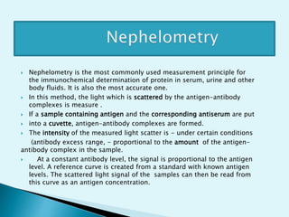
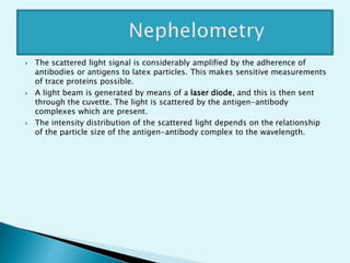


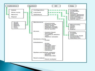
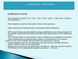
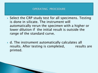
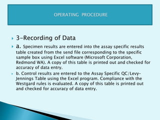
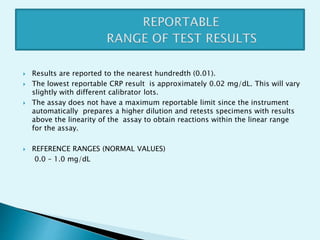


Nephelometry is commonly used to determine protein levels in body fluids like serum and urine. It works by measuring the light scattered by antigen-antibody complexes formed when a sample containing antigen is mixed with corresponding antiserum. The amount of light scattered is proportional to the antigen concentration in the sample. This document provides instructions for using a nephelometer to test samples for C-reactive protein (CRP) levels, including preparing reagents and standards, running samples, generating a calibration curve from standards, and recording results. CRP levels between 0.0-1.0 mg/dL are considered normal.


![Topic : Immunological Test [ C-Reactive Protein (CRP) ]](https://cdn.slidesharecdn.com/ss_thumbnails/c-reactiveproteincrp-241219194015-58e6b7e6-thumbnail.jpg?width=640&height=640&fit=bounds)


![ELISA results interpretation [Autosaved].pptx](https://cdn.slidesharecdn.com/ss_thumbnails/lab7elisaresultsinterpretationautosaved-240411174104-b696683f-thumbnail.jpg?width=640&height=640&fit=bounds)


