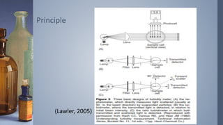
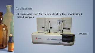
1. Joy Odia and Anneka Pierzga
College of Southern Maryland
Introduction to Nephelometry
2. › Measures the amount of light scattered by particles
suspended in a solution (usually a liquid)
Purpose
3. › Based on the principle of turbidity
– Light is scattered by small particles in the solution
› Nephelometry
– Degree of light scattering
Principle
4. › Factors that affect nephelometry
– Particle size and concentration
› Nephelometry preferred for small particles in low concentration
– Distance of light source
– Wavelength of light source
Principle
6. › Clinically, it is used to determine concentrations of
immunoglobins and serum proteins (ex. IgG, IgA, Albumin) in
serum and other biological samples.
Application
7. › It can also be used for therapeutic drug level monitoring in
blood samples
Application
(GMI, 2015)
8. Al-Mughales, J. A. (2015). Immunodiagnostic Significance of Anti-RA33 Autoantibodies in Saudi Patients
with Rheumatoid Arthritis. Journal of Immunology Research, 2015, 1-6.
Creveling, R. L., Deaton, C. D., Maxwell, K. W., & Smith, R. S. (1976). Use of Laser Nephelometry in the
Measurment of Serum Proteins. Clinical Chemistry, 22(9), 1465-1471.
Dye, J. A., Finley, P. R., LIchtI, D. A., & Williams, R. J. (1981). Rate-Nephelometric Inhibition Immunoassay
of Phenytoin and Phenobarbital. Clinical Chemistry, 27(3), 405-409.
GMI. (2015). Beckman Coulter IMMAGE Immunochemistry System. Retrieved from http://www.gmi-
inc.com/beckman-coulter-immage-immunochemistry-system.html
Hestridge, B., & Reynolds, A. P. (2012a). Nephelometry Basic Clinical Laboratory Techniques (6th ed., pp.
443-444): Cengage Learning.
Hestridge, B., & Reynolds, A. P. (2012b). Nephelometry and Turbidity Basic Clinical Laboratory
Techniques (6th ed., pp. 625-626): Cengage.
Khouja, H. Turbidimetry and Nephelometry. King Abdulaziz University.
Koivunen, M. E., & Krogsrud, R. L. (2006). Principles of Immunochemical Techniques Used in Clinical
Laboratories. LabMedicine, 37(8), 490-493.
Lawler, D. M. (2005). Spectrophotometry: Turbidity and Nephelometry. In C. F. Poole, Townshend, A.
and Worsfold, P.J. (Ed.), Encyclopedia of Analytical Science (2nd ed., pp. 343-351): Elsevier.
Morais, I. P. A., Rangel, A. O. S. S., & Tóth, I. V. (2006). Turbidimetric and Nephelometric Flow Analysis:
Concepts and Applications. Spectroscopy Letters: An International Journal for Rapid
Communication, 39(6), 547-579.
Neele, W. E., & Sung, E. (1985). A Cost-Effective System for Performing Therapeutic Drug Assays I.
Optimization of the Theophylline Assay. Clinical Chemistry, 31(7), 1210-1215.
Turgeon, M. L. (2015). Nephelometry Linné & Ringsrud's Clinical Laboratory Science: Concepts,
Procedures, and Clinical Applications (7th ed., pp. 187-188): Elsevier.
References
Editor's Notes
Nephelometry is the measurement of light scattered by particles suspended in a solution. (Hestridge & Reynolds, 2012a, 2012b; Koivunen & Krogsrud, 2006; Lawler, 2005; Turgeon, 2015)
Particles suspended in a solution will make the solution turbid (cloudy). When a beam of light is directed through the solution, some light will be blocked or absorbed by the particles, some light will be transmitted through the solution, and some light will be scattered by the particles in the solution (Hestridge & Reynolds, 2012a; Khouja; Koivunen & Krogsrud, 2006; Lawler, 2005)
Turbidity – property of solution that causes light to be scattered and absorbed, rather than transmitted straight through the sample
ISO defines turbidity as “reduction of transparency of a liquid caused by the presence of undissolved matter”
Opposite of clarity
(Lawler, 2005)
Nephelometry is measure of turbidity by evaluation of degree of light scattering taking place in the medium as light is being directed through it
The amount of light scattering is dependent upon many factors such as particle size, concentration of particles in solution, distance of the solution from the light source, wavelength of the light source (Hestridge & Reynolds, 2012b; Lawler, 2005)
- Nephelometry is preferred for use in samples of relatively low concertation/turbidity, as it is easier to measure a small amount of scattered light against a black background than it is to measure a small change in the intensity of transmitted light (Morais, Rangel, & Tóth, 2006)
- For this, turbidimetry is used to measure the intensity of a beam of light after it has passed through a sample i.e., measures the amount of light transmitted through the sample (Morais et al., 2006)
- Nephelometers usually provide better precision and sensitivity than turbidimeters, so are used for low turbidity samples containing very small particles (Lawler, 2005; Morais et al., 2006)
A beam of light is directed through a suspension of particles in a cuvette, and light scattered by the particles in the sample are measured by a photosensitive cell at an angle to the beam produced by the light source (usually 90°). (Creveling, Deaton, Maxwell, & Smith, 1976; Khouja; Lawler, 2005; Morais et al., 2006)
Often used in immunological testing to determine concentrations of immunoglobins and serum proteins in serum and other biological samples (Creveling et al., 1976; Hestridge & Reynolds, 2012a, 2012b; Khouja; Turgeon, 2015)
Antigen and antibody are combined to form antigen-antibody complexes that remain suspended in the sample (Hestridge & Reynolds, 2012a)
Amount of light scattered is proportional to the number of insoluble complexes formed in the solution and can be quantified by comparing patient values with standards of known protein concentrations (Turgeon, 2015)
Study of Saudi Arabian patients used Nephelometry to determine the immunodiagnostic significance of various antibodies for Rheumatoid Arthritis (Al-Mughales, 2015)
Also used to determine therapeutic drug levels of antiepileptic drugs, antibiotics, theophylline (Dye, Finley, LIchtI, & Williams, 1981; Neele & Sung, 1985)
Textbook mentions the IMMAGE Immunochemistry System (Beckman Coulter), which uses nephelometry for therapeutic drug level monitoring as well as immunoglobin and serum protein concentrations (GMI, 2015; Turgeon, 2015)