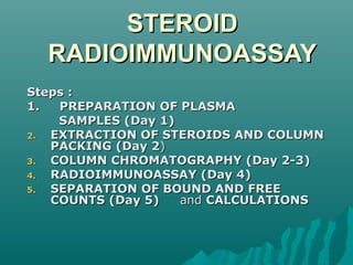
This document describes the steps for a radioimmunoassay to measure steroid levels in plasma samples. The process involves:
1. Preparing plasma samples and adding labeled steroid.
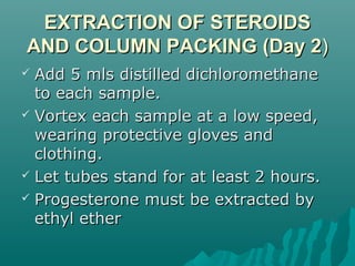
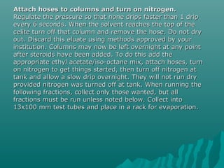
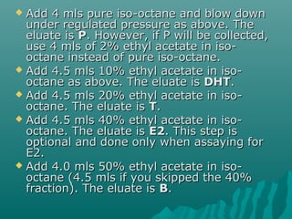
2. Extracting steroids from the plasma using dichloromethane and column packing.
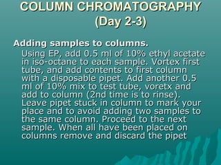
3. Performing column chromatography to separate the steroids.
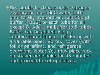
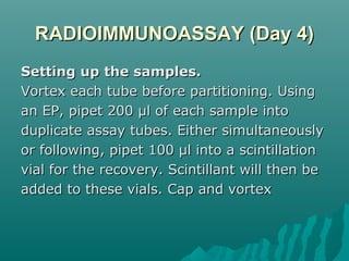
4. Setting up the separated steroids and standards for radioimmunoassay to measure steroid levels.
5. Using dextran-coated charcoal to separate bound and unbound steroid for counting.
This analysis deals with finding the residues of Endosulfan in blood. Researchers, Atmakuru Ramesh and Perumal Elumalai Ravi tested the blood samples of workers and people exposed to Endosulfan for a long time. The study concludes the absence of endosulfan residues in the blood reports.
For HPLC, sample solvents that adequately dissolve target compounds are required. Therefore, sample solvents that contain a high concentration of organic solvent are often used for reversed phase chromatography. The problem is that these solvents sometimes cause peak broadening.
This presentation discusses techniques for reducing the effects of sample solvents on UHPLC analyses.
Serine/threonine-protein kinase that acts downstream of mTOR signaling in response to growth factors and nutrients to promote cell proliferation, cell growth and cell cycle progression. Regulates protein synthesis through phosphorylation of EIF4B, RPS6 and EEF2K, and contributes to cell survival by repressing the pro-apoptotic function of BAD. Under conditions of nutrient depletion, the inactive form associates with the EIF3 translation initiation complex. Upon mitogenic stimulation, phosphorylation by the mammalian target of rapamycin complex 1 (mTORC1) leads to dissociation from the EIF3 complex and activation.
Anti-p70 S6 kinase α -http://www.stjohnslabs.com/p70-s6-kinase-a-antibody-p-93776
Join our Antibody Validation Project - http://www.stjohnslabs.com/services/antibody-validation
Physiology and Biochemistry-I (Marks-25)
a) Hematology
i) Study of compound microscope ii) Microscopically study of blood cells iii) Different types of WBC, erythrocytes and platelets iv) Examination of hemoglobin v) Differential count of WBC vi) Total count of RBC and WBC vii) Determination of clotting and bleeding time viii) Examination of clot under the microscope ix) Effect of chemical agents of RBC x) Fragility test of RBC. xi) Determination of erythrocytes sedimentation rate xii) Examination of haemin crystals
b) Histology: Histology of muscle, liver, spleen, stomach, duodenum, pancreas, lung, kidney, skin and endocrine glands.
c)Chemical physiology:
i) Qualitative test of carbohydrates, proteins and fats ii) Qualitative and quantitative experiments on digestive juice. iii) Examination of urine, estimation of main constituents and detection of abnormal constituents.
Serine/threonine-protein kinase that acts downstream of mTOR signaling in response to growth factors and nutrients to promote cell proliferation, cell growth and cell cycle progression. Regulates protein synthesis through phosphorylation of EIF4B, RPS6 and EEF2K, and contributes to cell survival by repressing the pro-apoptotic function of BAD. Under conditions of nutrient depletion, the inactive form associates with the EIF3 translation initiation complex. Upon mitogenic stimulation, phosphorylation by the mammalian target of rapamycin complex 1 (mTORC1) leads to dissociation from the EIF3 complex and activation. The active form then phosphorylates and activates several substrates in the pre-initiation complex, including the EIF2B complex and the cap-binding complex component EIF4B. Also controls translation initiation by phosphorylating a negative regulator of EIF4A, PDCD4, targeting it for ubiquitination and subsequent proteolysis.
Anti-p70 S6 kinase α -http://www.stjohnslabs.com/p70-s6-kinase-a-antibody-p-99095
Join our Antibody Validation Project - http://www.stjohnslabs.com/services/antibody-validation
Probably plays a role in facilitating the assembly of multimeric protein complexes inside the endoplasmic reticulum. Involved in the correct folding of proteins and degradation of misfolded proteins via its interaction with DNAJC10, probably to facilitate the release of DNAJC10 from its substrate
Anti-HSP A5 -http://www.stjohnslabs.com/hsp-a5-antibody
Join our Antibody Validation Project - http://www.stjohnslabs.com/services/antibody-validation
Radioimmunoassay allows for the measurement of wide range of materials of clinical and biological importance. This technique has a significant impact on medical diagnosis due to the ease with which the tests can be carried out, while assuring precision, specificity and sensitivity.
The radioimmunoassay technique, as the name implies, achieves sensitivity through the use of radionuclides and specificity that is uniquely associated with immunochemical reactions. It can detect substance from a range of Nano gram(ng) to Pico gram(pg).
Radioimmunoassay (RIA) is a very sensitive in vitro assay technique used to measure concentrations of antigens (for example, hormone levels in blood) by use of antibodies. As such, it can be seen as the inverse of a radiobinding assay, which quantifies an antibody by use of corresponding antigens.
- Lab 9 Preparation for Protein PurificationObjectivesGrow tra.docxoswald1horne84988
- Lab 9 Preparation for Protein Purification
Objectives
Grow transformed E. coli in preparation for isolation and separation of green fluorescent protein.
Backgrounds
When E. coli containing the pGLO plasmid is grown in a medium containing arabinose the green fluorescent protein (GFP) is expressed. GFP can then be Isolated and separated on a SDS PAGE gel.
Cultures will be grown at 32C as it is the optimum temperature for folding of the GFP.
Methods
Before the lab
· Use aseptic technique during set up and inoculation
· Select 3 tubes of broth if using 2ml: Label two ara/amp and one amp. If using 5 ml tubes of LB broth use 2 tubes label one ara/amp and one amp.
· Add (9ul /1ml) each of arabinose and ampicillin to two tubes (ara/amp)
· Add (9ul/1ml) of ampicillin to the third tube (amp)
· Thaw one of your frozen culture tubes and mix
· Add 100ul of frozen cultures to each tube
· Place tubes in a rack and incubate in a shaker overnight at 32oC. Be sure to shake vigorously.
During the lab
· WEAR SAFETY GLASSES - Use a UV lamp to view each tube – remark on your observations
· Transfer 2ml of culture from an ara/amp tube to a 2ml microfuge tube and spin for 5 minutes
· Remove the supernatant into a waste beaker by either pouring or using a pipette – be careful not to disturb the pellet.
· Add 250ul of TE buffer to the tubes and mix in the vortex mixer until the pellet is suspended
· Add 50ul of lysozyme to this tube and mix
· The tube will be collected and placed in the freezer for at least 24hr to lyse the cells
· Select the remaining ara/amp and amp tube. Label two microfuge tubes.
· Transfer 600ul of the ara/amp tube to a microfuge tube and 300 ul of the amp tube to the another microfuge tube.
· Spin for 5 minutes and discard the supernatant.
· Freeze these tubes and their pellets
LAB 10 Topic: Protein Purification
Objective
· Isolate and separate green fluorescent protein
Background
· When E.coli containing the pGLO plasmid is grown in a medium containing arabinose the green fluorescent protein (GFP) is expressed. GFP can then be isolated and separated on a SDS PAGE gel.
· The hydrophobic interaction column (HIC) allows the separation of hydrophobic proteins. GFP has several stretches of hydrophobic amino acids: this will enable it to stick to the beads in the HIC.
·
Materials
Lab
· Samples of lysed cells from lab 9
· Eppendorf tubes (sterile)
· Test tube racks
· P-1000 (200-1000 ul) – sterile
· P-200 (20 to 200 ul) – sterile
· Microfuge
· UV light
· HIC column (Biorad)
· Elution tubes
· Binding buffer (4M NH4SO4/TE pH 8)
· Equilibration buffer
· Wash buffer
· TE buffer (elution)
_____________________________________________________________
Lab Procedure
· Wear safety glasses and gloves.
· Remove the Eppendorf tube containing cells from Ara/amp broth and lysozyme from freezer.
· Thaw on the benchtop.
· Centrifuge the tubes for 10 minutes to bring down the bacterial debris.
· While the .
study Content about Aerosol Formulation and Packaging.
If you want to increase your knowledge and want to crack pharma related exam so please contact on below links:-
1. Join Facebook Group :- Target Pharmacy Competitive Exam
Group ( Public Group )
2. Like My Page :- #DeepakJoshi
This poster is an overview of the four main projects I worked on at my summer at Streck. This board was created as a summary for a all-employee showcase.
Biol 390 – Lab 8 Restriction Digest and Gel Electrophoresis .docxmoirarandell
Biol 390 – Lab 8 Restriction Digest and Gel Electrophoresis
2
Objective
· Digest DNA of pGLO plasmid using restriction endonuclease enzymes.
· Run an agarose gel to separate the DNA fragments.
Background
Restriction enzymes cut DNA at specific sites generating a number of different sized fragments. The size of the fragments will depend on the number of sites the plasmid has and the specific enzyme used. The number of fragments can be predicted by viewing the map of the plasmid
Gel electrophoresis is a means of separating DNA in an electrical field. DNA is negatively charged and so will move to the anode (+). Larger fragments will move slower through the agarose matrix than the smaller molecules. Agarose is a polysaccharide polymer derived from seaweed: it is a purified from agar by removing the agaropectin component. Fragments are visualized using ethidium bromide, which will glow orange when exposed to UV light.
Materials
Restriction digest
· Restriction enzymes: Nhe1 and EcoR1 (New England Biolabs) – (KEEP ON ICE)
· Plasmid prepared in lab 7
· NanoDrop Lite spectrophotometer
· Microfuge tubes – Sterile
· 37 C degree bath – block heater
· Sterile 10ul and 200ul tips
· Bleach bottles for cleaning bench
· 10X NE Cut Smart Buffer – comes with enzyme
· Nitrile gloves
· Sterile DI water
· Shaved ice
· Ice block for enzymes
Gel Electrophoresis
· Agarose
· Sterile miliQ Water
· 15 well comb
· 50x TAE buffer
· DNA ladder – diluted in sample buffer (1 KB)
· Gel loading dye
· Gel electrophoresis chamber
· Power supply
· Ethidium bromide
· Gel Sys – visualization system
_______________________________________________
Procedure
Restriction Digest of plasmid DNA
· Safety: Wear nitrile gloves – prevent DNAase from your hands affecting the reaction and protect yourself from ethidium bromide
· Clean the bench with bleach - prevents exogenous enzymes interfering you’re your digests.
· Use the NanoDrop to determine the amount of DNA in your plasmid prep. Use this information to calculate how much sample you need to pipette into the reaction mix.
· Label an Eppendorf tube ‘+’ and another ‘-‘
· Make up a reaction mix in both tubes as follows for one of your plasmid samples
· add 1ug of DNA from your plasmid prep
· 5ul of 10X NE Cut Smart Buffer
· Sterile DI water to make the reaction mix to 50ul
For the + tube
· DNA
x ul
· 10X NE Cut Smart Buffer
5ul
· Nhe1 (add last to + tube)
1ul
· EcoR1 (add last to + tube)
1ul
· Sterile DI water
To make final volume to 50ul
· Add the restriction enzymes last to the + tube ONLY
· Repeat with the other two plasmid samples
For the – tube
· DNA
x ul
· 10X NE Buffer
5ul
· Nhe1
None
· EcoR1
None
· Sterile DI water
To make final volume to 50ul
· Do not add any enzyme to the ‘-‘ tube
· Repeat with the other two plasmid samples
· Mix the tubes by flicking – DO NOT VORTEX
· Give a 5 second spin in the centrifuge to bring the contents to the bottom
· Incu.
Post-lab 1- Myths in Science (10 pts)Read the remaining myths” .docxChantellPantoja184
Post-lab 1- Myths in Science (10 pts)
Read the remaining “myths” in the article, The Principle Elements of the Nature of Science: Dispelling the Myths, by W.F. McComas. Then, reflect on your own understanding of science both before and after having read the article. Do not exceed one full page, double spaced, but use as much room as is necessary to address the following topics: Identify some of the myths you had believed to be true and why you had those misconceptions. How did the clarifications in this article change how you view science? Were those changes for better or worse? What are some aspects of the scientific process that have become more confusing, or unclear, after reading this article? Does a more full understanding of the scientific process make you optimistic, pessimistic, or indifferent to the prospects of being a scientist?
1
Edited 8/26/15 Biology 111 Lab Page
LAB 2- MOLECULAR BIOLOGY LAB TECHNIQUES
INTRODUCTION
This week’s lab will introduce you to three molecular biology techniques that you will use in future labs. During the course of this activity, you will be learning and practicing micropipetting, polymerase chain reaction (PCR), and DNA gel electrophoresis. Each topic below provides, or refers you to, background information on the technique prior to the hands-on activity where you will learn the technique.
Learning Objectives:
1. Be able to properly select and utilize micropipettes for the manipulation of small volumes of liquid.
2. Be able to explain how PCR amplifies DNA and be able to perform a PCR protocol.
3. Understand how gel electrophoresis is able to separate DNA fragments, be able to pour an agarose gel, load samples, and interpret results.
Lab notebooks:
Look over the notebook guidelines posted in the general Lab Materials content folder. Begin this lab by writing a summary of the lab’s objectives.
I. Micropipettes
Pre-lab Introduction:
A micropipette is a kind of fancy eyedropper – one that comes in many different models and volume ranges. But while an eyedropper dispenses drops, micropipettes transfer microliters of fluid. Recall that ‘micro-’ is a prefix in the metric system which means “one-millionth” of the base unit (in this case, a liter, “L”). It may be easier for you to picture one milliliter (mL or ml) of water. If you mentally subdivide that milliliter of water into 1000 tiny equal-sized volumes, each volume is one microliter (abbreviated μL or μl). Watch the 2 pipetting videos posted in the lab 2 content folder (https://www.youtube.com/watch?v=p-OPOYbeZP0 & https://www.youtube.com/watch?v=NgosWmRjjAo) , then continue from here.
Micropipette Anatomy:
1. Examine the figures to the right to familiarize yourself with the anatomy of a micropipette.
2. Micropipette plungers have 3 positions:
a. Rest position- no pressure on plunger
b. First stop- position that will draw desired volume into tip
c. Second stop- position that will fully expel a sample from the tip
3. Pipette tips are pressed.
Escozine for Pets™ has 4 major production steps.
1. Collection of Scorpions from the Scorpion Reservation. 2. Extraction of venom, purification and therapeutic dose preparation. 3. Polarization of extract and quality control of Polarization 4. Manufacturing, quality control, warehouse and shipment.
Tom Selleck Health: A Comprehensive Look at the Iconic Actor’s Wellness Journeygreendigital
Tom Selleck, an enduring figure in Hollywood. has captivated audiences for decades with his rugged charm, iconic moustache. and memorable roles in television and film. From his breakout role as Thomas Magnum in Magnum P.I. to his current portrayal of Frank Reagan in Blue Bloods. Selleck's career has spanned over 50 years. But beyond his professional achievements. fans have often been curious about Tom Selleck Health. especially as he has aged in the public eye.
Follow us on: Pinterest
Introduction
Many have been interested in Tom Selleck health. not only because of his enduring presence on screen but also because of the challenges. and lifestyle choices he has faced and made over the years. This article delves into the various aspects of Tom Selleck health. exploring his fitness regimen, diet, mental health. and the challenges he has encountered as he ages. We'll look at how he maintains his well-being. the health issues he has faced, and his approach to ageing .
Early Life and Career
Childhood and Athletic Beginnings
Tom Selleck was born on January 29, 1945, in Detroit, Michigan, and grew up in Sherman Oaks, California. From an early age, he was involved in sports, particularly basketball. which played a significant role in his physical development. His athletic pursuits continued into college. where he attended the University of Southern California (USC) on a basketball scholarship. This early involvement in sports laid a strong foundation for his physical health and disciplined lifestyle.
Transition to Acting
Selleck's transition from an athlete to an actor came with its physical demands. His first significant role in "Magnum P.I." required him to perform various stunts and maintain a fit appearance. This role, which he played from 1980 to 1988. necessitated a rigorous fitness routine to meet the show's demands. setting the stage for his long-term commitment to health and wellness.
Fitness Regimen
Workout Routine
Tom Selleck health and fitness regimen has evolved. adapting to his changing roles and age. During his "Magnum, P.I." days. Selleck's workouts were intense and focused on building and maintaining muscle mass. His routine included weightlifting, cardiovascular exercises. and specific training for the stunts he performed on the show.
Selleck adjusted his fitness routine as he aged to suit his body's needs. Today, his workouts focus on maintaining flexibility, strength, and cardiovascular health. He incorporates low-impact exercises such as swimming, walking, and light weightlifting. This balanced approach helps him stay fit without putting undue strain on his joints and muscles.
Importance of Flexibility and Mobility
In recent years, Selleck has emphasized the importance of flexibility and mobility in his fitness regimen. Understanding the natural decline in muscle mass and joint flexibility with age. he includes stretching and yoga in his routine. These practices help prevent injuries, improve posture, and maintain mobilit
New Directions in Targeted Therapeutic Approaches for Older Adults With Mantl...i3 Health
i3 Health is pleased to make the speaker slides from this activity available for use as a non-accredited self-study or teaching resource.
This slide deck presented by Dr. Kami Maddocks, Professor-Clinical in the Division of Hematology and
Associate Division Director for Ambulatory Operations
The Ohio State University Comprehensive Cancer Center, will provide insight into new directions in targeted therapeutic approaches for older adults with mantle cell lymphoma.
STATEMENT OF NEED
Mantle cell lymphoma (MCL) is a rare, aggressive B-cell non-Hodgkin lymphoma (NHL) accounting for 5% to 7% of all lymphomas. Its prognosis ranges from indolent disease that does not require treatment for years to very aggressive disease, which is associated with poor survival (Silkenstedt et al, 2021). Typically, MCL is diagnosed at advanced stage and in older patients who cannot tolerate intensive therapy (NCCN, 2022). Although recent advances have slightly increased remission rates, recurrence and relapse remain very common, leading to a median overall survival between 3 and 6 years (LLS, 2021). Though there are several effective options, progress is still needed towards establishing an accepted frontline approach for MCL (Castellino et al, 2022). Treatment selection and management of MCL are complicated by the heterogeneity of prognosis, advanced age and comorbidities of patients, and lack of an established standard approach for treatment, making it vital that clinicians be familiar with the latest research and advances in this area. In this activity chaired by Michael Wang, MD, Professor in the Department of Lymphoma & Myeloma at MD Anderson Cancer Center, expert faculty will discuss prognostic factors informing treatment, the promising results of recent trials in new therapeutic approaches, and the implications of treatment resistance in therapeutic selection for MCL.
Target Audience
Hematology/oncology fellows, attending faculty, and other health care professionals involved in the treatment of patients with mantle cell lymphoma (MCL).
Learning Objectives
1.) Identify clinical and biological prognostic factors that can guide treatment decision making for older adults with MCL
2.) Evaluate emerging data on targeted therapeutic approaches for treatment-naive and relapsed/refractory MCL and their applicability to older adults
3.) Assess mechanisms of resistance to targeted therapies for MCL and their implications for treatment selection
NVBDCP.pptx Nation vector borne disease control programSapna Thakur
NVBDCP was launched in 2003-2004 . Vector-Borne Disease: Disease that results from an infection transmitted to humans and other animals by blood-feeding arthropods, such as mosquitoes, ticks, and fleas. Examples of vector-borne diseases include Dengue fever, West Nile Virus, Lyme disease, and malaria.
ARTIFICIAL INTELLIGENCE IN HEALTHCARE.pdfAnujkumaranit
Artificial intelligence (AI) refers to the simulation of human intelligence processes by machines, especially computer systems. It encompasses tasks such as learning, reasoning, problem-solving, perception, and language understanding. AI technologies are revolutionizing various fields, from healthcare to finance, by enabling machines to perform tasks that typically require human intelligence.
The prostate is an exocrine gland of the male mammalian reproductive system
It is a walnut-sized gland that forms part of the male reproductive system and is located in front of the rectum and just below the urinary bladder
Function is to store and secrete a clear, slightly alkaline fluid that constitutes 10-30% of the volume of the seminal fluid that along with the spermatozoa, constitutes semen
A healthy human prostate measures (4cm-vertical, by 3cm-horizontal, 2cm ant-post ).
It surrounds the urethra just below the urinary bladder. It has anterior, median, posterior and two lateral lobes
It’s work is regulated by androgens which are responsible for male sex characteristics
Generalised disease of the prostate due to hormonal derangement which leads to non malignant enlargement of the gland (increase in the number of epithelial cells and stromal tissue)to cause compression of the urethra leading to symptoms (LUTS
New Drug Discovery and Development .....NEHA GUPTA
The "New Drug Discovery and Development" process involves the identification, design, testing, and manufacturing of novel pharmaceutical compounds with the aim of introducing new and improved treatments for various medical conditions. This comprehensive endeavor encompasses various stages, including target identification, preclinical studies, clinical trials, regulatory approval, and post-market surveillance. It involves multidisciplinary collaboration among scientists, researchers, clinicians, regulatory experts, and pharmaceutical companies to bring innovative therapies to market and address unmet medical needs.
Title: Sense of Taste
Presenter: Dr. Faiza, Assistant Professor of Physiology
Qualifications:
MBBS (Best Graduate, AIMC Lahore)
FCPS Physiology
ICMT, CHPE, DHPE (STMU)
MPH (GC University, Faisalabad)
MBA (Virtual University of Pakistan)
Learning Objectives:
Describe the structure and function of taste buds.
Describe the relationship between the taste threshold and taste index of common substances.
Explain the chemical basis and signal transduction of taste perception for each type of primary taste sensation.
Recognize different abnormalities of taste perception and their causes.
Key Topics:
Significance of Taste Sensation:
Differentiation between pleasant and harmful food
Influence on behavior
Selection of food based on metabolic needs
Receptors of Taste:
Taste buds on the tongue
Influence of sense of smell, texture of food, and pain stimulation (e.g., by pepper)
Primary and Secondary Taste Sensations:
Primary taste sensations: Sweet, Sour, Salty, Bitter, Umami
Chemical basis and signal transduction mechanisms for each taste
Taste Threshold and Index:
Taste threshold values for Sweet (sucrose), Salty (NaCl), Sour (HCl), and Bitter (Quinine)
Taste index relationship: Inversely proportional to taste threshold
Taste Blindness:
Inability to taste certain substances, particularly thiourea compounds
Example: Phenylthiocarbamide
Structure and Function of Taste Buds:
Composition: Epithelial cells, Sustentacular/Supporting cells, Taste cells, Basal cells
Features: Taste pores, Taste hairs/microvilli, and Taste nerve fibers
Location of Taste Buds:
Found in papillae of the tongue (Fungiform, Circumvallate, Foliate)
Also present on the palate, tonsillar pillars, epiglottis, and proximal esophagus
Mechanism of Taste Stimulation:
Interaction of taste substances with receptors on microvilli
Signal transduction pathways for Umami, Sweet, Bitter, Sour, and Salty tastes
Taste Sensitivity and Adaptation:
Decrease in sensitivity with age
Rapid adaptation of taste sensation
Role of Saliva in Taste:
Dissolution of tastants to reach receptors
Washing away the stimulus
Taste Preferences and Aversions:
Mechanisms behind taste preference and aversion
Influence of receptors and neural pathways
Impact of Sensory Nerve Damage:
Degeneration of taste buds if the sensory nerve fiber is cut
Abnormalities of Taste Detection:
Conditions: Ageusia, Hypogeusia, Dysgeusia (parageusia)
Causes: Nerve damage, neurological disorders, infections, poor oral hygiene, adverse drug effects, deficiencies, aging, tobacco use, altered neurotransmitter levels
Neurotransmitters and Taste Threshold:
Effects of serotonin (5-HT) and norepinephrine (NE) on taste sensitivity
Supertasters:
25% of the population with heightened sensitivity to taste, especially bitterness
Increased number of fungiform papillae
Ozempic: Preoperative Management of Patients on GLP-1 Receptor Agonists Saeid Safari
Preoperative Management of Patients on GLP-1 Receptor Agonists like Ozempic and Semiglutide
ASA GUIDELINE
NYSORA Guideline
2 Case Reports of Gastric Ultrasound
Explore natural remedies for syphilis treatment in Singapore. Discover alternative therapies, herbal remedies, and lifestyle changes that may complement conventional treatments. Learn about holistic approaches to managing syphilis symptoms and supporting overall health.
These lecture slides, by Dr Sidra Arshad, offer a quick overview of physiological basis of a normal electrocardiogram.
Learning objectives:
1. Define an electrocardiogram (ECG) and electrocardiography
2. Describe how dipoles generated by the heart produce the waveforms of the ECG
3. Describe the components of a normal electrocardiogram of a typical bipolar leads (limb II)
4. Differentiate between intervals and segments
5. Enlist some common indications for obtaining an ECG
Study Resources:
1. Chapter 11, Guyton and Hall Textbook of Medical Physiology, 14th edition
2. Chapter 9, Human Physiology - From Cells to Systems, Lauralee Sherwood, 9th edition
3. Chapter 29, Ganong’s Review of Medical Physiology, 26th edition
4. Electrocardiogram, StatPearls - https://www.ncbi.nlm.nih.gov/books/NBK549803/
5. ECG in Medical Practice by ABM Abdullah, 4th edition
6. ECG Basics, http://www.nataliescasebook.com/tag/e-c-g-basics
Prix Galien International 2024 Forum ProgramLevi Shapiro
June 20, 2024, Prix Galien International and Jerusalem Ethics Forum in ROME. Detailed agenda including panels:
- ADVANCES IN CARDIOLOGY: A NEW PARADIGM IS COMING
- WOMEN’S HEALTH: FERTILITY PRESERVATION
- WHAT’S NEW IN THE TREATMENT OF INFECTIOUS,
ONCOLOGICAL AND INFLAMMATORY SKIN DISEASES?
- ARTIFICIAL INTELLIGENCE AND ETHICS
- GENE THERAPY
- BEYOND BORDERS: GLOBAL INITIATIVES FOR DEMOCRATIZING LIFE SCIENCE TECHNOLOGIES AND PROMOTING ACCESS TO HEALTHCARE
- ETHICAL CHALLENGES IN LIFE SCIENCES
- Prix Galien International Awards Ceremony
How STIs Influence the Development of Pelvic Inflammatory Disease.pptx
Steroid radioimmunoassay
1. STEROIDSTEROID
RADIOIMMUNOASSAYRADIOIMMUNOASSAY
Steps :Steps :
1. PREPARATION OF PLASMA1. PREPARATION OF PLASMA
SAMPLES (Day 1)SAMPLES (Day 1)
2.2. EXTRACTION OF STEROIDS AND COLUMNEXTRACTION OF STEROIDS AND COLUMN
PACKING (Day 2PACKING (Day 2))
3.3. COLUMN CHROMATOGRAPHY (Day 2-3)COLUMN CHROMATOGRAPHY (Day 2-3)
4.4. RADIOIMMUNOASSAY (Day 4)RADIOIMMUNOASSAY (Day 4)
5.5. SEPARATION OF BOUND AND FREESEPARATION OF BOUND AND FREE
COUNTS (Day 5)COUNTS (Day 5) andand CALCULATIONSCALCULATIONS
2. PREPARATION OF PLASMAPREPARATION OF PLASMA
SAMPLESSAMPLES
Set up plasma samples in glass centrifuge tubes (pointedSet up plasma samples in glass centrifuge tubes (pointed
bottoms, 12 ml). The first and last tubes are always blanksbottoms, 12 ml). The first and last tubes are always blanks
and contain only distilled water (dH2O). The second tube isand contain only distilled water (dH2O). The second tube is
always aalways a standardstandard into which measured amounts of theinto which measured amounts of the
steroids being assayed are placed. This measures thesteroids being assayed are placed. This measures the
accuracy of the current assay as well as interassayaccuracy of the current assay as well as interassay
variation, when samples may be spread over more thanvariation, when samples may be spread over more than
one assay. When all plasma samples have been measured,one assay. When all plasma samples have been measured,
a small amount of labelled steroid (20 µl of each steroida small amount of labelled steroid (20 µl of each steroid
being tested=2,000cpm) is added to all tubes except thebeing tested=2,000cpm) is added to all tubes except the
blanks. Total cpm is measured at the end to determineblanks. Total cpm is measured at the end to determine
what percentage of the labelled steroid is recovered, and,what percentage of the labelled steroid is recovered, and,
assuming that the unknown steroid behaves the same, theassuming that the unknown steroid behaves the same, the
final dose of the unknown steroid is adjusted according tofinal dose of the unknown steroid is adjusted according to
thisthis recoveryrecovery value.value.
3. EXTRACTION OF STEROIDSEXTRACTION OF STEROIDS
AND COLUMN PACKING (Day 2AND COLUMN PACKING (Day 2))
Add 5 mls distilled dichloromethaneAdd 5 mls distilled dichloromethane
to each sample.to each sample.
Vortex each sample at a low speed,Vortex each sample at a low speed,
wearing protective gloves andwearing protective gloves and
clothing.clothing.
Let tubes stand for at least 2 hours.Let tubes stand for at least 2 hours.
Progesterone must be extracted byProgesterone must be extracted by
ethyl etherethyl ether
4. COLUMN CHROMATOGRAPHYCOLUMN CHROMATOGRAPHY
(Day 2-3)(Day 2-3)
Adding samples to columns.Adding samples to columns.
Using EP, add 0.5 ml of 10% ethyl acetateUsing EP, add 0.5 ml of 10% ethyl acetate
in iso-octane to each sample. Vortex firstin iso-octane to each sample. Vortex first
tube, and add contents to first columntube, and add contents to first column
with a disposable pipet. Add another 0.5with a disposable pipet. Add another 0.5
ml of 10% mix to test tube, voretx andml of 10% mix to test tube, voretx and
add to column (2nd time is to rinse).add to column (2nd time is to rinse).
Leave pipet stuck in column to mark yourLeave pipet stuck in column to mark your
place and to avoid adding two samples toplace and to avoid adding two samples to
the same column. Proceed to the nextthe same column. Proceed to the next
sample. When all have been placed onsample. When all have been placed on
columns remove and discard the pipetcolumns remove and discard the pipet
5. Attach hoses to columns and turn on nitrogen.Attach hoses to columns and turn on nitrogen.
Regulate the pressure so that none drips faster than 1 dripRegulate the pressure so that none drips faster than 1 drip
every 6 seconds. When the solvent reaches the top of theevery 6 seconds. When the solvent reaches the top of the
celite turn off that column and remove the hose. Do not drycelite turn off that column and remove the hose. Do not dry
out. Discard this eluate using methods approved by yourout. Discard this eluate using methods approved by your
institution. Columns may now be left overnight at any pointinstitution. Columns may now be left overnight at any point
after steroids have been added. To do this add theafter steroids have been added. To do this add the
appropriate ethyl acetate/iso-octane mix, attach hoses, turnappropriate ethyl acetate/iso-octane mix, attach hoses, turn
on nitrogen to get things started, then turn off nitrogen aton nitrogen to get things started, then turn off nitrogen at
tank and allow a slow drip overnight. They will not run drytank and allow a slow drip overnight. They will not run dry
provided nitrogen was turned off at tank. When running theprovided nitrogen was turned off at tank. When running the
following fractions, collect only those wanted, but allfollowing fractions, collect only those wanted, but all
fractions must be run unless noted below. Collect intofractions must be run unless noted below. Collect into
13x100 mm test tubes and place in a rack for evaporation.13x100 mm test tubes and place in a rack for evaporation.
6. Add 4 mls pure iso-octane and blow downAdd 4 mls pure iso-octane and blow down
under regulated pressure as above. Theunder regulated pressure as above. The
eluate iseluate is PP. However, if P will be collected,. However, if P will be collected,
use 4 mls of 2% ethyl acetate in iso-use 4 mls of 2% ethyl acetate in iso-
octane instead of pure iso-octane.octane instead of pure iso-octane.
Add 4.5 mls 10% ethyl acetate in iso-Add 4.5 mls 10% ethyl acetate in iso-
octane as above. The eluate isoctane as above. The eluate is DHTDHT..
Add 4.5 mls 20% ethyl acetate in iso-Add 4.5 mls 20% ethyl acetate in iso-
octane. The eluate isoctane. The eluate is TT..
Add 4.5 mls 40% ethyl acetate in iso-Add 4.5 mls 40% ethyl acetate in iso-
octane. The eluate isoctane. The eluate is E2E2. This step is. This step is
optional and done only when assaying foroptional and done only when assaying for
E2.E2.
Add 4.0 mls 50% ethyl acetate in iso-Add 4.0 mls 50% ethyl acetate in iso-
octane (4.5 mls if you skipped the 40%octane (4.5 mls if you skipped the 40%
fraction). The eluate isfraction). The eluate is BB..
7. Dry purified extracts under nitrogenDry purified extracts under nitrogen
evaporator in a 40oC water bathevaporator in a 40oC water bath
until totally evaporated. Add 550 µluntil totally evaporated. Add 550 µl
buffer (PBSG) to each tube for allbuffer (PBSG) to each tube for all
except B. Add 1 ml buffer to B tubes.except B. Add 1 ml buffer to B tubes.
Buffer can be added using aBuffer can be added using a
combination of tips on the ER or withcombination of tips on the ER or with
a variable pipet. Vortex, cover (witha variable pipet. Vortex, cover (with
foil or parafilm), and refrigeratefoil or parafilm), and refrigerate
overnight. Note: You may place rackovernight. Note: You may place rack
on shaker and shake for 45 minuteson shaker and shake for 45 minutes
and proceed to set up curves.and proceed to set up curves.
8. RADIOIMMUNOASSAY (Day 4)RADIOIMMUNOASSAY (Day 4)
Setting up the samples.Setting up the samples.
Vortex each tube before partitioning. UsingVortex each tube before partitioning. Using
an EP, pipet 200 µl of each sample intoan EP, pipet 200 µl of each sample into
duplicate assay tubes. Either simultaneouslyduplicate assay tubes. Either simultaneously
or following, pipet 100 µl into a scintillationor following, pipet 100 µl into a scintillation
vial for the recovery. Scintillant will then bevial for the recovery. Scintillant will then be
added to these vials. Cap and vortexadded to these vials. Cap and vortex
9. Setting up the standard curve:Setting up the standard curve:
The standard curve will be used toThe standard curve will be used to
determine the dose of steroid in thedetermine the dose of steroid in the
unknown samples. The first three tubes areunknown samples. The first three tubes are
B1-B3. B1 measures total cpm, B2 non-B1-B3. B1 measures total cpm, B2 non-
specific binding (or background), and B3specific binding (or background), and B3
maximum binding with the antiserum. Themaximum binding with the antiserum. The
remaining 9 pairs of tubes (S1-S9) generateremaining 9 pairs of tubes (S1-S9) generate
the curve.the curve.
10. SEPARATION OF BOUND ANDSEPARATION OF BOUND AND
FREE COUNTS Day 5FREE COUNTS Day 5
Dextran-coated charcoal will be addedDextran-coated charcoal will be added
to allto all
tubes (except B1) and allowed totubes (except B1) and allowed to
adsorb alladsorb all
unbound steroid. Tubes will beunbound steroid. Tubes will be
centrifuged,centrifuged,
and the supernatant will be decantedand the supernatant will be decanted
into 7into 7
ml scintillation vials for counting cpm.ml scintillation vials for counting cpm.