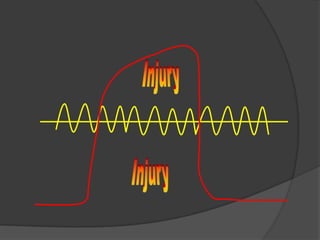
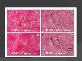
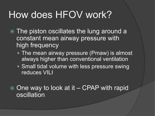
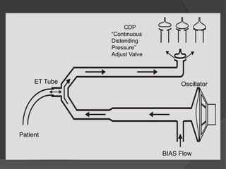
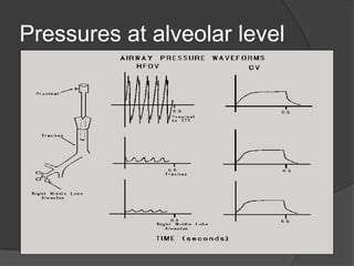
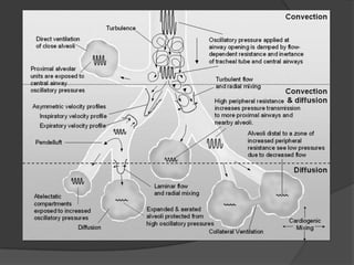
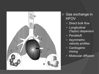
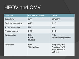
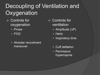
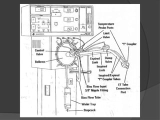
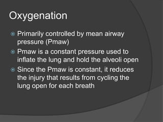
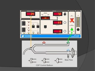
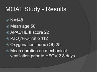
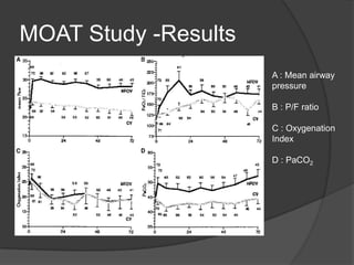
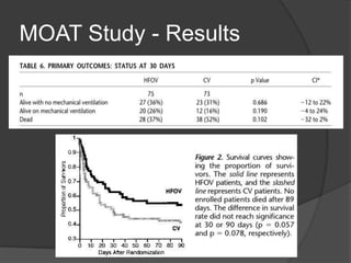
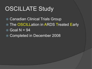
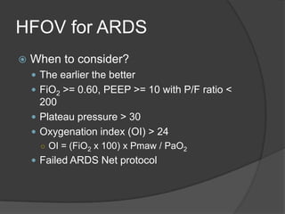
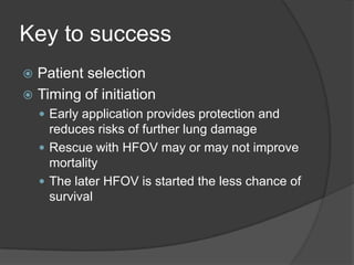
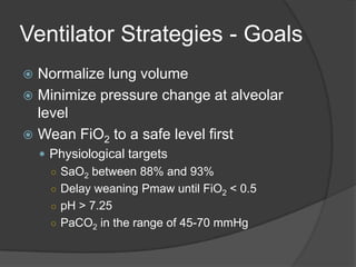
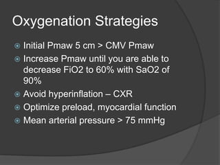
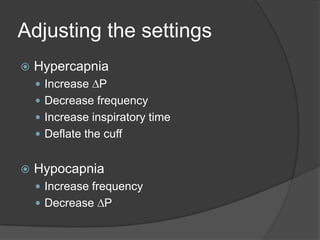
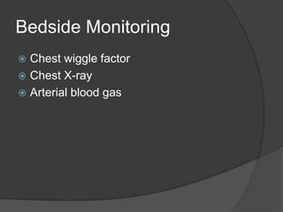
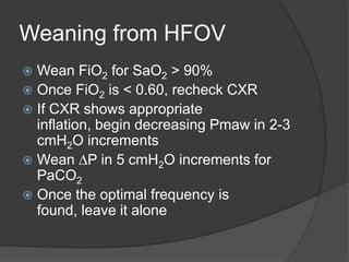
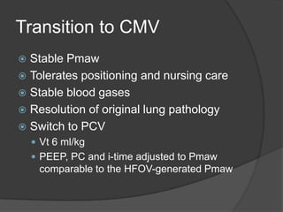
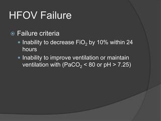
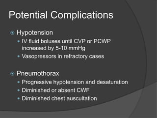
High Frequency Oscillatory VentilationPeteyLaohaburanakit, MD, FCCPCritical Care ServicesRogue Valley Medical Center
OutlineWhat is HFOV? Ventilator-induced lung injury (VILI)How does HFOV work?Basic concept and gas exchangeOxygenation and ventilation in HFOVClinical studiesInitiation and adjustmentCare for patients on HFOVPotential complications
What is HFOV?Not to be confused with high frequency jet ventilation (HFJV), which is rarely usedFirst established use in neonatal ARDSHFOV’s claim to fame is reduction of ventilator-induced lung injury (VILI)
What is VILI?Two major causesAlveolar distensionHigh plateau pressureCyclical opening and closing of atelectatic lungLarge pressure swing at the alveolar level from large tidal volumes
 V P V P V P
ZoneofOverdistensionInjury“Safe”WindowVolumeZone ofDerecruitmentand AtelectasisInjuryPressure
InjuryInjury
How does HFOV work?The piston oscillates the lung around a constant mean airway pressure with high frequencyThe mean airway pressure (Pmaw) is almost always higher than conventional ventilationSmall tidal volume with less pressure swing reduces VILIOne way to look at it – CPAP with rapid oscillation
CDP“Continuous DistendingPressure”Adjust Valve   ET TubeOscillatorPatientBIAS Flow
Pressures at alveolar level
Gas exchange in HFOVDirect bulk flowLongitudinal (Taylor) dispersionPendeluftAsymmetric velocity profilesCardiogenic mixingMolecular diffusion
HFOV and CMV
Decoupling of Ventilation and OxygenationControls for oxygenationPmawFiO2Alveolar recruitment maneuverControls for ventilationAmplitude (DP)HertzInspiratory timeCuff deflationPermissive hypercapnia
OxygenationPrimarily controlled by mean airway pressure (Pmaw)Pmaw is a constant pressure used to inflate the lung and hold the alveoli openSince the Pmaw is constant, it reduces the injury that results from cycling the lung open for each breath
xBias FlowCDP Control Balloon
VentilationControlled by the movement of pump/piston mechanismAlveolar ventilation during CMV is defined as f x VtAlveolar ventilation during HFOV is defined as f x Vt2Changes in volume delivery have the most significant effect on ventilation
Regulation of stroke volumeThe stroke volume will increase ifThe amplitude increases (higher DP)The frequency decreased (longer cycle time)There is an increase in inspiratory time
Amplitude (DP)The force created by piston movementDependent on the power settingResults in chest wiggle
Inspiratory timeControls the time for movement of the pistonIncreases inspiratory time increases CO2 eliminationIncreases inspiratory time increases delivered Pmaw
PaO2PaCO2
Clinical DataPilot studiesMehta S et al. Crit Care Med 2001Derdak S et al. Am J RespirCrit Care Med 2002Multicenter oscillatory ventilation for ARDS trial (MOAT)OSCILLATE – Canadian clinical trials group
Pilot StudiesHFOV was as effective as CMV for ARDSHFOV patients reached oxygenation goals earlierEarly implementation was associated with better outcomesCMV groups were not ventilated with ARDS Network protocol*
MOAT Study - Design13 university-affiliated medical centers, recruitment 1997-2000Eligibility: age >= 16 on mechanical ventilationPaO2/FiO2 < 200 while on PEEP >= 10Bilateral pulmonary infiltrates on CXRNo evidence of left atrialhypertension
MOAT Study - DesignExclusion:Weight < 35 KgSevere COPD or asthmaIntractable shockSevere airleakNonpulmonary terminal diagnosisFiO2 > 0.80 for more than 2 days
MOAT Study - ResultsN=148Mean age 50APACHE II score 22PaO2/FiO2 ratio 112Oxygenation index (OI) 25Mean duration on mechanical ventilation prior to HFOV 2.8 days
MOAT Study -ResultsA : Mean airwaypressureB : P/F ratioC : OxygenationIndexD : PaCO2
MOAT Study - Results
MOAT Study – OI for prognosis
MOAT Study - CriticismsNot powered to evaluate mortality (would need n=199)Control group did not comply with ARDS Network standardsHigher Vt (8 ml/kg measured wt, 10.6 ml/kg ideal wt)Peak Paw 38 cm H2O at 48 hours
OSCILLATE StudyCanadian Clinical Trials GroupThe OSCILLation in ARDS Treated EarlyGoal N = 94Completed in December 2008
HFOV for ARDSWhen to consider?The earlier the betterFiO2 >= 0.60, PEEP >= 10 with P/F ratio < 200Plateau pressure > 30Oxygenation index (OI) > 24OI = (FiO2 x 100) x Pmaw / PaO2Failed ARDS Net protocol
Key to successPatient selectionTiming of initiationEarly application provides protection and reduces risks of further lung damageRescue with HFOV may or may not improve mortalityThe later HFOV is started the less chance of survival
Initial settingsRecruitment maneuverPmaw 5 cmH2O above CMV PmawFiO2 1.0Frequency 5-6 HertzPower 40, adjust for good chest wiggleInspiratory time at 33%Set bias flow at > 25 lpm, go higher if needed
Ventilator Strategies - GoalsNormalize lung volumeMinimize pressure change at alveolar levelWean FiO2 to a safe level firstPhysiological targetsSaO2 between 88% and 93%Delay weaning Pmaw until FiO2 < 0.5pH > 7.25PaCO2 in the range of 45-70 mmHg
Oxygenation StrategiesInitial Pmaw 5 cm > CMV PmawIncrease Pmaw until you are able to decrease FiO2 to 60% with SaO2 of 90%Avoid hyperinflation – CXROptimize preload, myocardial functionMean arterial pressure > 75 mmHg
Adjusting the settingsHypercapniaIncrease DPDecrease frequencyIncrease inspiratory timeDeflate the cuffHypocapniaIncrease frequencyDecrease DP
Bedside MonitoringChest wiggle factorChest X-rayArterial blood gas
Chest wiggle factor (CWF)Wiggling from clavicles to mid-thighsMonitor at initiation and closely thereafterReassess after any position changeAbsent or diminished CWFAirway or ET tube obstructionAsymmetrical CWFOne-lung intubationPneumothoraxUnilateral mucous plug
Chest X-rayFirst CXR at 1 hour, no later than 4 hoursChest inflation to 10-12th ribsGet CXR if unsure whether is patient is hyperinflated or derecruitedDo not stop the piston or disconnect the patient from HFOV for CXRThe purpose of CXR is to assess lung inflation while the patient is on HFOV
Physical ExamHeart soundsStop the piston, listen to the heart sound quickly, re-start the pistonBreath soundsCannot be heard with HFOVIntensity of sound produced by the piston should be equal throughoutIf not, get CXR
Patient careSuctioningIndicated by decreased or absent CWF, decrease in SaO2 or increase in PaCO2Every time the patient is disconnected from HFOV, the lung is de-recruitedClosed suction catheter may mitigate de-recruitment, DP may need adjustment to compensate for attenuation of DP due to right angle adapterMay require temporary increase in Pmaw
Patient CareBronchodilator therapyRarely needed because HFOV is relatively contraindicated in active airflow obstructionOnly few ones with active bronchospasmAdministered via baggingIV Terbutaline for patients who do not tolerate disconnections
Patient CareHumidificationTraditional heated humidifierHeated wire humidifierCircuitLonger, flexible circuit allows patient positioning to prevent skin breakdown
Patient CarePositioningAvoid disconnectionAfter change of position, observe chest wiggle, SpO2 and PtcCO2Check ET tube positionReadjust HFOV parameters as needed
Patient CareSedationPatient often needs to be heavily sedated to avoid spontaneous breathingSpontaneous breathing leads to unstable, fluctatingPmawParalytics have become less popular
Weaning from HFOVWean FiO2 for SaO2 > 90%Once FiO2 is < 0.60, recheck CXRIf CXR shows appropriate inflation, begin decreasing Pmaw in 2-3 cmH2O incrementsWean DP in 5 cmH2O increments for PaCO2Once the optimal frequency is found, leave it alone
Transition to CMVStable PmawTolerates positioning and nursing careStable blood gasesResolution of original lung pathologySwitch to PCV Vt6 ml/kgPEEP, PC and i-time adjusted to Pmaw comparable to the HFOV-generated Pmaw
HFOV FailureFailure criteriaInability to decrease FiO2 by 10% within 24 hoursInability to improve ventilation or maintain ventilation with (PaCO2 < 80 or pH > 7.25)
Potential ComplicationsHypotensionIV fluid boluses until CVP or PCWP increased by 5-10 mmHgVasopressors in refractory casesPneumothoraxProgressive hypotension and desaturationDiminished or absent CWFDiminished chest auscultation
Potential ComplicationsEndotracheal tube obstructionRise in PaCO2 in otherwise stable patientInability to pass suction catheter

High Frequency Oscillatory Ventilation

  • 1.
    High Frequency OscillatoryVentilationPeteyLaohaburanakit, MD, FCCPCritical Care ServicesRogue Valley Medical Center
  • 2.
    OutlineWhat is HFOV?Ventilator-induced lung injury (VILI)How does HFOV work?Basic concept and gas exchangeOxygenation and ventilation in HFOVClinical studiesInitiation and adjustmentCare for patients on HFOVPotential complications
  • 3.
    What is HFOV?Notto be confused with high frequency jet ventilation (HFJV), which is rarely usedFirst established use in neonatal ARDSHFOV’s claim to fame is reduction of ventilator-induced lung injury (VILI)
  • 5.
    What is VILI?Twomajor causesAlveolar distensionHigh plateau pressureCyclical opening and closing of atelectatic lungLarge pressure swing at the alveolar level from large tidal volumes
  • 6.
     V PV P V P
  • 7.
  • 8.
  • 10.
    How does HFOVwork?The piston oscillates the lung around a constant mean airway pressure with high frequencyThe mean airway pressure (Pmaw) is almost always higher than conventional ventilationSmall tidal volume with less pressure swing reduces VILIOne way to look at it – CPAP with rapid oscillation
  • 11.
    CDP“Continuous DistendingPressure”Adjust Valve ET TubeOscillatorPatientBIAS Flow
  • 12.
  • 14.
    Gas exchange inHFOVDirect bulk flowLongitudinal (Taylor) dispersionPendeluftAsymmetric velocity profilesCardiogenic mixingMolecular diffusion
  • 15.
  • 16.
    Decoupling of Ventilationand OxygenationControls for oxygenationPmawFiO2Alveolar recruitment maneuverControls for ventilationAmplitude (DP)HertzInspiratory timeCuff deflationPermissive hypercapnia
  • 18.
    OxygenationPrimarily controlled bymean airway pressure (Pmaw)Pmaw is a constant pressure used to inflate the lung and hold the alveoli openSince the Pmaw is constant, it reduces the injury that results from cycling the lung open for each breath
  • 19.
  • 20.
    VentilationControlled by themovement of pump/piston mechanismAlveolar ventilation during CMV is defined as f x VtAlveolar ventilation during HFOV is defined as f x Vt2Changes in volume delivery have the most significant effect on ventilation
  • 23.
    Regulation of strokevolumeThe stroke volume will increase ifThe amplitude increases (higher DP)The frequency decreased (longer cycle time)There is an increase in inspiratory time
  • 24.
    Amplitude (DP)The forcecreated by piston movementDependent on the power settingResults in chest wiggle
  • 26.
    Inspiratory timeControls thetime for movement of the pistonIncreases inspiratory time increases CO2 eliminationIncreases inspiratory time increases delivered Pmaw
  • 28.
  • 29.
    Clinical DataPilot studiesMehtaS et al. Crit Care Med 2001Derdak S et al. Am J RespirCrit Care Med 2002Multicenter oscillatory ventilation for ARDS trial (MOAT)OSCILLATE – Canadian clinical trials group
  • 30.
    Pilot StudiesHFOV wasas effective as CMV for ARDSHFOV patients reached oxygenation goals earlierEarly implementation was associated with better outcomesCMV groups were not ventilated with ARDS Network protocol*
  • 31.
    MOAT Study -Design13 university-affiliated medical centers, recruitment 1997-2000Eligibility: age >= 16 on mechanical ventilationPaO2/FiO2 < 200 while on PEEP >= 10Bilateral pulmonary infiltrates on CXRNo evidence of left atrialhypertension
  • 32.
    MOAT Study -DesignExclusion:Weight < 35 KgSevere COPD or asthmaIntractable shockSevere airleakNonpulmonary terminal diagnosisFiO2 > 0.80 for more than 2 days
  • 33.
    MOAT Study -ResultsN=148Mean age 50APACHE II score 22PaO2/FiO2 ratio 112Oxygenation index (OI) 25Mean duration on mechanical ventilation prior to HFOV 2.8 days
  • 34.
    MOAT Study -ResultsA: Mean airwaypressureB : P/F ratioC : OxygenationIndexD : PaCO2
  • 35.
  • 36.
    MOAT Study –OI for prognosis
  • 37.
    MOAT Study -CriticismsNot powered to evaluate mortality (would need n=199)Control group did not comply with ARDS Network standardsHigher Vt (8 ml/kg measured wt, 10.6 ml/kg ideal wt)Peak Paw 38 cm H2O at 48 hours
  • 38.
    OSCILLATE StudyCanadian ClinicalTrials GroupThe OSCILLation in ARDS Treated EarlyGoal N = 94Completed in December 2008
  • 39.
    HFOV for ARDSWhento consider?The earlier the betterFiO2 >= 0.60, PEEP >= 10 with P/F ratio < 200Plateau pressure > 30Oxygenation index (OI) > 24OI = (FiO2 x 100) x Pmaw / PaO2Failed ARDS Net protocol
  • 40.
    Key to successPatientselectionTiming of initiationEarly application provides protection and reduces risks of further lung damageRescue with HFOV may or may not improve mortalityThe later HFOV is started the less chance of survival
  • 41.
    Initial settingsRecruitment maneuverPmaw5 cmH2O above CMV PmawFiO2 1.0Frequency 5-6 HertzPower 40, adjust for good chest wiggleInspiratory time at 33%Set bias flow at > 25 lpm, go higher if needed
  • 42.
    Ventilator Strategies -GoalsNormalize lung volumeMinimize pressure change at alveolar levelWean FiO2 to a safe level firstPhysiological targetsSaO2 between 88% and 93%Delay weaning Pmaw until FiO2 < 0.5pH > 7.25PaCO2 in the range of 45-70 mmHg
  • 43.
    Oxygenation StrategiesInitial Pmaw5 cm > CMV PmawIncrease Pmaw until you are able to decrease FiO2 to 60% with SaO2 of 90%Avoid hyperinflation – CXROptimize preload, myocardial functionMean arterial pressure > 75 mmHg
  • 44.
    Adjusting the settingsHypercapniaIncreaseDPDecrease frequencyIncrease inspiratory timeDeflate the cuffHypocapniaIncrease frequencyDecrease DP
  • 45.
    Bedside MonitoringChest wigglefactorChest X-rayArterial blood gas
  • 46.
    Chest wiggle factor(CWF)Wiggling from clavicles to mid-thighsMonitor at initiation and closely thereafterReassess after any position changeAbsent or diminished CWFAirway or ET tube obstructionAsymmetrical CWFOne-lung intubationPneumothoraxUnilateral mucous plug
  • 47.
    Chest X-rayFirst CXRat 1 hour, no later than 4 hoursChest inflation to 10-12th ribsGet CXR if unsure whether is patient is hyperinflated or derecruitedDo not stop the piston or disconnect the patient from HFOV for CXRThe purpose of CXR is to assess lung inflation while the patient is on HFOV
  • 48.
    Physical ExamHeart soundsStopthe piston, listen to the heart sound quickly, re-start the pistonBreath soundsCannot be heard with HFOVIntensity of sound produced by the piston should be equal throughoutIf not, get CXR
  • 49.
    Patient careSuctioningIndicated bydecreased or absent CWF, decrease in SaO2 or increase in PaCO2Every time the patient is disconnected from HFOV, the lung is de-recruitedClosed suction catheter may mitigate de-recruitment, DP may need adjustment to compensate for attenuation of DP due to right angle adapterMay require temporary increase in Pmaw
  • 50.
    Patient CareBronchodilator therapyRarelyneeded because HFOV is relatively contraindicated in active airflow obstructionOnly few ones with active bronchospasmAdministered via baggingIV Terbutaline for patients who do not tolerate disconnections
  • 51.
    Patient CareHumidificationTraditional heatedhumidifierHeated wire humidifierCircuitLonger, flexible circuit allows patient positioning to prevent skin breakdown
  • 52.
    Patient CarePositioningAvoid disconnectionAfterchange of position, observe chest wiggle, SpO2 and PtcCO2Check ET tube positionReadjust HFOV parameters as needed
  • 53.
    Patient CareSedationPatient oftenneeds to be heavily sedated to avoid spontaneous breathingSpontaneous breathing leads to unstable, fluctatingPmawParalytics have become less popular
  • 54.
    Weaning from HFOVWeanFiO2 for SaO2 > 90%Once FiO2 is < 0.60, recheck CXRIf CXR shows appropriate inflation, begin decreasing Pmaw in 2-3 cmH2O incrementsWean DP in 5 cmH2O increments for PaCO2Once the optimal frequency is found, leave it alone
  • 55.
    Transition to CMVStablePmawTolerates positioning and nursing careStable blood gasesResolution of original lung pathologySwitch to PCV Vt6 ml/kgPEEP, PC and i-time adjusted to Pmaw comparable to the HFOV-generated Pmaw
  • 56.
    HFOV FailureFailure criteriaInabilityto decrease FiO2 by 10% within 24 hoursInability to improve ventilation or maintain ventilation with (PaCO2 < 80 or pH > 7.25)
  • 57.
    Potential ComplicationsHypotensionIV fluidboluses until CVP or PCWP increased by 5-10 mmHgVasopressors in refractory casesPneumothoraxProgressive hypotension and desaturationDiminished or absent CWFDiminished chest auscultation
  • 58.
    Potential ComplicationsEndotracheal tubeobstructionRise in PaCO2 in otherwise stable patientInability to pass suction catheter