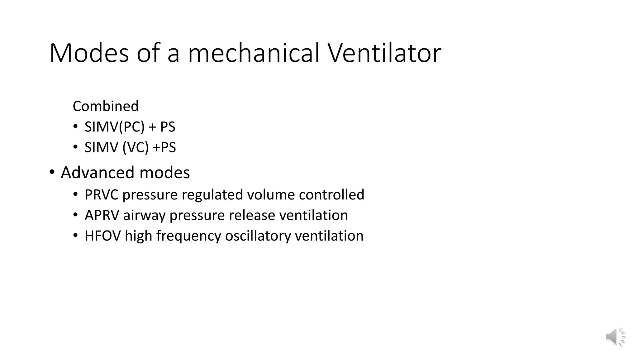
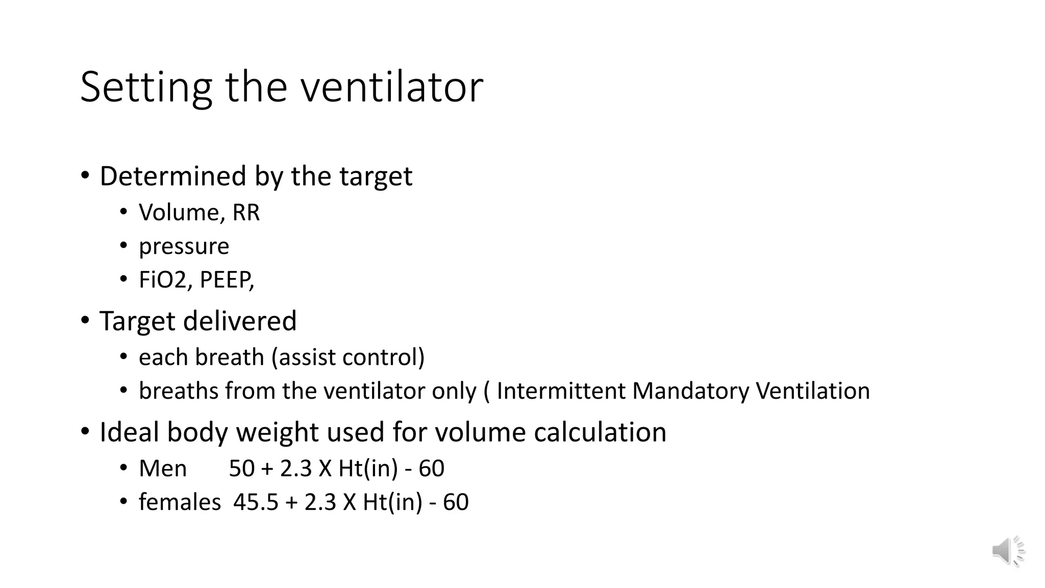
Mechanical ventilation and
pulmonary rehabilitation
DR CHINWE CHUKWUKA
UNTH ENUGU
Expected outcome
• Understand the function of mechanical ventilator and identify its components
• Identify patients that need mechanical Ventilation or Pulmonary rehabilitation
• Justify the need for different modes of ventilator
• Plan the care of patients on a mechanical ventilator
• Identify weaning parameters
• Identify Complications and prevention
Outline
• Introduce the equation of motion for respiratory system
• Indications for mechanical Ventilation
• Modes
• Care of the ventilated patient
• Complications of mechanical ventilation and prevention
• Pulmonary rehabilitation
Understanding function of mechanical
ventilation
• Mechanical ventilator is a machine used to augment or takeover the
function of the respiratory system.
• Interaction of airway pressure, respiratory muscle pressure, flow,
volume and respiratory mechanics cause the flow of air between the
atmosphere and the lungs
• The equation of motion
• Paw + Pmus = Flow X resist+ Vol /Compliance
• Pvent + Pmus = Flow X resist + Vol/ Compliance
Parameters that can be controlled in MV
• Tidal volume
• Respiratory rate
• Pressure
• PEEP
• FiO2
Indications for mechanical ventilation
• Treatment of hypoxemic respiratory failure
• Treatment of hypercapnic respiratory failure
• Treatment of combined hypoxemic and hypercapnic respiratory
failure
Mechanical ventilator components
• Ventilator interface: a set of flexible tubes thro the Ventilator delivers
gas to the patient
• Power source
• Control system
• Patient monitoring system
• Pressure
• Alarms
• Graphic displays
Phase variables of MV
• Trigger phase ……trigger variable
• Inspiration …….limit variable
• Ending or cycling phase ………cycling variable
• Expiration…….baseline variable
Modes of a mechanical Ventilator
• Method of inspiratory support during MV
• Controlled
• Assist control Pressure or
• Assist control volume: full ventilator control
• Supported
• Pressure support
Modes of a mechanical Ventilator
Combined
• SIMV(PC) + PS
• SIMV (VC) +PS
• Advanced modes
• PRVC pressure regulated volume controlled
• APRV airway pressure release ventilation
• HFOV high frequency oscillatory ventilation
Setting the ventilator
• Determined by the target
• Volume, RR
• pressure
• FiO2, PEEP,
• Target delivered
• each breath (assist control)
• breaths from the ventilator only ( Intermittent Mandatory Ventilation
• Ideal body weight used for volume calculation
• Men 50 + 2.3 X Ht(in) - 60
• females 45.5 + 2.3 X Ht(in) - 60
Setting the ventilator ..2
• Initial tidal vol 6-8ml/Kg IBW , RR 2/3 pre-intubation rate or IBW X100
= Minute ventilation
• Pressure start at 20cmH2O
• FiO2 start at 100 and titrate based on PaO2 from ABG or SPO2
• PEEP start at 5cmH2O
• Flow rate 60L/min
• I :E
Setting trade offs
• FiO2 fraction of inspired oxygen; use the lowest that achieves
targeted oxygenation. Avoid prolonged use of high FiO2 (0.60)
• Respiratory rate 10-20/min
• (Hypo..Resp acidosis
• Hyper ..resp alka)
• Tidal vol(6-8ml/kg IBW)vol of air exchanged at each breath , reduce if
lungs are stiff
• Trigger may be
• ventilator-initiated prompted by timer from set RR or frequency
• patient initiated
• Inspiration: Expiration (I:E) ratio
• normal 1:2
• Inverse ratio 1:1, 2:1, 3:1, 4:1;
• Reduced I:E in COPD with resp acidosis to encourage longer expiration
• Peak inspiratory pressure (PIP) < 35 cmH2O
• low PIP may cause hypoventilation
• high PIP may cause lung damage
• Minute ventilation 5-10L/minute;
• volume of gas exchanged per minute
• VE = RR X VT
• Plateau pressure (Pplat) reflects lung compliance (brief inspiratory
pause after ventilation)
• normal Pplat and high PIP indicates resistance to air flow as in bronchospasm
or endotrachael tube obstruction
• high Pplat and high PIP ..reduced compliance pulmonary edema, ARDs ;
• Positive End Expiratory Pressure (PEEP)
• keeps alveoli open and recruits more alveoli to improve oxygenation.
• High levels may cause barotrauma, increased ICP and decreased cardiac
output
• Pressure support (PS) 8-20cmH2O provides extra pressure during
inspiration to increase ventilation with minimal patient effort. It is
used to reduce the work of breathing through ventilator tubing
Clinical settings
• COPD
• low tidal vol 6-8ml/Kg IBW, RR 6-20,
• low FiO2, low PEEP, permissive hypercapnia,
• target SPO2 88-92% and normal PH not PaCO2
• Asthma
• low tidal vol 6-10ml/Kg IBW,
• flow 80-100L/min,
• RR <20/min, Low PEEP,
• PIP 40-45cmH2O, Pplat <25cmH2O
• ARDS permissive hypercapnia may be accepted to minimize lung
injury,
• PH >7.25, low tidal volume 6mL/Kg IBW,
• low Pplat <30cmH2O, low driving P <15cmH2O,
• optimize PEEP gently by gradual increment to prevent atelectasis and recruit
alveoli
• Proning when PaO2:FiO2 <150 persistently, watch ETT
Ventilator patient care
• Daily assessment (team work):
• status,
• full clinical examination,
• sedation interruption,
• assess for readiness for SBT
• Be systematic, document, ETT position, connections, humidifier in
use, water level, control system
• PEEP
Spontaneous breath trial SBT
• Patient stable and improving medically
• FiO2 </= 0.5
• PEEP <10cmH2O
• Patient can make spontaneous effort
• 2 minute test of readiness for SBT
• Reduce all pressure settings to 0 for 2 minutes
• If RSBI < 105 (rapid shallow breath index)
• RR/Tidal vol (L)
SBT
• Minimum time 30 minutes max 2hours
• With pt on pressure support Ventilation, remove all support from
ventilator but allow RR and minute ventilation to be monitored.
Monitor RR, oxygen saturation, HR, Systolic BP
• SpO2 > 90%, hemodynamic stability, sufficient cough, alert mark
success
Extubation/ weaning parameters
• Adequate oxygenation
• PaO2 >60-70 @ FiO2 0.4-0.5
• PaO2/FiO2 ratio >150-200
• Adequate ventilation
• PaCO2 35-45mmHg
• PH 7.3 – 7.45
• Adequate respiratory mechanics
• Tidal vol, minute ventilation
• Hemodynamic stability
Complications of Mechanical ventilation
• Larnygeal edema, lacerations, vocal cord injury correct cuff pressure
• Aspiration of oral and gastric secretions suction, head at angle 30-45
• Auto PEEP Baro and volutrauma avoid high auto peep, pressure,
reduce tidal volume, keep Pplat<30cmH20
• Asynchrony with ventilator identify early and correct
• Hemodynamic compromise identify early and correct
Complications of Mechanical ventilation
• Ventilator ass pneumonia nurse head up, daily interruption of
sedation, suction of secretions,early mobility, infrequent change of
ventilator tubes
• Gastritis, ulcers, malnutrition, constipation nutrition plan, early
mobility, gastrointestinal prophylaxis,
• Sleep deprivation, anxiety,
• Muscle deconditioning, ventilator dependence plan for early
weaning:assess SBT daily
• Renal, hepatic dysfunction assess function clinically and chemistry
Pulmonary rehabilitation
• Inter disciplinary program of care for patients with chronic respiratory
impairment that is individually designed to optimize patient’s physical
and social performance and autonomy. Use of exercise, education
and behavioral intervention to improve symptoms, restore functional
ability and improve quality of life in patients with chronic lung
disease.
• Evaluation: med hx, exam, review of medical records, lab results: lung
function tests, EKG,6 minutes walk
• Components of PR
• Inspiratory muscle training
Pulmonary rehabilitation
• Evaluation: med hx, exam, review of medical records, lab results: lung
function tests, EKG,6 minutes walk
• Components of PR
• Inspiratory muscle training
• Education
Inspiratory muscle training
• Control breathing
• Breathing exercise,
• pursed lip, slow deep, diaphragmatic, segmental breathing
• Diaphragmatic breathing -/+ pursed lip breathing significantly improved tidal
volume, reduced breathing frequency without causing dyspnea
Liliane et al.
Inspiratory muscle training
• Clearance of secretion: postural drainage, chest percussion &
vibration, control cough
• Trunk flexibility
• Upper extremity exercise: strengthening upper back, shoulder girdle
muscles
• Reconditioning: aerobic @ least 30 minutes, 3-4times a week
Education
• Smoking cessation
• Disease prognosis, information on the medications
• Lifestyle change to adapt to the disease
• Goals and benefit of the PR program
Duration
• 6- 12 weeks @ least 12 supervised sessions
• Encouraged to continue the exercises at the end of the sessions
Indication
• Motivated patients
• Functional limitation due to the disease
• Symptomatic patients who are stable on their medications.
• Pre or post thoracic surgery
Assessment
• Clinical improvement in
• exercise capacity
• Dyspnea
• Psychological wellbeing
• Patient satisfaction and feedback
References
• Pierson DJ. Complications associated with mechanical ventilation. Crit
Care Clin. 1990 Jul; 6(3): 711-24
• Strieter RM, Lynch JP 3rd. Complications in the ventilated patient.
Clin. Chest Med. 1988Mar; 9(1): 127-39
• Haribhai S, Mahboobi SK. Ventilator complications( updated 2021
October) In: StatPearls (internet). Treasure Island (Fl): StatPearls
Publishing: 2022 Jan.available at ncbi.nlm.nih.gov
• Moied S, Athar M. Mechanical ventilation in patients with chronic
obstructive pulmonary disease and asthma. Indian J Anaesth. 2015
Sep; 59(9): 589-598.
• Bolton CE, Bevan-Smith EF, Blakkey JD, Crowe P, Elkin SL Garrod R et
al. British Thoracic Society guideline on pulmonary rehabilitation in
adults: accredited by NICE. Thorax 68(2) suppl2.
http://dx.doi.org/10.1136/thoraxjnl-2013-203808.
• National Institute for Health and Clinical Excellence. Chronic
obstructive pulmonary disease. Management of chronic of chronic
obstructive lung disease in adults in primary and secondary care
(partial update). 2010.
http://www.nice.org.uk/nicemedia/live/13029/4

Mechanical ventilation & Pulmonary Rehabilitation -1.pdf

  • 1.
    Mechanical ventilation and pulmonaryrehabilitation DR CHINWE CHUKWUKA UNTH ENUGU
  • 2.
    Expected outcome • Understandthe function of mechanical ventilator and identify its components • Identify patients that need mechanical Ventilation or Pulmonary rehabilitation • Justify the need for different modes of ventilator • Plan the care of patients on a mechanical ventilator • Identify weaning parameters • Identify Complications and prevention
  • 3.
    Outline • Introduce theequation of motion for respiratory system • Indications for mechanical Ventilation • Modes • Care of the ventilated patient • Complications of mechanical ventilation and prevention • Pulmonary rehabilitation
  • 4.
    Understanding function ofmechanical ventilation • Mechanical ventilator is a machine used to augment or takeover the function of the respiratory system. • Interaction of airway pressure, respiratory muscle pressure, flow, volume and respiratory mechanics cause the flow of air between the atmosphere and the lungs • The equation of motion • Paw + Pmus = Flow X resist+ Vol /Compliance • Pvent + Pmus = Flow X resist + Vol/ Compliance
  • 5.
    Parameters that canbe controlled in MV • Tidal volume • Respiratory rate • Pressure • PEEP • FiO2
  • 6.
    Indications for mechanicalventilation • Treatment of hypoxemic respiratory failure • Treatment of hypercapnic respiratory failure • Treatment of combined hypoxemic and hypercapnic respiratory failure
  • 7.
    Mechanical ventilator components •Ventilator interface: a set of flexible tubes thro the Ventilator delivers gas to the patient • Power source • Control system • Patient monitoring system • Pressure • Alarms • Graphic displays
  • 8.
    Phase variables ofMV • Trigger phase ……trigger variable • Inspiration …….limit variable • Ending or cycling phase ………cycling variable • Expiration…….baseline variable
  • 9.
    Modes of amechanical Ventilator • Method of inspiratory support during MV • Controlled • Assist control Pressure or • Assist control volume: full ventilator control • Supported • Pressure support
  • 10.
    Modes of amechanical Ventilator Combined • SIMV(PC) + PS • SIMV (VC) +PS • Advanced modes • PRVC pressure regulated volume controlled • APRV airway pressure release ventilation • HFOV high frequency oscillatory ventilation
  • 11.
    Setting the ventilator •Determined by the target • Volume, RR • pressure • FiO2, PEEP, • Target delivered • each breath (assist control) • breaths from the ventilator only ( Intermittent Mandatory Ventilation • Ideal body weight used for volume calculation • Men 50 + 2.3 X Ht(in) - 60 • females 45.5 + 2.3 X Ht(in) - 60
  • 12.
    Setting the ventilator..2 • Initial tidal vol 6-8ml/Kg IBW , RR 2/3 pre-intubation rate or IBW X100 = Minute ventilation • Pressure start at 20cmH2O • FiO2 start at 100 and titrate based on PaO2 from ABG or SPO2 • PEEP start at 5cmH2O • Flow rate 60L/min • I :E
  • 13.
    Setting trade offs •FiO2 fraction of inspired oxygen; use the lowest that achieves targeted oxygenation. Avoid prolonged use of high FiO2 (0.60) • Respiratory rate 10-20/min • (Hypo..Resp acidosis • Hyper ..resp alka) • Tidal vol(6-8ml/kg IBW)vol of air exchanged at each breath , reduce if lungs are stiff • Trigger may be • ventilator-initiated prompted by timer from set RR or frequency • patient initiated
  • 14.
    • Inspiration: Expiration(I:E) ratio • normal 1:2 • Inverse ratio 1:1, 2:1, 3:1, 4:1; • Reduced I:E in COPD with resp acidosis to encourage longer expiration • Peak inspiratory pressure (PIP) < 35 cmH2O • low PIP may cause hypoventilation • high PIP may cause lung damage • Minute ventilation 5-10L/minute; • volume of gas exchanged per minute • VE = RR X VT
  • 15.
    • Plateau pressure(Pplat) reflects lung compliance (brief inspiratory pause after ventilation) • normal Pplat and high PIP indicates resistance to air flow as in bronchospasm or endotrachael tube obstruction • high Pplat and high PIP ..reduced compliance pulmonary edema, ARDs ; • Positive End Expiratory Pressure (PEEP) • keeps alveoli open and recruits more alveoli to improve oxygenation. • High levels may cause barotrauma, increased ICP and decreased cardiac output
  • 16.
    • Pressure support(PS) 8-20cmH2O provides extra pressure during inspiration to increase ventilation with minimal patient effort. It is used to reduce the work of breathing through ventilator tubing
  • 17.
    Clinical settings • COPD •low tidal vol 6-8ml/Kg IBW, RR 6-20, • low FiO2, low PEEP, permissive hypercapnia, • target SPO2 88-92% and normal PH not PaCO2 • Asthma • low tidal vol 6-10ml/Kg IBW, • flow 80-100L/min, • RR <20/min, Low PEEP, • PIP 40-45cmH2O, Pplat <25cmH2O
  • 18.
    • ARDS permissivehypercapnia may be accepted to minimize lung injury, • PH >7.25, low tidal volume 6mL/Kg IBW, • low Pplat <30cmH2O, low driving P <15cmH2O, • optimize PEEP gently by gradual increment to prevent atelectasis and recruit alveoli • Proning when PaO2:FiO2 <150 persistently, watch ETT
  • 19.
    Ventilator patient care •Daily assessment (team work): • status, • full clinical examination, • sedation interruption, • assess for readiness for SBT • Be systematic, document, ETT position, connections, humidifier in use, water level, control system • PEEP
  • 20.
    Spontaneous breath trialSBT • Patient stable and improving medically • FiO2 </= 0.5 • PEEP <10cmH2O • Patient can make spontaneous effort • 2 minute test of readiness for SBT • Reduce all pressure settings to 0 for 2 minutes • If RSBI < 105 (rapid shallow breath index) • RR/Tidal vol (L)
  • 21.
    SBT • Minimum time30 minutes max 2hours • With pt on pressure support Ventilation, remove all support from ventilator but allow RR and minute ventilation to be monitored. Monitor RR, oxygen saturation, HR, Systolic BP • SpO2 > 90%, hemodynamic stability, sufficient cough, alert mark success
  • 22.
    Extubation/ weaning parameters •Adequate oxygenation • PaO2 >60-70 @ FiO2 0.4-0.5 • PaO2/FiO2 ratio >150-200 • Adequate ventilation • PaCO2 35-45mmHg • PH 7.3 – 7.45 • Adequate respiratory mechanics • Tidal vol, minute ventilation • Hemodynamic stability
  • 23.
    Complications of Mechanicalventilation • Larnygeal edema, lacerations, vocal cord injury correct cuff pressure • Aspiration of oral and gastric secretions suction, head at angle 30-45 • Auto PEEP Baro and volutrauma avoid high auto peep, pressure, reduce tidal volume, keep Pplat<30cmH20 • Asynchrony with ventilator identify early and correct • Hemodynamic compromise identify early and correct
  • 24.
    Complications of Mechanicalventilation • Ventilator ass pneumonia nurse head up, daily interruption of sedation, suction of secretions,early mobility, infrequent change of ventilator tubes • Gastritis, ulcers, malnutrition, constipation nutrition plan, early mobility, gastrointestinal prophylaxis, • Sleep deprivation, anxiety, • Muscle deconditioning, ventilator dependence plan for early weaning:assess SBT daily • Renal, hepatic dysfunction assess function clinically and chemistry
  • 25.
    Pulmonary rehabilitation • Interdisciplinary program of care for patients with chronic respiratory impairment that is individually designed to optimize patient’s physical and social performance and autonomy. Use of exercise, education and behavioral intervention to improve symptoms, restore functional ability and improve quality of life in patients with chronic lung disease. • Evaluation: med hx, exam, review of medical records, lab results: lung function tests, EKG,6 minutes walk • Components of PR • Inspiratory muscle training
  • 26.
    Pulmonary rehabilitation • Evaluation:med hx, exam, review of medical records, lab results: lung function tests, EKG,6 minutes walk • Components of PR • Inspiratory muscle training • Education
  • 27.
    Inspiratory muscle training •Control breathing • Breathing exercise, • pursed lip, slow deep, diaphragmatic, segmental breathing • Diaphragmatic breathing -/+ pursed lip breathing significantly improved tidal volume, reduced breathing frequency without causing dyspnea Liliane et al.
  • 28.
    Inspiratory muscle training •Clearance of secretion: postural drainage, chest percussion & vibration, control cough • Trunk flexibility • Upper extremity exercise: strengthening upper back, shoulder girdle muscles • Reconditioning: aerobic @ least 30 minutes, 3-4times a week
  • 29.
    Education • Smoking cessation •Disease prognosis, information on the medications • Lifestyle change to adapt to the disease • Goals and benefit of the PR program
  • 30.
    Duration • 6- 12weeks @ least 12 supervised sessions • Encouraged to continue the exercises at the end of the sessions
  • 31.
    Indication • Motivated patients •Functional limitation due to the disease • Symptomatic patients who are stable on their medications. • Pre or post thoracic surgery
  • 32.
    Assessment • Clinical improvementin • exercise capacity • Dyspnea • Psychological wellbeing • Patient satisfaction and feedback
  • 33.
    References • Pierson DJ.Complications associated with mechanical ventilation. Crit Care Clin. 1990 Jul; 6(3): 711-24 • Strieter RM, Lynch JP 3rd. Complications in the ventilated patient. Clin. Chest Med. 1988Mar; 9(1): 127-39 • Haribhai S, Mahboobi SK. Ventilator complications( updated 2021 October) In: StatPearls (internet). Treasure Island (Fl): StatPearls Publishing: 2022 Jan.available at ncbi.nlm.nih.gov • Moied S, Athar M. Mechanical ventilation in patients with chronic obstructive pulmonary disease and asthma. Indian J Anaesth. 2015 Sep; 59(9): 589-598.
  • 34.
    • Bolton CE,Bevan-Smith EF, Blakkey JD, Crowe P, Elkin SL Garrod R et al. British Thoracic Society guideline on pulmonary rehabilitation in adults: accredited by NICE. Thorax 68(2) suppl2. http://dx.doi.org/10.1136/thoraxjnl-2013-203808. • National Institute for Health and Clinical Excellence. Chronic obstructive pulmonary disease. Management of chronic of chronic obstructive lung disease in adults in primary and secondary care (partial update). 2010. http://www.nice.org.uk/nicemedia/live/13029/4