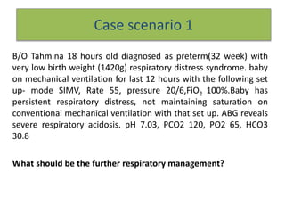
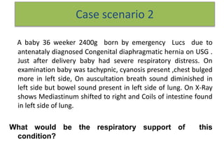
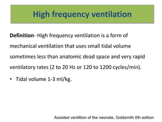
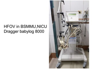
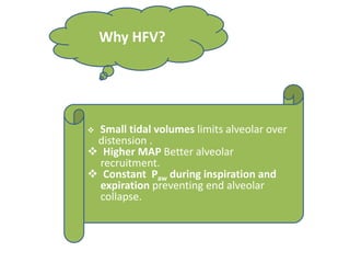
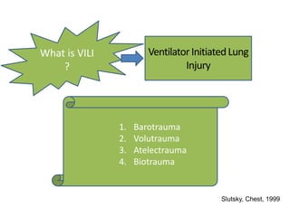
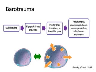
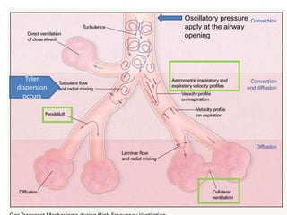
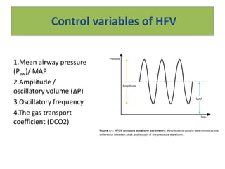
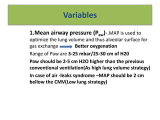
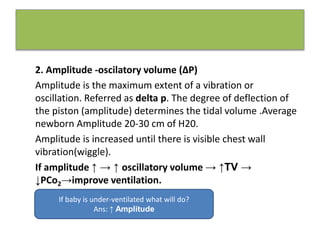
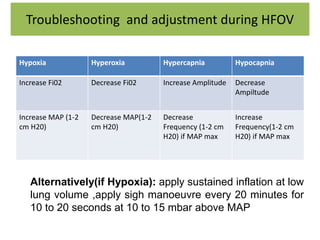
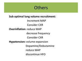
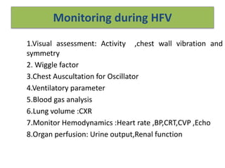
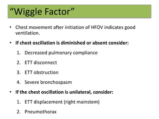
Call Girls Kanakapura Road Just Call 7001305949 Top Class Call Girl Service A...
seminar on hfv - high frequency ventilation
1. Case scenario 1
B/O Tahmina 18 hours old diagnosed as preterm(32 week) with
very low birth weight (1420g) respiratory distress syndrome. baby
on mechanical ventilation for last 12 hours with the following set
up- mode SIMV, Rate 55, pressure 20/6,FiO2 100%.Baby has
persistent respiratory distress, not maintaining saturation on
conventional mechanical ventilation with that set up. ABG reveals
severe respiratory acidosis. pH 7.03, PCO2 120, PO2 65, HCO3
30.8
What should be the further respiratory management?
2. Case scenario 2
A baby 36 weeker 2400g born by emergency Lucs due to
antenataly diagnosed Congenital diaphragmatic hernia on USG .
Just after delivery baby had severe respiratory distress. On
examination baby was tachypnic, cyanosis present ,chest bulged
more in left side, On auscultation breath sound diminished in
left side but bowel sound present in left side of lung. On X-Ray
shows Mediastinum shifted to right and Coils of intestine found
in left side of lung.
What would be the respiratory support of this
condition?
4. High frequency ventilation
Definition- High frequency ventilation is a form of
mechanical ventilation that uses small tidal volume
sometimes less than anatomic dead space and very rapid
ventilatory rates (2 to 20 Hz or 120 to 1200 cycles/min).
• Tidal volume 1-3 ml/kg.
Assisted ventiltion of the neonate, Goldsmith 6th edition
5. 1915,
Dr. Henderson studied
small tidal volumes and
rapid rates
1950-1970
Dr. Emerson,
Dr. Bird, Dr. Bunnell studied
HFV
1970’s
Success with animal
studies
1980’s
Four received FDA
approval
1990’s
HFOV
emerged
2000’s
HFOV for Adults
1991
1995 2001
???
HistoryofHigh FrequencyVentilation
6. Types of high frequency ventilation
1.High frequency flow interruption
(HFFI)
2.High frequency jet ventilation
(HFJV)
3.High frequency oscillator ventilation
(HFOV)
Assisted ventiltion of the neonate, Goldsmith 6th edition
7. HFOV HFJV HFFI
High flow
generated by
Electromagnet
ic piston or
diaphragm
oscillator
Pince valve or
injector
cannula
Solenoid valve
Expiration Active passive passive
Inspiration Active Active Active
Frequency 5 to15 HZ 5 to 10HZ 8 to 12 HZ
Sigh
superimpose
yes yes yes
Basic differences
16. HFOV is used to prevent
ventilator induced lung
injury
Critical opening
pressure
Critical maximum
pressure
Critical closing
pressure
17. Pressure and Volume Swings
•During CMV, there are swings
between the zones of injury from
inspiration to expiration.
•During HFOV, the entire cycle
operates in the “safe window” and
avoids the injury zones.
18.
19.
20.
21. Advantages of HFV
• Small tidal volumes prevents alveolar over distension and
Volutrauma .
• Higher mean airway pressure (MAP )-improved alveolar
recruitment leading to better oxygenation.
• Smaller gradient between inspiratory and expiratory
pressure- prevent cyclical distention and alveolar collapse—
hence less atelectrauma
•Low peak pressure –reduce barotrauma
22. Indications of HFV
1.When conventional ventilation fails
– reduced compliance,RDS/ARDS,meconium aspiration ,BPD
,pneumonia,atelectasis, lung hypoplasia
Other: PPHN
2.Primary mode of ventilation in extreme prematurity.
3.Air leak syndromes (pneumothorax, PIE)
4.Congenital diaphragmatic hernia.
Assisted ventiltion of the neonate, Goldsmith 6th edition
Dragger manuals – high frequency ventilation basics and
practical application
24. Mechanism of gas exchange
• HFV Provides augmented gas distribution by means of
numerous gas transport mechanisms.
Assisted ventiltion of the neonate, Goldsmith 6th edition
Convection ventilation( bulk flow)
Taylor dispersion
Pendelluft effect
Asymmetric velocity profiles
Cardiogenic Mixing
Molecular diffusion
Collateral Ventilation
25. Convection ventilation( bulk flow)
Mechanism of gas exchange
In HFOV, where the "tidal volume" is negligible, bulk convection is represented
by the continual entrainment of fresh gas, as oxygen is absorbed at the
alveolus. The decreased pressure generated by continual gas removal makes
space for more gas.
26. Mechanism of gas exchange
• Taylor dispersion- in which augmented diffusion occurs because of turbulant air
currents that results from interection between axial velocity and the radial
concentration gradient.
High frequency ventilation:current status, AAP ,Drager
27. Taylor dispersion
Secondary gas movements occur at airway bends and bifurcations creating
turbulent eddies that enhance radial gas mixing (movement of gas particles
from the centre of flow to the stationary boundary layer along the wall) at the
expense of longitudinal gas transport
28. Mechanism of gas exchange
Pendelluft effect
Not all regions of the lung have the
same compliance and resistance.
Therefore, neighboring units with
different time constants are ventilated
out of phase, filling and emptying at
different rates. Due to this asynchrony
these units can mutually exchange by
blowing gas into each other when one
is collapsing and the other still
remains open, an effect known as
pendelluft. By way of this mechanism
even very small fresh-gas volumes can
reach a large number of alveoli and
regions.
Pendelluft effect - in which regional
differences in time constants for inflation
and deflation cause gas to recirculate
among lung units and improve gas
exchange.
29. Asymmetrical velocity
Airflow moving through the airways moves in a u-shape formation. At the
center of the lumen air will move at a faster velocity, than air that is closest
to the wall.
30. Mechanism of gas exchange
As the heart beats the heart provides additional peripheral mixing
by exerting pressure against the lungs during contraction of the
heart. This pressure promotes the movement of gas flow through
the neighboring parenchymal regions.
31. Molecular diffusion
Maintaining a constant
distending pressure with
HFV within the lungs
along with movement of
gas molecules promotes
gas diffusion across the
alveolar membrane, at a
faster rate.
34. Control variables of HFV
1.Mean airway pressure
(Paw)/ MAP
2.Amplitude /
oscillatory volume (∆P)
3.Oscillatory frequency
4.The gas transport
coefficient (DCO2)
35. Variables
1.Mean airway pressure (Paw)-.MAP is used to
optimize the lung volume and thus alveolar surface for
gas exchange Better oxygenation
Range of Paw are 3-25 mbar/25-30 cm of H20
Paw should be 2-5 cm H2O higher than the previous
conventional ventilation(As high lung volume strategy)
In case of air -leaks syndrome –MAP should be 2 cm
bellow the CMV(Low lung strategy)
36. 2. Amplitude -oscilatory volume (∆P)
Amplitude is the maximum extent of a vibration or
oscillation. Referred as delta p. The degree of deflection of
the piston (amplitude) determines the tidal volume .Average
newborn Amplitude 20-30 cm of H20.
Amplitude is increased until there is visible chest wall
vibration(wiggle).
If amplitude ↑ → ↑ oscillatory volume → ↑TV →
↓PCo2→improve ventilation.
If baby is under-ventilated what will do?
Ans: ↑ Amplitude
37. 3.Frequency- Number of cycles per unit of time.
measured in units of Hertz. ( 1 Hz = 60 breaths/min)
Usually start as 10 -12 HZ but set as 15 Hz for
premature infants with RDS.
↓ Oscillatory frequency → ↑ oscillatory amplitude,
↑oscillatory volume → ↑TV → ↓ Pco2
and vice versa
38.
39. 4. Gas transport co efficient (DCo2)
In conventional ventilation the product of tidal volume and
frequency, known as minute volume or minute ventilation, aptly
describes pulmonary gas exchange. Different study groups have
found that CO2 elimination in HFO however correlates well with
VT2 x f
Here, VT and f stand for oscillatory volume and frequency, re
spectively. This parameter is called ‘gas transport coefficient’,
DCO2.
An increase in DCO2 will decrease pCO2
40. Variables in oxygenation
• The two primarily variables that control oxygenation are:
– FiO2
– Mean airway pressure (Paw)
41. Variables in ventilation
• The two primarily variables that control ventilation are:
– Tidal volume (P or amplitude)
• Controlled by the force with which the oscillatory
piston moves. (represented as stroke volume or P)
– Frequency ()
• Referenced in Hertz (1 Hz = 60 breaths/second)
• Range: 3 - 15 Hz
43. MAP: 2-5mbar above MAP of CMV
Frequency: 10 Hz
Amplitude: 50- 100% watch thorax
vibrations
Volume: about 2 to 2.5 ml/kg
Fi02:100%
Start HFV
44. Oxygenation
Initiate Fio2 100% and Set MAP 2 cm above or bellow (air leak syndrome) than
CMV to give spo2 90-95%
Increase MAP 1-2 cm of H20 in every 2-5 minutes and reduce Fio2 stepwise 5-
10% to maintain Spo2 90-95% ( MAP increased until Fio2 less than 0.3)
Once baby Stable in an Fio2 less than 0.3 then MAP should be cautiously
reduced 1-2 cm H20 as allowed the oxygenation
Dragger manuals – high frequency ventilation basics and practical application
RPA Newborn Care Guidelines
45. Ventilation
Set Amplitude (ďP) 50%
observe good wiggle and adjust Amplitude accordingly
Adjust Amplitude increment 10%(3-5 cm of H20) for optimum PaCo2
If PaCo2 fails to fall even after increased Amplitude then decrease
Frequency 0.5-1 HZ
46. Troubleshooting and adjustment during HFOV
Hypoxia Hyperoxia Hypercapnia Hypocapnia
Increase Fi02 Decrease Fi02 Increase Amplitude Decrease
Ampiltude
Increase MAP (1-2
cm H20)
Decrease MAP(1-2
cm H20)
Decrease
Frequency (1-2 cm
H20) if MAP max
Increase
Frequency(1-2 cm
H20) if MAP max
Alternatively(if Hypoxia): apply sustained inflation at low
lung volume ,apply sigh manoeuvre every 20 minutes for
10 to 20 seconds at 10 to 15 mbar above MAP
48. Monitoring during HFV
1.Visual assessment: Activity ,chest wall vibration and
symmetry
2. Wiggle factor
3.Chest Auscultation for Oscillator
4.Ventilatory parameter
5.Blood gas analysis
6.Lung volume :CXR
7.Monitor Hemodynamics :Heart rate ,BP,CRT,CVP ,Echo
8.Organ perfusion: Urine output,Renal function
49. “Wiggle Factor”
• Chest movement after initiation of HFOV indicates good
ventilation.
• If chest oscillation is diminished or absent consider:
1. Decreased pulmonary compliance
2. ETT disconnect
3. ETT obstruction
4. Severe bronchospasm
• If the chest oscillation is unilateral, consider:
1. ETT displacement (right mainstem)
2. Pneumothorax
50. Humidification
It is essential to adequately humidify (90%) the breathing
gas. Otherwise severe irreversible damage to the trachea
may result. Viscous secretion could obstruct bronchi and
deteriorate the pulmonary situation. Excessive
humidification on the other hand can lead to
condensation in the patient circuit, the ET tube and the
airways, completely undoing the effect of HFV.
51. Weaning
HFV: Weaning
1. Reduce FiO2 to 0.3 – 0.5
2. Reduce MAP by 1 to 2 mbar per hour until (8) to 9 mbar;
then increase IMV rate
3. Reduce amplitude
4. Continue ventilation with IMV/SIMV and weaning
5. Extubation from HFV is also possible if respiratory
activity is sufficient
52. Points Diffuse homogeneous lung
diseases
Inhomogeneous lung
diseases
Goals lung expansion less barotrauma improved oxygenation
and ventilation at
minimum MAP
Starts/initiation MAP 2 to 5 mbar above that of
CMV
MAP like or below that of
CMV and low
frequency(7HZ)
Changes & weaning increase MAP until pO2 rises by 20
to 30 mmHg, or signs of over-
inflation appear then reduce FiO2
to 0.3 – 0.5 then continue weaning
increase MAP until PO2
slightly rises; keep
MAP constant; if
respiratory situation
fails to improve return
to CMV.
Risk Overinflation partial overexpansion
Assesment Clinical and CXR Clinical and CXR
Strategies for various lung diseases
53. Points Air leaks diseases Pulmonary hypertension of
the newborn (PPHN)
Goals improved oxygenation and
ventilation at minimum MAP;
(accept lower pO2 and higher pCO2)
to optimize lung
volume and perfusion
to improve hypoxia
and hypercapnia
Starts/initiation MAP like or below that of CMV and
low frequency(7HZ)
-Frequency: <10 Hz
– amplitude: 100%
– MAP: on the level of
CMV
Changes & weaning Reduce pressure prior to FiO2
– Continue HFV for 24 to 48
hours after improvement
reduce O2 prior to
MAP and Maintain
HFV for 24 to 48 hours
after recovery
Risk Over-distention Avoid hypovolemia
Assesment Clinical and CXR Clinical and CVP
54. Complications
1. Irritation- require more sedation.
2. hemodynamics- high MAP can jeopardies venous
return and cardiac output and also increase
puolmonary vascular resistance, Hypotension
3.Air trapping
4.Overinflation
5.Necrotizing tracheobronchitis
6.Intracranial haemorrhages.
55. Evidences
High-frequency oscillatory ventilation versus conventional
mechanical ventilation for very-low-birth-weight infants.
N Engl J Med. 2002 Aug 29;347(9):643-52.
Courtney SE, Durand DJ, Asselin JM, Hudak ML, Aschner JL, Shoemaker
CT; Neonatal Ventilation Study Group.
Here was a small but significant benefit of high-frequency oscillatory
ventilation in terms of the pulmonary outcome for very-low-birth-weight
infants without an increase in the occurrence of other complications of
premature birth.
56. Evidence
As a primary mode
Elective high frequency ventilation compared to
conventional mechanical ventilation in the early stabilization
of infants with respiratory distress - Cochrane-March 2015
Insufficient evidence exists to support the routine use of high
frequency oscillatory ventilation instead of conventional ventilation
for preterm infants.
57. • High frequency oscillatory ventilation is a way of providing
artificial ventilation of the lungs that theoretically may produce
less injury to the lungs and therefore reduce the rate of chronic
lung disease. This review of the evidence from 19 randomized
controlled trials showed that although a small protective effect
towards the lungs can be seen, this moderate benefit is highly
variable between studies and should be weighed against
possible harm.
58. Authors’ conclusions :There are no data from randomized controlled trials
supporting the routine use of rescue HFOV in term or near term infants with
severe pulmonary dysfunction. The area is complicated by diverse pathology in
such infants and by the occurrence of other interventions (surfactant, inhaled
nitric oxide, inotropes). Randomized controlled trials are needed to establish the
role of rescue HFOV in near term and term infants with pulmonary dysfunction
before widespread use of this mode of ventilation in such infants.
59.
60.
61. • Conclusions. The results of this study support the idea that rescue
HFOV may increase survival of CDH patients, when conventional
mechanical ventilation fails .We suggest that all deceased CDH patients
should have a necropsy study in order to identify other congenital
malformations that may pass undetected on prenatal and postnatal
evaluation, and the lung histological findings that may explain the failure
of the respiratory mode.
62. Looking towards the future
• A great deal remains unknown about HFOV:
– the exact mechanism of gas exchange
– the most effective strategy to manipulate ventilator settings
– the safest approach to manipulate ventilator settings
– a reliable method to measure tidal volume
– the appropriate use of sedation and neuromuscular blockade
to optimize patient-ventilator interactions
• Additional research in these and other issues related to HFOV
are necessary to maximize the benefit and minimize the
potential risks associated with HFOV.
63. References
• Goldsmith: Assisted ventilation of the neonate
• Dragger manuals – high frequency ventilation basics and practical
application
• High frequency ventilation: current status, pediatrics in review
• Priebe GP, Arnold JH: High-frequency oscillatory ventilation in
pediatric patients. Respir Care Clin N Am 2001; 7(4):633-645
• RPA newborn care guideline
• Arnold JH: High-frequency ventilation in the pediatric intensive
care unit. Pediatr Crit Care Med 2000; 1(2):93-99