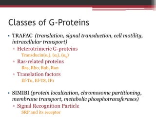
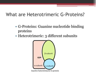
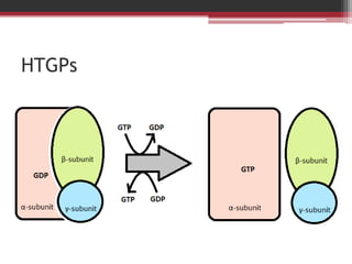
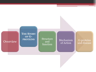
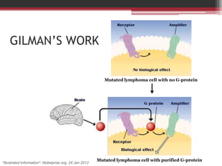
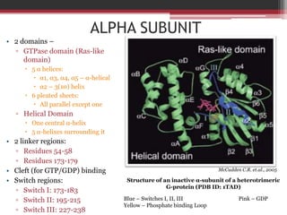
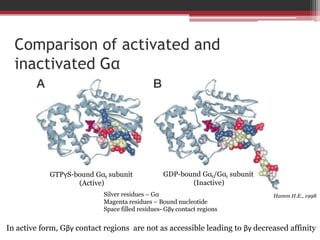
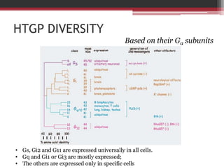
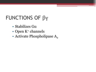
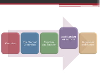
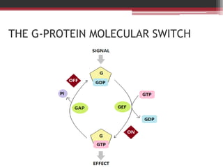
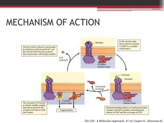
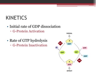
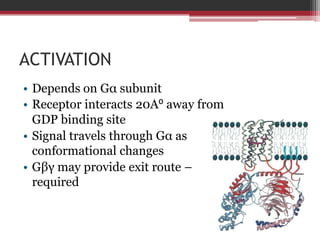
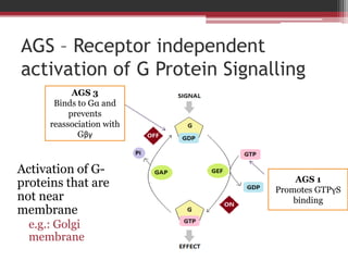
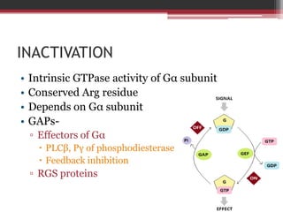
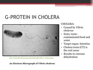
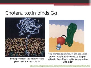
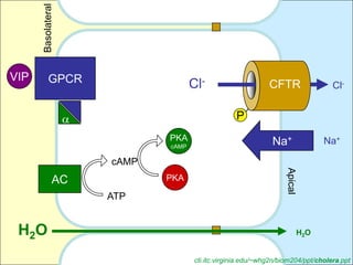
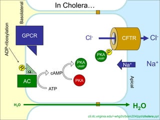
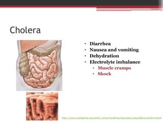
10. Nobel Prize in Physiology or Medicine, 1994
ALFRED G. GILMAN MARTIN RODBELL
"for their discovery of G-proteins and the role of these proteins in
signal transduction in cells"
12. Effect of GTP on glucagon dissociation
It is possible that the effects of Guanyl nucleotides on glucagon have some
relationship to its action on adenyl cyclase in plasma membrane
Rodbell M. et.al., 1971
13. Effect of GTP
on cAMP
synthesis
Rodbell M. et.al., 1971
14. GILMAN’S WORK
Mutated lymphoma cell with no G-protein
Mutated lymphoma cell with purified G-protein
“Illustrated Information". Nobelprize.org. 24 Jan 2012
15. STRUCTURE
AND
The Story of FUNCTION Mechanism G-proteins
Overview
G-proteins of Action and disease
16. ALPHA SUBUNIT
• 2 domains –
▫ GTPase domain (Ras-like
domain)
5 α helices:
α1, α3, α4, α5 – α-helical
α2 – 3(10) helix
6 pleated sheets:
All parallel except one
▫ Helical Domain
One central α-helix
5 α-helixes surrounding it
• 2 linker regions:
▫ Residues 54-58
▫ Residues 173-179
• Cleft (for GTP/GDP) binding McCudden C.R. et.al., 2005
• Switch regions: Structure of an inactive α-subunit of a heterotrimeric
G-protein (PDB ID: 1TAD)
▫ Switch I: 173-183
▫ Switch II: 195-215 Blue – Switches I, II, III Pink – GDP
Yellow – Phosphate binding Loop
▫ Switch III: 227-238
17. Comparison of activated and
inactivated Gα
GTPγS-bound Gαt subunit GDP-bound Gαt/Gαi subunit
(Active) (Inactive)
Silver residues – Gα Hamm H.E., 1998
Magenta residues – Bound nucleotide
Space filled residues- Gβγ contact regions
In active form, Gβγ contact regions are not as accessible leading to βγ decreased affinity
18. βγ SUBUNIT
• β Subunit
▫ 7-bladed propeller
▫ WD-40 repeats
▫ 7 small, antiparallel β strands
▫ N-terminus: α-helix
• γ Subunit
▫ 2 helical coils
• N termini of β and γ subunits form McCudden C.R. et.al., 2005
a helical coiled coil Strucure of a βγ subunit of a
heterotrimeric G-protein (PDB ID: 1TBG)
• No change in conformation on
addition of GTP/GDP Yellow: β-subunit
Red: γ-subunit
19. • Gβγ make
multiple contacts
in switch regions
of Gα
• N terminal of Gα
palmitylated to
make contacts
Strucure of an inactive heterotrimeric G-protein (PDB ID: 1GOT)
http://www.pdb.org/pdb/explore/jmol.do?structureId=1GOT&bionumber=1
20. INTERACTIONS WITH MEMBRANE
• Gα has regions of
contact on N-
terminal of its ras-like
domain
• C terminus of Gγ
prenylated to contact
plasma membrane
Hamm H.E., 1998
22. HTGP DIVERSITY
Based on their Gα subunits
• Gs, Gi2 and G11 are expressed universally in all cells.
• Gq and Gi1 or Gi3 are mostly expressed;
• The others are expressed only in specific cells
23. HTGPs are highly conserved!
Well, I and We have
you have just the same
an amino acid αs !
different in
our αi1 !
28. MECHANISM OF ACTION
The Cell - A Molecular Approach, 4th ed, Cooper G., Hausman R.
29. KINETICS
• Initial rate of GDP dissociation
▫ G-Protein Activation
• Rate of GTP hydrolysis
▫ G-Protein Inactivation
30. ACTIVATION
• Depends on Gα subunit
• Receptor interacts 20A⁰ away from
GDP binding site
• Signal travels through Gα as
conformational changes
• Gβγ may provide exit route –
required
31. AGS – Receptor independent
activation of G Protein Signalling
AGS 3
Binds to Gα and
prevents
reassociation with
Gβγ
Activation of G-
AGS 1
proteins that are Promotes GTPγS
not near binding
membrane
e.g.: Golgi
membrane
32. INACTIVATION
• Intrinsic GTPase activity of Gα subunit
• Conserved Arg residue
• Depends on Gα subunit
• GAPs-
▫ Effectors of Gα
PLCβ, Pγ of phosphodiesterase
Feedback inhibition
▫ RGS proteins
33. RGS
• RGS box- 125 aa domain
• GAPs
• Bind to Switch regions on Gα
▫ Stabilizes Gα transition state to GTP hydrolysis
• Pγ may enhance RGS9
34. G-
PROTEINS
AND
Structure DISEASE
The Story of Mechanism
Overview and
G-proteins of Action
function
35. G-PROTEIN IN CHOLERA
CHOLERA:
• Caused by Vibrio
cholerae
• Entry route:
contaminated food and
water
• Target organ: Intestine
• Cholera toxin (CT) is
the real cause
• Results in extreme
http://microbewiki.kenyon.edu/index.php/File:V_cholerae.jpg dehydration
An Electron Micrograph of Vibrio cholerae
36. Cholera toxin binds Gα
The enzymatic activity of cholera toxin
ADP-ribosylates the G-protein alpha
Some portion of the cholera toxin subunit, thus, blocking its reassociation
penetrates the membrane with GTP
http://www.nobelprize.org/nobel_prizes/medicine/laureates/1994/illpres/cholera.html
37. Basolateral
VIP GPCR Cl- CFTR Cl-
P
PKA Na+ Na+
cAMP
cAMP
Apical
AC PKA
ATP
H 2O H2O
cti.itc.virginia.edu/~whg2n/biom204/ppt/cholera.ppt
38. In Cholera…
Basolateral
ADP-ribosylation
GPCR Cl- CFTR Cl-
PKA P
cAMP
PKA
cAMP Na+ Na+
cAMP
Apical
AC PKA
ATP
H2O
H 2O
cti.itc.virginia.edu/~whg2n/biom204/ppt/cholera.ppt
First, let’s understand what are Heterotrimeric G-protein? Let’s break it up - what is a G-protein? A protein that binds to guanine nucleotides is a G-protein. What are guanine nucleotides? GTP, GMP, GDP are guanine nucleotides. Any protein that likes to bind to these is called a G-protein.
However, G-proteins are a huge class of proteins, that include molecules with varied functions. Some relevant families are shown here. There are 2 main classes – TRAFAC and SIMIBI. TRAFAC are the G-proteins involved in translation, signal transduction, cell motility, intracellular transport. These include- And then we have SIMIBI Class which includes the G-proteins involved in protein localization, chromosome partitioning, membrane transport, metabolic phosphotransferases. There are over 60 families of G-proteins and with this slide, I wanted to emphasize that we are studying a very small portion of an entire class of G-proteins, so please do not ever say just ‘G-protein’ for a heterotrimeric G-protein. There is a lot of difference.
Now, let’s get back to heterotrimeric G-proteins. We already know that they are G-proteins. And let’s also understand that they are heterotrimeric, which implies that they are 3 different subunits, as we can see here.
For years, scientists had known that hormones and neurotransmitters bind to receptors on the cell surface and this leads to the amplification of a signal, leading to the desired effect. However, the link between the receptor and amplifier was unknown.
Rodbell found that there is a transducer molecule that transfers the signal from the receptor to the amplifier. And Gilman found the first G-protein and named it transducin.For this achievement, they received the nobel prize in 1994.
Rodbell has done a lot of work on the hormone glucagon, its effector, adenylyl cyclase on rat liver plasma membranes. I’m going to present a small portion of his work. This is a flowchart showing how he checked the effect of GTP on the dissociation of glucagon from plasma membranes.In vivo conditions, Glucagon binds to a receptor which is present in the plasma membrane. It produces it’s effects and is then, expected to dissociate. However, in an invitro system, the glucagon would not dissociate from the plasma membrane. So, Rodbell worked on this aspect. He incubated rat liver membranes in a complete medium for 10 mins with glucagon. So glucagon would bind to the receptors.Then, he checked for any dissociation.In another set, he also added different nucleotides and incubated the mixture again. Again, he checked for glucagon dissociation.
And this is what he found… presence of GTP increased the rate of dissociation of glucagon (As depicted in the table and the graph). Well, the graph also showed that there was increase in dissociation due to addition of ATP, but that was probably just because ATP is required for various cellular processes in low concentrations. Also, the effect of other nucleotides could be due to contamination with GTP.With this study, Rodbell suggested that these effects of GTP on flucagon could be related to its action on adenylcyclase in plasma membrane.
This is another experiment that shows proves his earlier concept. In this case, plasma membranes are fed with a radioactive mix of AMP-PNP, which is a non-hydrolysable analog of ATP. So, we check the rate of formation of cAMP from this analog in the presence of GTP, and as you can see, it is pretty high compared to the basal rates. This was some significant research in terms of the discovery of G-proteins.
Gilman followed up on Rodbell’s work from a different perspective. He found the G-protein and characterized it, determined it’s properties and worked on its mechanism. Here’s how he found the protein.In a lymphoma cell line, he found a cell line which did not show any adenylatecyclase activity. Though, this mutant had adenylatecyclase, there was no activity. He hypothesized that this must be due to the absence of the transducer funtion, the G-protein. So, he tried a large number of proteins and finally found a protein which compensated for this activity. This was the G-protein. His further research is spent on characterizing various aspects of this protein.
All Gb subunits contain seven WD-40 repeats, a tryptophan-aspartic acid sequence that repeats about every 40 amino acids and forms small antiparallel b strands
This slide shows a classification of G-proteins on the basis of the G-alpha subunits. As you can see, there are 4 classes of HTGPs. Here, you can see their effector functions. These are important because the classification if based on these funcitons. So, Gs stimulates adenylcyclase. Remember the ‘S’. Simlarly, Gi inhibits ad. Cyclase. Whereas Gq/11 stimulate PLC-beta. And the last category, 12 and 13 modulate the activity of proteins in signal transduction pathways. 12 and 13 have opposing effects on rhoGEF. I’d also like to divert your attention to this part, that states the amino acid identity. As you can see, even the most unrelated HTGP has a sequence similarity of atleast 40%.
Well, that means that HTGPs are highly conserved. And here’s a fun fact. Cows and Humans have the same alpha-s subunit. And rats have only an amino acid difference in alpha i1! So that’s kinda cool
Ok besides those effector functions of alpha, beta gamma also has some important functions, such as these.
Now, we are going to di
Arg-174 in Gαt
Pγ may enhance RGS9- suggests that RGS may be regulated through pathways of signal transduction