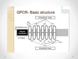
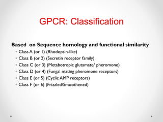
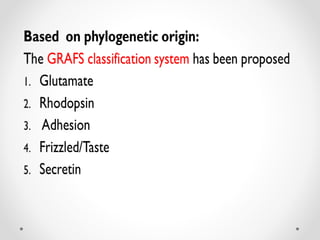
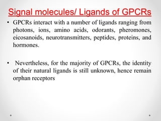
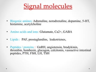
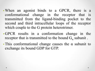
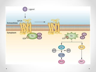
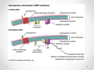
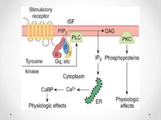
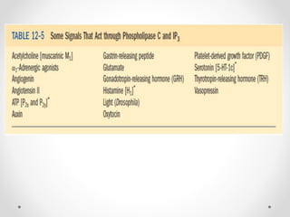
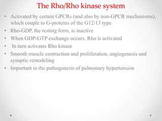
2. • G proteins, also known as guanine nucleotide-binding
proteins, involved in transmitting signals and function
as molecular switches.
• G protein-coupled receptors (GPCRs), also known
as seven-transmembrane domain receptors, 7T
receptors, serpentine receptor, and G protein-linked
receptors (GPLR),
• It constitute a large protein family of receptors that
sense molecules outside the cell and activate
inside signal transduction pathways and ultimately,
cellular responses.
3. • They are called seven-transmembrane receptors
because they pass through the cell membrane seven
times.
• Humans express over 800 GPCRs that make up the
third largest family of genes in humans.
• GPCRs are the targets for many drugs; perhaps half
of all non-antibiotic prescription drugs act at these
receptors.
4. Structure of G Protein
The structure of a GPCR can be divided into three
parts:
1.The extra-cellular region, consisting of the N terminus
and three extracellular loops (ECL1–ECL3);
2. The TM region, consisting of seven a-helices(TM1–
TM7)
3. The intracellular region, consisting of three intra-
cellular loops (ICL1–ICL3), an intracellular
amphipathic helix (H8), and the C terminus .
5.
6. G protein complexes are Made up
• 23 alpha (α)
• 7 beta (β)
• 12 gamma (γ) subunits.
• Beta and gamma subunits can form a stable dimeric
complex referred to as the beta-gamma complex
• In a broad sense, the extracellular region modulates
ligand access; the TM region forms the structural core,
binds ligands and transduces this information to the
intracellular region through conformational changes, and
the intracellular region interfaces with cytosolic signalling
proteins
• The α subunits fall into four families (Gs, Gi, Gq, and
G12/13) which are responsible for coupling GPCRs to
relatively distinct effectors.
7.
8. • GPCRs are responsible for every aspect of human biology
from vision, taste, sense of smell, sympathetic and
parasympathetic nervous functions, metabolism, and immune
regulation to reproduction.
• ~45% of all pharmaceutical drugs are known to target GPCRs
9. • Robert Lefkowitz and Brian Kobilka: the 2012 Nobel Prize in
Chemistry for groundbreaking discoveries that reveal the inner
workings of an important family of receptors: G-protein–
coupled receptors
12. Signal molecules/ Ligands of GPCRs
• GPCRs interact with a number of ligands ranging from
photons, ions, amino acids, odorants, pheromones,
eicosanoids, neurotransmitters, peptides, proteins, and
hormones.
• Nevertheless, for the majority of GPCRs, the identity
of their natural ligands is still unknown, hence remain
orphan receptors
18. • When an agonist binds to a GPCR, there is a
conformational change in the receptor that is
transmitted from the ligand-binding pocket to the
second and third intracellular loops of the receptor
which couple to the G protein heterotrimer.
• GPCR results in a conformation change in the
receptor that is transmitted to the bound Gα subunit .
• This conformational change causes the α subunit to
exchange its bound GDP for GTP.
19. • Binding of GTP activates the α subunit and causes it to
release both the βγ dimer and the receptor, and active
signaling molecules, both the GTP-bound α subunit and
the βγ heterodimer become and active signaling molecules
• The interaction of the agonist-bound GPCR with the G
protein is transient; following activation of one G protein,
the receptor is freed to interact with other G proteins.
20.
21. • There are two principal signal transduction pathways involving
the G protein–coupled receptors:
• the cAMP signal pathway and
• the phosphatidylinositol signal pathway
22. • Depending on the nature of the subunit, the active, GTP-bound
form binds to and regulates effectors such as adenylyl cyclase
(via Gs ) or phospholipase C (via Gq ).
• The subunit can regulate many effectors including ion
channels and enzymes such as PI3-K(phosphoinositide 3-
kinase).
• The G protein remains active until the GTP bound to the
subunit is hydrolyzed to GDP.
• The α subunit has a slow intrinsic rate of GTP hydrolysis that
is modulated by a family of proteins termed regulators of G
protein signaling (RGSs).
23. • The RGS proteins greatly accelerate the hydrolysis of GTP and are
potentially attractive drug targets.
• Once the GDP bound to the α subunit is hydrolyzed to GDP, the βγ
subunit and receptor recombine to form the inactive receptor-G
protein heterotrimer basal complex that can be reactivated by
another ligand-binding event
24. • The main targets for G-proteins, through which GPCRs
control different aspects of cell function are
• adenylyl cyclase, the enzyme responsible for cAMP
formation
• phospholipase C, the enzyme responsible for inositol
phosphate and diacylglycerol (DAG) formation
• ion channels, particularly calcium and potassium channels
• Rho A/Rho kinase, a system that controls the activity of
many signalling pathways controlling cell growth and
proliferation, smooth muscle contraction, etc.
25. The Adenylyl cyclase/cAMP system
• cAMP is a nucleotide
• Synthesized within the cell from ATP by membrane-bound,
adenylyl cyclase
• Produced continuously
• Inactivated by hydrolysis to 5´-AMP, by the
Phosphodiesterases
• Common mechanism, namely the activation of protein kinases
• Involved in:
Energy metabolism
Cell division and cell differentiation
Ion transport, ion channels
Contractile proteins in smooth muscle
26.
27.
28.
29. Nucleotide Exchange Factors (Gefs)
• Integrate extracellular signals from membrane receptors to
produce cytoskeletal changes
• this pathway provides an additional effector system for cAMP
signaling and drug action that can act independently or
cooperatively with PKA
• Activation of diverse signaling pathways, regulate :
Phagocytosis
Progression through the cell cycle
Cell adhesion
Gene expression
Apoptosis
30.
31. Phosphodiesterases
• Hydrolyze the cyclic 3',5'-phosphodiester bond in cAMP
and cGMP
• >50 different PDE proteins which are divided into 11
subfamilies
• Drug targets for :
Asthma
Cardiovascular diseases such as heart failure
Atherosclerotic coronary and peripheral arterial
disease
Neurological disorders
32. The Phospholipase C/ inositol phosphate
system
• 1950s by Hokin and Hokin
• PIP2 is the substrate for a membrane-bound enzyme,
phospholipase Cβ (PLCβ),
• Which splits it into DAG and inositol (1,4,5)
trisphosphate (IP3)
• Both function as second messengers
• IP3 receptor- a ligand-gated calcium channel present
on the membrane of the endoplasmic reticulum
33.
34. Diacylglycerol and protein kinase C
• DAG, unlike the inositol phosphates, is highly lipophilic and
remains within the membrane
• Binds to a specific site on the PKC molecule, which migrates from
the cytosol to the cell membrane in the presence of DAG, thereby
becoming activated
• 10 different mammalian PKC subtypes
• Kinases in general play a central role in signal transduction, and
control many different aspects of cell function
35.
36.
37. The Rho/Rho kinase system
• Activated by certain GPCRs (and also by non-GPCR mechanisms),
which couple to G-proteins of the G12/13 type
• Rho-GDP, the resting form, is inactive
• When GDP-GTP exchange occurs, Rho is activated
• In turn activates Rho kinase
• Smooth muscle contraction and proliferation, angiogenesis and
synaptic remodeling
• Important in the pathogenesis of pulmonary hypertension
38. Conclusion
• Nearly 40% of the drugs approved for marketing by the
FDA target GPCRs
• 800-1,000 different GPCRs and the drugs that are
marketed target less than 50 GPCRs
• GPCR will continue to be highly important in clinical
medicine because of their large number, wide expression
and role in physiologically important responses
• Future discoveries will reveal new GPCR drugs, in part
because it is relatively easy to screen for pharmacologic
agents that access these receptors and stimulate or block
receptor-mediated biochemical or physiological
responses