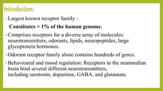
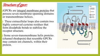
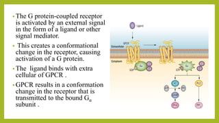
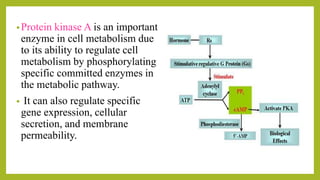
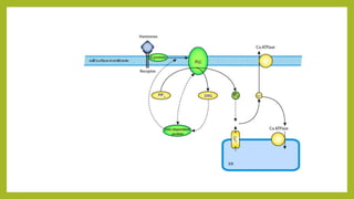
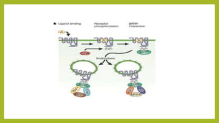
G-protein coupled receptors (GPCRs) are the largest family of membrane receptors found in humans. They have seven transmembrane domains and are activated by extracellular ligands binding, which causes a conformational change and activation of an intracellular G protein. This leads to second messenger signaling pathways involving cAMP or phosphatidylinositol. GPCRs regulate many physiological processes and are involved in behaviors, moods, and cellular responses. Their activity is modulated by phosphorylation, arrestins, and degradation of second messengers.
GPCRs are the most dynamic and most abundant all the receptors. The G protein-coupled receptor (GPCR) superfamily comprises the largest and most diverse group of proteins in mammals. GPCRs are responsible for every aspect of human biology from vision, taste, sense of smell, sympathetic and parasympathetic nervous functions, metabolism, and immune regulation to reproduction. GPCRs interact with a number of ligands ranging from photons, ions, amino acids, odorants, pheromones, eicosanoids, neurotransmitters, peptides, proteins, and hormones.
Nevertheless, for the majority of GPCRs, the identity of their natural ligands is still unknown, hence remain orphan receptors.
The simple dogma that underpins much of our current understanding of GPCRs, namely,
one GPCR gene− one GPCR protein− one functional GPCR− one G protein −one response
is showing distinct signs of wear.
General principles of signal transduction
G Protein-coupled Receptors (GPCRs): Structure and Mechanism.
GPCRs that Regulate Adenylyl Cyclase.
GPCRs that Activate Phospholipase C.
GPCRs that Regulate Ion Channels.
GPCRs that Regulate Gene Transcription.
Cell signaling / Signal Transduction / Transmembrane signaling.
It is the process by which cells communicate with their environment and respond to external stimuli.
When a signaling molecule(ligand) binds to its receptor, it alters the shape or activity of the receptor, triggering a change inside of the cell such as alteration in the activity of a gene / cell division. Thus the original Intercellular Signal is converted into an Intracellular Signal that triggers as a response.
This Presentation provides an outline knowledge about Cellular Communication, Steps involved, Its Types, Signal Transduction, Secondary Messenger , Receptors with some Interesting Facts and Current Trends. An assignment for the subject, Cellular and Molecular Pharmacology, 1st year M.Pharm, 1st semester.
GPCRs are the largest and most diverse group of integral membrane proteins. These proteins are used by cells to convert extracellular signals into intracellular responses and mediate most of our physiological responses to hormones, neurotransmitters as well as responses to vision, olfaction and taste signal. They mediate most of our and environmental stimulants, and so have a great potential as therapeutic targets for a broad spectrum of diseases. At the most basic level, all GPCRS are characterized by the presence of seven membrane-spanning alpha helical segments separated by alternating intracellular and extracellular loop regions. Coupling with G proteins, they are called seven-transmembrane receptors because they pass through the cell membrane seven times.
1. INTRODUCTION
2. WHAT IS A RECEPTOR
3. HISTORY
4. CONCEPT OF CELL SIGNALLING
5. RECEPTOR SUPER FAMILIES
6. GPCRs- SIGNAL TRANSDUCTION & ITS SECOND MESSENGERS
This presentation is about the functioning of G-Protein coupled receptors. It also gives necessary information about the G-protein and it functions. It ends by explaining some of the faults associated with GPCR (G-PROTEIN COUPLED RECEPTORS).
New Directions in Targeted Therapeutic Approaches for Older Adults With Mantl...i3 Health
i3 Health is pleased to make the speaker slides from this activity available for use as a non-accredited self-study or teaching resource.
This slide deck presented by Dr. Kami Maddocks, Professor-Clinical in the Division of Hematology and
Associate Division Director for Ambulatory Operations
The Ohio State University Comprehensive Cancer Center, will provide insight into new directions in targeted therapeutic approaches for older adults with mantle cell lymphoma.
STATEMENT OF NEED
Mantle cell lymphoma (MCL) is a rare, aggressive B-cell non-Hodgkin lymphoma (NHL) accounting for 5% to 7% of all lymphomas. Its prognosis ranges from indolent disease that does not require treatment for years to very aggressive disease, which is associated with poor survival (Silkenstedt et al, 2021). Typically, MCL is diagnosed at advanced stage and in older patients who cannot tolerate intensive therapy (NCCN, 2022). Although recent advances have slightly increased remission rates, recurrence and relapse remain very common, leading to a median overall survival between 3 and 6 years (LLS, 2021). Though there are several effective options, progress is still needed towards establishing an accepted frontline approach for MCL (Castellino et al, 2022). Treatment selection and management of MCL are complicated by the heterogeneity of prognosis, advanced age and comorbidities of patients, and lack of an established standard approach for treatment, making it vital that clinicians be familiar with the latest research and advances in this area. In this activity chaired by Michael Wang, MD, Professor in the Department of Lymphoma & Myeloma at MD Anderson Cancer Center, expert faculty will discuss prognostic factors informing treatment, the promising results of recent trials in new therapeutic approaches, and the implications of treatment resistance in therapeutic selection for MCL.
Target Audience
Hematology/oncology fellows, attending faculty, and other health care professionals involved in the treatment of patients with mantle cell lymphoma (MCL).
Learning Objectives
1.) Identify clinical and biological prognostic factors that can guide treatment decision making for older adults with MCL
2.) Evaluate emerging data on targeted therapeutic approaches for treatment-naive and relapsed/refractory MCL and their applicability to older adults
3.) Assess mechanisms of resistance to targeted therapies for MCL and their implications for treatment selection
TEST BANK for Operations Management, 14th Edition by William J. Stevenson, Ve...kevinkariuki227
TEST BANK for Operations Management, 14th Edition by William J. Stevenson, Verified Chapters 1 - 19, Complete Newest Version.pdf
Lung Cancer: Artificial Intelligence, Synergetics, Complex System Analysis, S...Oleg Kshivets
RESULTS: Overall life span (LS) was 2252.1±1742.5 days and cumulative 5-year survival (5YS) reached 73.2%, 10 years – 64.8%, 20 years – 42.5%. 513 LCP lived more than 5 years (LS=3124.6±1525.6 days), 148 LCP – more than 10 years (LS=5054.4±1504.1 days).199 LCP died because of LC (LS=562.7±374.5 days). 5YS of LCP after bi/lobectomies was significantly superior in comparison with LCP after pneumonectomies (78.1% vs.63.7%, P=0.00001 by log-rank test). AT significantly improved 5YS (66.3% vs. 34.8%) (P=0.00000 by log-rank test) only for LCP with N1-2. Cox modeling displayed that 5YS of LCP significantly depended on: phase transition (PT) early-invasive LC in terms of synergetics, PT N0—N12, cell ratio factors (ratio between cancer cells- CC and blood cells subpopulations), G1-3, histology, glucose, AT, blood cell circuit, prothrombin index, heparin tolerance, recalcification time (P=0.000-0.038). Neural networks, genetic algorithm selection and bootstrap simulation revealed relationships between 5YS and PT early-invasive LC (rank=1), PT N0—N12 (rank=2), thrombocytes/CC (3), erythrocytes/CC (4), eosinophils/CC (5), healthy cells/CC (6), lymphocytes/CC (7), segmented neutrophils/CC (8), stick neutrophils/CC (9), monocytes/CC (10); leucocytes/CC (11). Correct prediction of 5YS was 100% by neural networks computing (area under ROC curve=1.0; error=0.0).
CONCLUSIONS: 5YS of LCP after radical procedures significantly depended on: 1) PT early-invasive cancer; 2) PT N0--N12; 3) cell ratio factors; 4) blood cell circuit; 5) biochemical factors; 6) hemostasis system; 7) AT; 8) LC characteristics; 9) LC cell dynamics; 10) surgery type: lobectomy/pneumonectomy; 11) anthropometric data. Optimal diagnosis and treatment strategies for LC are: 1) screening and early detection of LC; 2) availability of experienced thoracic surgeons because of complexity of radical procedures; 3) aggressive en block surgery and adequate lymph node dissection for completeness; 4) precise prediction; 5) adjuvant chemoimmunoradiotherapy for LCP with unfavorable prognosis.
These simplified slides by Dr. Sidra Arshad present an overview of the non-respiratory functions of the respiratory tract.
Learning objectives:
1. Enlist the non-respiratory functions of the respiratory tract
2. Briefly explain how these functions are carried out
3. Discuss the significance of dead space
4. Differentiate between minute ventilation and alveolar ventilation
5. Describe the cough and sneeze reflexes
Study Resources:
1. Chapter 39, Guyton and Hall Textbook of Medical Physiology, 14th edition
2. Chapter 34, Ganong’s Review of Medical Physiology, 26th edition
3. Chapter 17, Human Physiology by Lauralee Sherwood, 9th edition
4. Non-respiratory functions of the lungs https://academic.oup.com/bjaed/article/13/3/98/278874
These lecture slides, by Dr Sidra Arshad, offer a quick overview of physiological basis of a normal electrocardiogram.
Learning objectives:
1. Define an electrocardiogram (ECG) and electrocardiography
2. Describe how dipoles generated by the heart produce the waveforms of the ECG
3. Describe the components of a normal electrocardiogram of a typical bipolar leads (limb II)
4. Differentiate between intervals and segments
5. Enlist some common indications for obtaining an ECG
Study Resources:
1. Chapter 11, Guyton and Hall Textbook of Medical Physiology, 14th edition
2. Chapter 9, Human Physiology - From Cells to Systems, Lauralee Sherwood, 9th edition
3. Chapter 29, Ganong’s Review of Medical Physiology, 26th edition
4. Electrocardiogram, StatPearls - https://www.ncbi.nlm.nih.gov/books/NBK549803/
5. ECG in Medical Practice by ABM Abdullah, 4th edition
6. ECG Basics, http://www.nataliescasebook.com/tag/e-c-g-basics
Ozempic: Preoperative Management of Patients on GLP-1 Receptor Agonists Saeid Safari
Preoperative Management of Patients on GLP-1 Receptor Agonists like Ozempic and Semiglutide
ASA GUIDELINE
NYSORA Guideline
2 Case Reports of Gastric Ultrasound
Report Back from SGO 2024: What’s the Latest in Cervical Cancer?bkling
Are you curious about what’s new in cervical cancer research or unsure what the findings mean? Join Dr. Emily Ko, a gynecologic oncologist at Penn Medicine, to learn about the latest updates from the Society of Gynecologic Oncology (SGO) 2024 Annual Meeting on Women’s Cancer. Dr. Ko will discuss what the research presented at the conference means for you and answer your questions about the new developments.
Ethanol (CH3CH2OH), or beverage alcohol, is a two-carbon alcohol
that is rapidly distributed in the body and brain. Ethanol alters many
neurochemical systems and has rewarding and addictive properties. It
is the oldest recreational drug and likely contributes to more morbidity,
mortality, and public health costs than all illicit drugs combined. The
5th edition of the Diagnostic and Statistical Manual of Mental Disorders
(DSM-5) integrates alcohol abuse and alcohol dependence into a single
disorder called alcohol use disorder (AUD), with mild, moderate,
and severe subclassifications (American Psychiatric Association, 2013).
In the DSM-5, all types of substance abuse and dependence have been
combined into a single substance use disorder (SUD) on a continuum
from mild to severe. A diagnosis of AUD requires that at least two of
the 11 DSM-5 behaviors be present within a 12-month period (mild
AUD: 2–3 criteria; moderate AUD: 4–5 criteria; severe AUD: 6–11 criteria).
The four main behavioral effects of AUD are impaired control over
drinking, negative social consequences, risky use, and altered physiological
effects (tolerance, withdrawal). This chapter presents an overview
of the prevalence and harmful consequences of AUD in the U.S.,
the systemic nature of the disease, neurocircuitry and stages of AUD,
comorbidities, fetal alcohol spectrum disorders, genetic risk factors, and
pharmacotherapies for AUD.
MANAGEMENT OF ATRIOVENTRICULAR CONDUCTION BLOCK.pdfJim Jacob Roy
Cardiac conduction defects can occur due to various causes.
Atrioventricular conduction blocks ( AV blocks ) are classified into 3 types.
This document describes the acute management of AV block.
3. Introduction:
•Largest known receptor family –
Constitutes > 1% of the human genome.
•Comprises receptors for a diverse array of molecules:
neurotransmitters, odorants, lipids, neuropeptides, large
glycoprotein hormones.
•Odorant receptor family alone contains hundreds of genes.
•Behavioural and mood regulation: Receptors in the mammalian
brain bind several different neurotransmitters,
including serotonin, dopamine, GABA, and glutamate.
4. Structureof gpcr:
GPCRs are integral membrane proteins that
possess seven membrane-spanning domains
or transmembrane helices.
These extracellular loops also contain two
highly conserved cysteine residues that
form disulphide bonds to stabilize the
receptor structure.
Some seven-transmembrane helix proteins
(channel rhodopsin) that resemble GPCRs
may contain ion channels, within their
protein.
6. • The G protein-coupled receptor
is activated by an external signal
in the form of a ligand or other
signal mediator.
• This creates a conformational
change in the receptor, causing
activation of a G protein.
• The ligand binds with extra
cellular of GPCR .
• GPCR results in a conformation
change in the receptor that is
transmitted to the bound Gα
subunit .
7.
8. • There are two principal signal transduction pathways involving the G
protein–coupled receptors:
•the cAMP signal pathway and
•the phosphatidylinositol signal pathway
9. • The cAMP signal transduction contains:
• Stimulative hormone receptor (Rs) is a receptor that can bind with
stimulative signal molecules, while inhibitory hormone receptor (Ri)
is a receptor that can bind with inhibitory signal molecules.
• Adenylyl cyclase is a 12-transmembrane glycoprotein that catalyzes
ATP to form cAMP with the help of cofactor Mg2+ or Mn2+.
• The cAMP produced is a second messenger in cellular metabolism
and is an allosteric activator of protein kinase A
10. •Protein kinase A is an important
enzyme in cell metabolism due
to its ability to regulate cell
metabolism by phosphorylating
specific committed enzymes in
the metabolic pathway.
• It can also regulate specific
gene expression, cellular
secretion, and membrane
permeability.
11. • In the phosphatidylinositol signal pathway, the extracellular signal
molecule binds with the G-protein receptor (Gq) on the cell surface
and activates phospholipase C, which is located on the plasma
membrane.
• IP3 binds to IP3 receptor in the membrane of the smooth ER and
mitochondria to open Ca2+ channels.
• DAG helps activate protein kinase C (PKC), which phosphorylates
many other proteins, changing their catalytic activities, leading to
cellular responses.
12.
13. GPCR functions :
a) generation of second messengers including cGMP and IP3, which
stimulate phosphorylation reactions, causing release of second-
messenger calcium ions from storage in ER.
b) generation of cAMP and activation of the transcription factor,
cAMP response element binding protein (CREB) to stimulate gene
transcription.
c) regulation of gene transcription
d) chemotaxis
e) ion channel opening (conformational change) in response to
neurotransmitters
14. Regulationof gpcr:
•Members of arrestin/beta-arrestin protein family are thought
to participate in agonist-mediated desensitization of G
protein-coupled receptors and cause specific dampening of
cellular responses to stimuli such as hormones,
neurotransmitters, or sensory signals.
•Arrestin beta 1 is a cytosolic protein and acts as a cofactor in
the beta-adrenergic receptor kinase (BARK) mediated
desensitization of beta-adrenergic receptors
15.
16. •The Gs alpha subunit slowly catalyses the hydrolysis of GTP to
GDP, which in turn deactivates the Gs protein, shutting off the
cAMP pathway.
•The pathway may also be deactivated downstream by directly
inhibiting adenylyl cyclase or dephosphorylating the proteins
phosphorylated by PKA.
Molecules that inhibit the cAMP pathway include:
•cAMP phosphodiesterase dephosphorylates cAMP into AMP,
reducing the cAMP levels