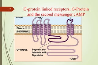
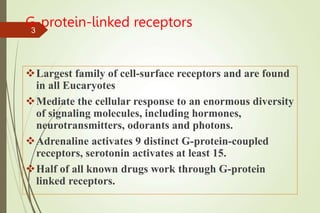
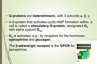
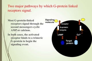
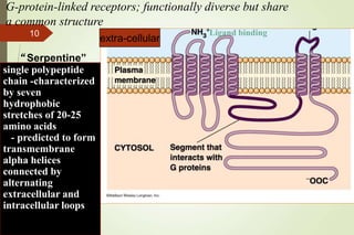
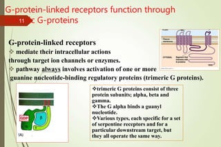
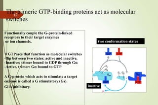
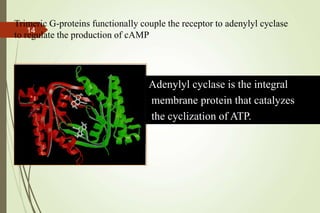
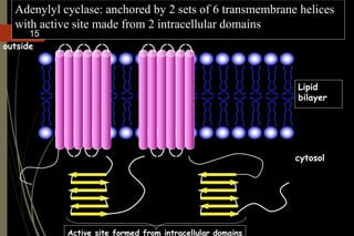
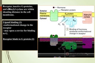
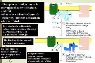
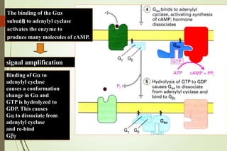
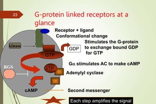
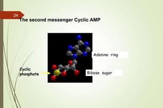
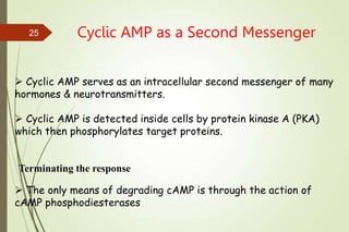
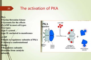
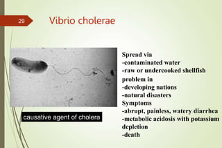
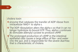
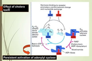
Shri Sharda Bhavan Education Society’s
Nanded Pharmacy College
Lecture on,
G-Protein Coupled
Receptors
By
Dr. Shriniwas K. Sarje
Asso. Professor & HOD Department of Pharmacology
1
G-protein linked receptors, G-Protein
and the second messenger cAMP
2
G-protein-linked receptors
Largest family of cell-surface receptors and are found
in all Eucaryotes
Mediate the cellular response to an enormous diversity
of signaling molecules, including hormones,
neurotransmitters, odorants and photons.
Adrenaline activates 9 distinct G-protein-coupled
receptors, serotonin activates at least 15.
Half of all known drugs work through G-protein
linked receptors.
3
Rhodopsin PDB 1F88
G Protein Signal Cascade
Most signal molecules targeted to a cell bind at the cell surface to receptors
embedded in the plasma membrane.
Only signal molecules able to cross the
plasma membrane (e.g., steroid hormones)
interact with intracellular receptors.
A large family of cell surface receptors
have a common structural motif, 7
transmembrane a-helices.
Rhodopsin was the first of these to have its
7-helix structure confirmed by X-ray
crystallography.
4
 Rhodopsin is unique.
It senses light, via a bound
chromophore, retinal.
 Most 7-helix receptors have domains
facing the extracellular side of the
plasma membrane that recognize & bind
signal molecules (ligands).
E.g., the b-adrenergic receptor is
activated by epinephrine &
norepinephrine.
b-Adrenergic
Receptor
PDB 2RH1
Lysozyme
insert
ligand 
5
The signal is usually passed from a 7-helix receptor to an
intracellular G-protein.
 Seven-helix receptors are thus called GPCR, or G-
Protein-Coupled Receptors.
 Approx. 800 different GPCRs are encoded in the human
genome.
6
G-protein-Coupled Receptors may dimerize or form oligomeric
complexes within the membrane.
Ligand binding may promote oligomerization, which may in turn
affect activity of the receptor.
Various GPCR-interacting proteins (GIPs) modulate receptor
function. Effects of GIPs may include:
 altered ligand affinity
 receptor dimerization or oligomerization
 control of receptor localization, including transfer to or removal
from the plasma membrane
 promoting close association with other signal proteins 7
 G-proteins are heterotrimeric, with 3 subunits a, b, g.
 A G-protein that activates cyclic-AMP formation within a
cell is called a stimulatory G-protein, designated Gs
with alpha subunit Gsa.
 Gs is activated, e.g., by receptors for the hormones
epinephrine and glucagon.
The b-adrenergic receptor is the GPCR for
epinephrine.
8
Two major pathways by which G-protein linked
receptors signal.
Most G-protein-linked
receptors signal through the
second messengers cyclic
AMP or calcium.
In both cases, the activated
receptor binds to a trimeric
G-protein to begin the
signaling event.
Signaling
molecule
receptor
G-protein
enzyme
cAMP Ca2+
Target
protein
9
extra-cellular
G-protein-linked receptors; functionally diverse but share
a common structure
single polypeptide
chain -characterized
by seven
hydrophobic
stretches of 20-25
amino acids
- predicted to form
transmembrane
alpha helices
connected by
alternating
extracellular and
intracellular loops
“Serpentine”
Ligand binding
10
G-protein-linked receptors function through
trimeric G-proteins
G-protein-linked receptors
 mediate their intracellular actions
through target ion channels or enzymes.
 pathway always involves activation of one or more
guanine nucleotide-binding regulatory proteins (trimeric G proteins).
trimeric G proteins consist of three
protein subunits; alpha, beta and
gamma.
The G alpha binds a guanyl
nucleotide.
Various types, each specific for a set
of serpentine receptors and for a
particular downstream target, but
they all operate the same way.
11
Functionally couple the G-protein-linked
receptors to their target enzymes
or ion channels.
GTPases that function as molecular switches
-flip between two states: active and inactive.
-Inactive; trimer bound to GDP through Ga
-Active, trimer: Ga bound to GTP
A G-protein which acts to stimulate a target
enzyme is called a G stimulatory (Gs).
Gi is inhibitory.
The trimeric GTP-binding proteins act as molecular
switches
active
inactive
two conformation states
12
The structure of a trimeric G-protein bound to GDP based
on x-ray crystallographic analysis
Alpha subunit
Gamma subunit
Beta subunit
Binding of GTP induces large
conformational changes
in the switch regions leading to
dissociation of the subunits.
13
Adenylyl cyclase is the integral
membrane protein that catalyzes
the cyclization of ATP.
Trimeric G-proteins functionally couple the receptor to adenylyl cyclase
to regulate the production of cAMP
14
Adenylyl cyclase: anchored by 2 sets of 6 transmembrane helices
with active site made from 2 intracellular domains
cytosol
outside
Lipid
bilayer
Active site formed from intracellular domains
15
Ligand binding (1)
-conformational change in the
receptor
- may open a crevice for binding
to G.
Receptor binds to G protein (2)
Receptor, inactive G-proteins,
and adenylyl cyclase are within
shouting distance in the cell
membrane.
16
Receptor activation results in
activation of adenylyl cyclase.
-indirect
-stimulates a trimeric G-protein
-trimeric G-proteins dissasemble
when activated.
Receptor binds to G-protein
induces conformational change (3)
GDP is replaced by GTP
Ga dissociates from Gbg
Ga then binds to
adenylyl cyclase (4),
activating synthesis
of cAMP
The binding site for adenylyl
cyclase is unmasked.
A single hormone/
receptor complex
stimulates the production
of many molecules of Gsa
17
Binding of Ga to
adenylyl cyclase
causes a conformation
change in Ga and
GTP is hydrolyzed to
GDP. This causes
Ga to dissociate from
adenylyl cyclase
and re-bind
Gbg
The binding of the Gas
subunit to adenylyl cyclase
activates the enzyme to
produce many molecules of cAMP.
signal amplification
18
Terminating the response
The hormone/receptor complex must be deactivated to return to
the unstimulated state.
-phosphorylation events on the carboxy terminal tail of the receptor
lead to the inactivation of the receptor.
Hydrolysis of GTP leads to inactivation of the trimeric G-protein.
-enhanced by RGS proteins (regulator of G-protein signaling).
19
Mammalian RGS proteins activate the GTPase
activities of G-protein alpha subunits
RGS proteins are GAPs (GTPase activating proteins).
-no effect on the time course of nucleotide binding
-but they stimulate the rate of GTP hydrolysis.
MODEL: RGS proteins accelerate GTP hydrolysis by preferentially
binding to and stabilizing G proteins in their transition state for the
hydrolysis reaction.
GTP hydrolysis
RGS protein
GDP
a
b g
GTP
b/g
a
20
Inhibitory G proteins
While the beta-adrenergic
receptors are functionally
coupled to G-stimulatory
proteins, the alpha-2
adrenergic receptors are
coupled to inhibitory G
proteins.
G i can contain the same beta/gamma
subunits as Gs, but the alpha
subunits are different.
G i inhibits adenylyl cyclase in an
indirect manner.
21
Hormone-induced activation and inhibition of adenylyl cyclase is
mediated by G-sa and G-ia
22
G-protein linked receptors at a
glance
GTP
Receptor + ligand
Conformational change
GDP
GTP
Adenylyl cyclase
cAMP Second messenger
Stimulates the G-protein
to exchange bound GDP
for GTP
Ga stimulates AC to make cAMP
kinase
Each step amplifies the signal
RGS
23
Adenine ring
Ribose sugar
Cyclic
phosphate
The second messenger Cyclic AMP
24
Cyclic AMP as a Second Messenger
 Cyclic AMP serves as an intracellular second messenger of many
hormones & neurotransmitters.
 Cyclic AMP is detected inside cells by protein kinase A (PKA)
which then phosphorylates target proteins.
 The only means of degrading cAMP is through the action of
cAMP phosphodiesterases
Terminating the response
25
The activation of PKA
inactive
cAMP
Binds to regulatory subunits of PKA
 induces a conformational
change
Regulatory subunits
dissociate from catalytic
subunits.
inactive
PKA
Serine/threonine kinase
Accounts for the effects
of cAMP in most cell types
Two types
-type I: cytosol
-type II: anchored to membranes
PKA
26
Triglyceride breakdown in fat mediated by adrenaline.
Increase in heart rate and force of contraction in heart
mediated by adrenaline.
Glycogen breakdown in muscle mediated by adrenaline
Changes in gene transcription
Some Hormone-induced Cellular Responses Mediated by cAMP
27
How does a rise in cAMP levels lead to
altered gene transcription?
Increase in cAMP activates PKA
PKA catalytic subunits move into
the nucleus
PKA phosphorylates the transcription
factor CREB (cAMP response
element binding protein).
Phosphorylated CREB binds to the co-
activator CBP
Transcription is activated.
28
Vibrio cholerae
causative agent of cholera
Spread via
-contaminated water
-raw or undercooked shellfish
problem in
-developing nations
-natural disasters
Symptoms
-abrupt, painless, watery diarrhea
-metabolic acidosis with potassium
depletion
-death
29
Cholera Toxin
cholera toxin
enzyme that catalyzes the transfer of ADP ribose from
intracellular NAD+ to alpha s.
The ADP ribosylation alters the alpha s so that it can no
longer hydrolyze its bound GTP. Thus, alpha s continues
to stimulate adenylyl cyclase to produce cAMP.
The prolonged production of cAMP in the intestinal
epithelial cells causes a large efflux of Na+ and water
into the gut, and is responsible for the severe diarrhea
that is characteristic of cholera.
30
Effect of cholera
toxin
Persistent activation of adenylyl cyclase
31
Summary
Gprotein-linked receptors
Mediate the cellular response to an enormous diversity of signaling
molecules, including hormones, neurotransmitters, odorants and photons
Signal through trimeric G-proteins
Trimeric G-proteins functionally couple their receptors to target enzymes
Some trimeric G-proteins activate adenylyl cyclase to synthesize cAMP
cAMP is an important second messenger that functions through
PKA to regulate metabolism and transcription.
32
THANK YOU
33

G-protein for lecture.ppt

  • 1.
    Shri Sharda BhavanEducation Society’s Nanded Pharmacy College Lecture on, G-Protein Coupled Receptors By Dr. Shriniwas K. Sarje Asso. Professor & HOD Department of Pharmacology 1
  • 2.
    G-protein linked receptors,G-Protein and the second messenger cAMP 2
  • 3.
    G-protein-linked receptors Largest familyof cell-surface receptors and are found in all Eucaryotes Mediate the cellular response to an enormous diversity of signaling molecules, including hormones, neurotransmitters, odorants and photons. Adrenaline activates 9 distinct G-protein-coupled receptors, serotonin activates at least 15. Half of all known drugs work through G-protein linked receptors. 3
  • 4.
    Rhodopsin PDB 1F88 GProtein Signal Cascade Most signal molecules targeted to a cell bind at the cell surface to receptors embedded in the plasma membrane. Only signal molecules able to cross the plasma membrane (e.g., steroid hormones) interact with intracellular receptors. A large family of cell surface receptors have a common structural motif, 7 transmembrane a-helices. Rhodopsin was the first of these to have its 7-helix structure confirmed by X-ray crystallography. 4
  • 5.
     Rhodopsin isunique. It senses light, via a bound chromophore, retinal.  Most 7-helix receptors have domains facing the extracellular side of the plasma membrane that recognize & bind signal molecules (ligands). E.g., the b-adrenergic receptor is activated by epinephrine & norepinephrine. b-Adrenergic Receptor PDB 2RH1 Lysozyme insert ligand  5
  • 6.
    The signal isusually passed from a 7-helix receptor to an intracellular G-protein.  Seven-helix receptors are thus called GPCR, or G- Protein-Coupled Receptors.  Approx. 800 different GPCRs are encoded in the human genome. 6
  • 7.
    G-protein-Coupled Receptors maydimerize or form oligomeric complexes within the membrane. Ligand binding may promote oligomerization, which may in turn affect activity of the receptor. Various GPCR-interacting proteins (GIPs) modulate receptor function. Effects of GIPs may include:  altered ligand affinity  receptor dimerization or oligomerization  control of receptor localization, including transfer to or removal from the plasma membrane  promoting close association with other signal proteins 7
  • 8.
     G-proteins areheterotrimeric, with 3 subunits a, b, g.  A G-protein that activates cyclic-AMP formation within a cell is called a stimulatory G-protein, designated Gs with alpha subunit Gsa.  Gs is activated, e.g., by receptors for the hormones epinephrine and glucagon. The b-adrenergic receptor is the GPCR for epinephrine. 8
  • 9.
    Two major pathwaysby which G-protein linked receptors signal. Most G-protein-linked receptors signal through the second messengers cyclic AMP or calcium. In both cases, the activated receptor binds to a trimeric G-protein to begin the signaling event. Signaling molecule receptor G-protein enzyme cAMP Ca2+ Target protein 9
  • 10.
    extra-cellular G-protein-linked receptors; functionallydiverse but share a common structure single polypeptide chain -characterized by seven hydrophobic stretches of 20-25 amino acids - predicted to form transmembrane alpha helices connected by alternating extracellular and intracellular loops “Serpentine” Ligand binding 10
  • 11.
    G-protein-linked receptors functionthrough trimeric G-proteins G-protein-linked receptors  mediate their intracellular actions through target ion channels or enzymes.  pathway always involves activation of one or more guanine nucleotide-binding regulatory proteins (trimeric G proteins). trimeric G proteins consist of three protein subunits; alpha, beta and gamma. The G alpha binds a guanyl nucleotide. Various types, each specific for a set of serpentine receptors and for a particular downstream target, but they all operate the same way. 11
  • 12.
    Functionally couple theG-protein-linked receptors to their target enzymes or ion channels. GTPases that function as molecular switches -flip between two states: active and inactive. -Inactive; trimer bound to GDP through Ga -Active, trimer: Ga bound to GTP A G-protein which acts to stimulate a target enzyme is called a G stimulatory (Gs). Gi is inhibitory. The trimeric GTP-binding proteins act as molecular switches active inactive two conformation states 12
  • 13.
    The structure ofa trimeric G-protein bound to GDP based on x-ray crystallographic analysis Alpha subunit Gamma subunit Beta subunit Binding of GTP induces large conformational changes in the switch regions leading to dissociation of the subunits. 13
  • 14.
    Adenylyl cyclase isthe integral membrane protein that catalyzes the cyclization of ATP. Trimeric G-proteins functionally couple the receptor to adenylyl cyclase to regulate the production of cAMP 14
  • 15.
    Adenylyl cyclase: anchoredby 2 sets of 6 transmembrane helices with active site made from 2 intracellular domains cytosol outside Lipid bilayer Active site formed from intracellular domains 15
  • 16.
    Ligand binding (1) -conformationalchange in the receptor - may open a crevice for binding to G. Receptor binds to G protein (2) Receptor, inactive G-proteins, and adenylyl cyclase are within shouting distance in the cell membrane. 16
  • 17.
    Receptor activation resultsin activation of adenylyl cyclase. -indirect -stimulates a trimeric G-protein -trimeric G-proteins dissasemble when activated. Receptor binds to G-protein induces conformational change (3) GDP is replaced by GTP Ga dissociates from Gbg Ga then binds to adenylyl cyclase (4), activating synthesis of cAMP The binding site for adenylyl cyclase is unmasked. A single hormone/ receptor complex stimulates the production of many molecules of Gsa 17
  • 18.
    Binding of Gato adenylyl cyclase causes a conformation change in Ga and GTP is hydrolyzed to GDP. This causes Ga to dissociate from adenylyl cyclase and re-bind Gbg The binding of the Gas subunit to adenylyl cyclase activates the enzyme to produce many molecules of cAMP. signal amplification 18
  • 19.
    Terminating the response Thehormone/receptor complex must be deactivated to return to the unstimulated state. -phosphorylation events on the carboxy terminal tail of the receptor lead to the inactivation of the receptor. Hydrolysis of GTP leads to inactivation of the trimeric G-protein. -enhanced by RGS proteins (regulator of G-protein signaling). 19
  • 20.
    Mammalian RGS proteinsactivate the GTPase activities of G-protein alpha subunits RGS proteins are GAPs (GTPase activating proteins). -no effect on the time course of nucleotide binding -but they stimulate the rate of GTP hydrolysis. MODEL: RGS proteins accelerate GTP hydrolysis by preferentially binding to and stabilizing G proteins in their transition state for the hydrolysis reaction. GTP hydrolysis RGS protein GDP a b g GTP b/g a 20
  • 21.
    Inhibitory G proteins Whilethe beta-adrenergic receptors are functionally coupled to G-stimulatory proteins, the alpha-2 adrenergic receptors are coupled to inhibitory G proteins. G i can contain the same beta/gamma subunits as Gs, but the alpha subunits are different. G i inhibits adenylyl cyclase in an indirect manner. 21
  • 22.
    Hormone-induced activation andinhibition of adenylyl cyclase is mediated by G-sa and G-ia 22
  • 23.
    G-protein linked receptorsat a glance GTP Receptor + ligand Conformational change GDP GTP Adenylyl cyclase cAMP Second messenger Stimulates the G-protein to exchange bound GDP for GTP Ga stimulates AC to make cAMP kinase Each step amplifies the signal RGS 23
  • 24.
    Adenine ring Ribose sugar Cyclic phosphate Thesecond messenger Cyclic AMP 24
  • 25.
    Cyclic AMP asa Second Messenger  Cyclic AMP serves as an intracellular second messenger of many hormones & neurotransmitters.  Cyclic AMP is detected inside cells by protein kinase A (PKA) which then phosphorylates target proteins.  The only means of degrading cAMP is through the action of cAMP phosphodiesterases Terminating the response 25
  • 26.
    The activation ofPKA inactive cAMP Binds to regulatory subunits of PKA  induces a conformational change Regulatory subunits dissociate from catalytic subunits. inactive PKA Serine/threonine kinase Accounts for the effects of cAMP in most cell types Two types -type I: cytosol -type II: anchored to membranes PKA 26
  • 27.
    Triglyceride breakdown infat mediated by adrenaline. Increase in heart rate and force of contraction in heart mediated by adrenaline. Glycogen breakdown in muscle mediated by adrenaline Changes in gene transcription Some Hormone-induced Cellular Responses Mediated by cAMP 27
  • 28.
    How does arise in cAMP levels lead to altered gene transcription? Increase in cAMP activates PKA PKA catalytic subunits move into the nucleus PKA phosphorylates the transcription factor CREB (cAMP response element binding protein). Phosphorylated CREB binds to the co- activator CBP Transcription is activated. 28
  • 29.
    Vibrio cholerae causative agentof cholera Spread via -contaminated water -raw or undercooked shellfish problem in -developing nations -natural disasters Symptoms -abrupt, painless, watery diarrhea -metabolic acidosis with potassium depletion -death 29
  • 30.
    Cholera Toxin cholera toxin enzymethat catalyzes the transfer of ADP ribose from intracellular NAD+ to alpha s. The ADP ribosylation alters the alpha s so that it can no longer hydrolyze its bound GTP. Thus, alpha s continues to stimulate adenylyl cyclase to produce cAMP. The prolonged production of cAMP in the intestinal epithelial cells causes a large efflux of Na+ and water into the gut, and is responsible for the severe diarrhea that is characteristic of cholera. 30
  • 31.
    Effect of cholera toxin Persistentactivation of adenylyl cyclase 31
  • 32.
    Summary Gprotein-linked receptors Mediate thecellular response to an enormous diversity of signaling molecules, including hormones, neurotransmitters, odorants and photons Signal through trimeric G-proteins Trimeric G-proteins functionally couple their receptors to target enzymes Some trimeric G-proteins activate adenylyl cyclase to synthesize cAMP cAMP is an important second messenger that functions through PKA to regulate metabolism and transcription. 32
  • 33.