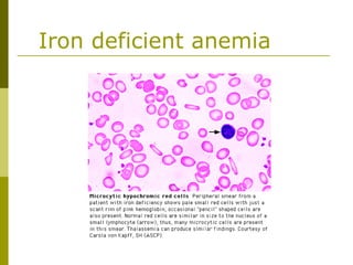
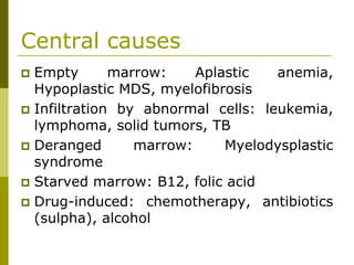
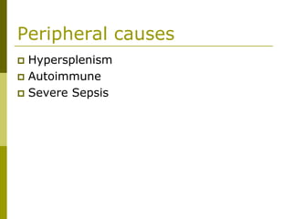
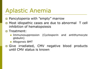
This document provides an overview of hematology and approaches to common blood disorders. It defines anemia and describes how to classify it based on mean corpuscular volume. Common causes of microcytic, macrocytic, and normocytic anemia are outlined. Diagnostic criteria and treatments for iron deficiency anemia, anemia of chronic disease, and other specific anemias are summarized. The document also reviews approaches to thrombocytopenia, neutropenia, pancytopenia, and transfusion reactions.
Immuno-Oncology: An Evolving Approach to Cancer Care
Review a downloadable slide deck by Thomas F. Gajewski, MD, PhD, covering the most clinically relevant new data reported from Immuno-Oncology: An Evolving Approach to Cancer Care.
Target Audience
This activity is designed to meet the educational needs of oncologists and other healthcare professionals involved in cancer care.
Format: Microsoft PowerPoint (.ppt) | File size: 26.2 MB | Date posted: 6/20/2012
Slide Deck Disclaimer
This slide deck in its original and unaltered format is for educational purposes and is current as of June 2012. All materials contained herein reflect the views of the faculty, and not those of IMER, the CE provider, or the commercial supporter. These materials may discuss therapeutic products that have not been approved by the US Food and Drug Administration and off-label uses of approved products. Readers should not rely on this information as a substitute for professional medical advice, diagnosis, or treatment. The use of any information provided is solely at your own risk, and readers should verify the prescribing information and all data before treating patients or employing any therapeutic products described in this educational activity.
Usage Rights
This slide deck is provided for educational purposes and individual slides may be used for personal, non-commercial presentations only if the content and references remain unchanged. No part of this slide deck may be published in print or electronically as a promotional or certified educational activity without prior written permission from IMER. Additional terms may apply. See Terms of Service on IMERonline.com for details.
Plasma cell disorders is a difficult topic where most residents and students confuse with regarding to differentiating between various types of para-proteinemias or plasma cell dyscrasias. This simple presentation will highlight the key points in differentiating, diagnosing these orders. Initial management principles are discussed as well.
Co-Chairs Srdan Verstovsek, MD, PhD, and Ruben A. Mesa, MD, FACP, prepared useful Practice Aids pertaining to myelofibrosis for this CME activity titled “Understanding the Clinical Spectrum of Myelofibrosis: Expert Perspectives on Molecular Biology, JAK Inhibitors, and Emerging Therapeutics.” For the full presentation, downloadable Practice Aids, and complete CME information, and to apply for credit, please visit us at https://bit.ly/3wzK6zG. CME credit will be available until October 9, 2022.
CAR-T cells (Chimeric Antigen Receptor- T cells) are T cells that have been genetically engineered to produce an artificial T cell receptor for use in immunotherapy. Chimeric antigen receptors are receptor proteins that have been engineered to give T cells the new ability to target a specific protein.
This therapy use to treat several type of cancer but significantly treat leukemia. And this therapy is very effective than other.
Types of immunotherapy
Oncology
cancer vaccines
adoptive T cell transfer
oncolytic viruses
monoclonal antibodies
cytokine
treatment of cancer with immunotherapy
This slideshare conatins detailed overview of immunotheraphy,humanisation of antibodies and its clinical application
this is the topic from cellular and molecular pharmacology of m pharmacy first year
immunotheraphy is further classified to its various types which has been discussed individually
its also conatins various immunotheraphy drugs which has other clinical advantages
In the work-up of a disease, the peripheral smear gives a very good information of what disease the patient may be suffering from. It also gives direction for further evaluation, all impacting the treatment and well being of the patient.
Slideshow is from the University of Michigan Medical
School's M1 Infectious Disease / Microbiology sequence
View additional course materials on Open.Michigan:
openmi.ch/med-M1IDM
Chapter 19- el filibusterismo published in ghent(1891)Alvin Bugaoisan
El filibusterismo summary:
Ang nobela ay nagsisimula sa isang paglalakbay ng isang bapor, ang Bapor Tabo. Doon ipinakilala ang ilang tauhan ng nobela na si Simoun, Isagani, at Basilio. Si Crisostomo Ibarra, ang bida sa Noli Me Tangere, ay nagbalik sa Pilipinas at nagbalatkayo bilang isang mayamang alahero na nagngangalang Simoun. Taglay ang poot at layong makapaghiganti at iligtas si Maria Clara sa kumbento, naglunsad si Simoun ng mga plano upang bulukin at pahinain ang pamahalaan upang maging sanhi ng himagsikan.
Lihim at masinop siyang nagbalak at nakipagkuntsaba sa iba't ibang tauhan sa nobela, kabilang na si Basilio. Una, binalak niyang manghimagsik at manggulo sa isang pulutong na sapilitang magbubukas sa kumbento ng Santa Clara upang agawin si Maria Clara. Ngunit hindi natuloy ang planong ito sapagkat namatay nang hapong iyon si Maria Clara. Pangalawa, nagkaroon ng pagkakataon si Simoun sa kasal ni Paulita Gomez at Juanito Pelaez kung saan dadalo ang lahat ng makapangyarihan sa pamahalaan. Niregaluhan ni Simoun ang ikinasal ng isang magarang lamparang may hugis granada na kasinlaki ng ulo ng tao.Lingid sa kaalaman ng lahat, ang ilawang ito ay nagtataglay ng granada na kapag itataas ang mitsa upang paliwanagin ay sasabog ito. Sa kasawiang palad at sa pangalawang pagkakataon, hindi natuloy ang balak na ito ni Simoun sapagkat nalaman ni Isagani ang maitim na balak na ito at mabilis na inihagis ang ilawan sa ilog.
Matapos ang pangyayari, namundok si Simoun dala ang kaniyang mga alahas at nakipagkita kay Padre Florentino. Nangumpisal si Simoun at pinatawad naman ng pari. Uminom si Simoun ng lason upang hindi mahuli ng mga guardia sibil na buhay. Nagwakas ang nobela nang ihagis ng pari ang kayamanan ni Simoun sa dagat at umasang matatagpuan iyon at magagamit para sa kabutihan ng taumbayan.
Instructions for Submissions thorugh G- Classroom.pptxJheel Barad
This presentation provides a briefing on how to upload submissions and documents in Google Classroom. It was prepared as part of an orientation for new Sainik School in-service teacher trainees. As a training officer, my goal is to ensure that you are comfortable and proficient with this essential tool for managing assignments and fostering student engagement.
A Strategic Approach: GenAI in EducationPeter Windle
Artificial Intelligence (AI) technologies such as Generative AI, Image Generators and Large Language Models have had a dramatic impact on teaching, learning and assessment over the past 18 months. The most immediate threat AI posed was to Academic Integrity with Higher Education Institutes (HEIs) focusing their efforts on combating the use of GenAI in assessment. Guidelines were developed for staff and students, policies put in place too. Innovative educators have forged paths in the use of Generative AI for teaching, learning and assessments leading to pockets of transformation springing up across HEIs, often with little or no top-down guidance, support or direction.
This Gasta posits a strategic approach to integrating AI into HEIs to prepare staff, students and the curriculum for an evolving world and workplace. We will highlight the advantages of working with these technologies beyond the realm of teaching, learning and assessment by considering prompt engineering skills, industry impact, curriculum changes, and the need for staff upskilling. In contrast, not engaging strategically with Generative AI poses risks, including falling behind peers, missed opportunities and failing to ensure our graduates remain employable. The rapid evolution of AI technologies necessitates a proactive and strategic approach if we are to remain relevant.
Palestine last event orientationfvgnh .pptxRaedMohamed3
An EFL lesson about the current events in Palestine. It is intended to be for intermediate students who wish to increase their listening skills through a short lesson in power point.
Welcome to TechSoup New Member Orientation and Q&A (May 2024).pdfTechSoup
In this webinar you will learn how your organization can access TechSoup's wide variety of product discount and donation programs. From hardware to software, we'll give you a tour of the tools available to help your nonprofit with productivity, collaboration, financial management, donor tracking, security, and more.
4. Approach to anemia
Definition (The WHO criteria):
Men: Hb <13.0 g/dL or Ht<40%
Women: Hb <12.0 g/dL or Ht<36%
Useful measurements:
Mean corpuscular volume (MCV): 80 to 100 fl
RDW (red cell distribution width): increased
RDW indicates the presence of cells of widely
differing sizes
Reticulocytes: suggestive of regeneration
5. Approach to Anemia
Microcytic:
MCV low, RDW high - iron deficiency
MCV low, RDW normal – thalassemia
Macrocytic:
MCV very high - B12 or folate deficiency
Normocytic:
MCV normal
6. Morphologic approach
Microcytic anemia – MCV < 80 fl
Reduced iron availability — severe iron deficiency, the
anemia of chronic disease, copper deficiency
Reduced heme synthesis — lead poisoning, congenital or
acquired sideroblastic anemia
Reduced globin production — thalassemic states, other
hemoglobinopathies
The three most common causes of microcytosis in
clinical practice are
iron deficiency (↓ iron stores)
alpha or beta thalassemia minor (often N or ↑ iron stores)
anemia of chronic disease (hepatoma, RCC)
7. Morphologic approach
Macrocytic anemia — MCV > 100 fL
Reticulocytosis
Abnormal nucleic acid metabolism of erythroid
precursors (eg, folate or vitamin 12 deficiency and
drugs interfering with nucleic acid synthesis, such
as zidovudine, hydroxyurea and Septra)
Abnormal RBC maturation (eg, myelodysplastic
syndrome, acute leukemia, LGL leukemia)
Other common causes:
alcohol abuse
liver disease
hypothyroidism
8. Morphologic approach
Normocytic anemia – MCV normal
Increased distruction
Reduced production (bone marrow
supression):
Bone marrow invasion (myelofibrosis, multiple
myeloma)
Myelodysplastic syndromes
Aplastic anemia
Acute blood loss
Chronic renal failure
9. Iron deficient anemia
Diagnosis:
↓ serum iron
↑ TIBC
↓ Transferrin saturation <20%
↓ Ferritin < 10
Iron replacement requires normalization of
the hemoglobin and the body stores
ALWAYS A SYMPTOM – LOOK FOR THE
CAUSE
11. Anemia of Chronic Disease
Common in patients with infection, cancer,
inflammatory and rheumatologic diseases
Iron can not be remobilized from storage
Blunted production of erythropoietin and
response to erythropoietin
Usually normocytic and normochromic but may
be microcytic if severe
TIBC ↓, Iron ↓, Transferrin saturation ↓, Ferritin↑
13. Sideroblastic Anemia
Two common features:
Ring sideroblasts in the bone marrow (abnormal
normoblasts with excessive accumulation of iron in the
mitochondria)
Impaired heme biosynthesis
Produces a “dimorphic” blood film with
microcytes and macrocytes
Usually acquired:
Myelodysplastic syndrome
Drugs (Ethanol, INH)
Toxins (Lead, zinc)
Nutritional (Pyridoxine deficiency, copper deficiency)
15. Thalassemia
Anemia 2º reduced or absent production
of one or more globin chains
Poikilocytosis (variation in shape) and
basophilic stippling may be seen in the
blood film
Hemoglobin electrophoresis is only
diagnostic for beta-thalassemia and may
not be diagnostic if iron deficiency is also
present
Hb H (β-4) prep or DNA analysis is needed
to diagnose alpha-thalassemia
17. Sickle Cell Disease
Inherited anemia (HbS)
Acute crisis management includes fluids,
oxygen and pain control +/- transfusion
Transfusion therapy, hydroxyurea, magnesium
and clotrimazole may reduce the frequency of
vaso-occlusive crises
Full vaccination program essential before
functional hyposplenism develops
Transplant may be curative
19. B12 Deficiency
Megaloblastic anemia (macrocytosis,
hypersegmented neutrophils, abnormal
megakaryocytes)
Be aware of the neurological complications
Never treat possible B12 deficiency with
folate - the CNS lesions may progress
Schilling test distinguishes pernicious
anemia from other causes
21. Folic Acid Deficiency
The peripheral blood film and bone
marrow are identical to B12 deficiency
Women of childbearing age should take
supplemental folate to prevent neural tube
defects in their children
Folate supplementation lowers
homocysteine levels leading to less heart
disease and stroke
25. Approach to Bruising & Bleeding
Family history - bleeding or transfusion
Drugs - ASA, NSAIDS and alcohol, steroids
Other diseases - myeloma, renal or liver disease
Pattern - lifelong or recent, deep seated bleeds or
superficial bruising and petechiae
Check for the spleen, petechiae, purpura and
telangiectasia
26. Hemostasis Investigations
Platelet count and platelet function studies
INR, PTT, fibrinogen, FDP, bleeding time
Thrombin time and reptilase time
Euglobulin lysis time
Inhibitor studies
Factor assays
made in the liver, vitamin K dependent - 7
made in the liver, not vitamin K dependant - 5
made in endothelial cells -8
27. Platelets
Acquired dysfunction is common in ill patients
and DDAVP is often a valuable treatment
The blood film helps differentiate ITP from the
early phases of TTP. RBC fragments are present
in TTP but not in ITP.
Consider a bone marrow if platelet count is very
low
28. Approach to thrombocytopenia
Increase distruction (ITP, TTP, HIT)
Decreased production (amegakaryocytic
thrombocytopenia, aplastic anemia, acute
leukemia, etc)
Idiopathic (ITP)
Secondary:
Drug toxicity
Connective tissue diseases
Infections: HIV
Hypersplenism
31. Immune Thrombocytopenic
Purpura
Acquired: postviral infections in children
(history of infection in the several weeks
preceding the illness)
Immune/Idiopathic:
Mainstem of treatment: steroids (response in
up to 2 weeks)
If no response to steroids after 2 weeks
consider splenectomy
In severe bleeding treat with IvIg (rapid
response)
32. Disorders of Secondary Hemostasis
Hereditary
Hemophilia A (factor VIII deficiency) and
Hemophilia B (factor IX deficiency) are X-
linked and produce hemarthroses and
hematomas and are treated with
recombinant factor concentrates and
DDAVP (prolonged PTT)
von Willebrand’s disease (prolonged
bleeding time and prolonged PTT)
33. Disorders of Secondary Hemostasis
Acquired
Vitamin K deficiency (factors II, VII, IX
and X)
Liver disease (all factors other than VIII)
Circulating anticoagulants (lupus
anticoagulant)
DIC
34. DIC
DIC = uncontrolled THROMBIN and
PLASMIN
Excess thrombin leads to clotting, excess
plasmin leads to bleeding
Numerous disorders can trigger DIC ie.
Sepsis, trauma, cancer, fat or amniotic
fluid emboli, acute promyelocytic leukemia
35. DIC
Can be acute or chronic
Produces RBC fragmentation, confusion or
coma, focal necrosis in the skin, ARDS,
renal failure, bleeding and
hypercoagulability
36. Management of DIC
Treat the underlying cause
Give cautious replacement therapy with
FFP, cryoprecipitate and platelets
Avoid products with activated factors
In exceptional cases use low dose heparin
37. Approach to Neutropenia
History - drugs, toxins, recurring mouth
sores
Physical - splenomegaly, bone pain
Blood film - are granulocytic precursors or
blasts present
Bone marrow
40. Leukemoid Reactions
CML mimicked by acute bacterial infection
inflammatory reactions, severe marrow
stress such as bleeding, underlying tumors
and treatment with G-CSF and GM-CSF
CLL mimicked by pertussis, TB and mono
CMML and acute monoblastic leukemia
mimicked by TB
41. Febrile Transfusion Reactions
The most common reaction, non-immune
Within 1-6 hours of transfusion
Most often due to cytokines in the product
Become more common as the product
ages
Treatment - Acetaminophen, Demerol and
the transfusion of young products, washed
products or leukodepleted products. The
value of corticosteroids is less clear
42. Other Transfusion Reactions
Urticaria (soluble plasma substances react with
donor IgE) - treat with antihistamines
Anaphylaxis (sec-min) 2o IgA deficiency -
HISTORY! – only transfuse washed blood
products
Acute hemolytic (ABO mismatch)- recheck blood
group and crossmatch - usually due to clerical
error
Delayed hemolytic (2-10 days) - mimics AIHA
Citrate toxicity (hypocalcemia)- give calcium
gluconate
46. Aplastic Anemia
Pancytopenia with ”empty” marrow
Most idiopathic cases are due to abnormal T cell
inhibition of hematopoiesis
Treatment:
Immunosuppression (Cyclosporin and antithymocyte
globulin)
Allogeneic BMT
Give irradiated, CMV negative blood products
until CMV status is known
47. Myeloproliferative Disorders
All are disorders of the pluripotent stem
cell
Acute Myelogenous Leukemia and Acute
Lymphoblastic Leukemia
Chronic Myeloproliferative Disorders
Myelodysplastic Syndromes
48. Acute Myelogenous Leukemia
Usually seen in adults
Intensive, toxic treatment is needed to
produce a complete remission
Marrow transplantation may be curative
Blasts are large with abundant cytoplasm
Auer rods are diagnostic and granules are
common
50. Acute Lymphoblastic Leukemia
Usually seen in children and adolescents
Complete remission and cure rates are
high
Clinically: lymphadenopathy, hepato-
splenomegaly
Blasts are small, have scant cytoplasm, no
granules and few nucleoli
Some cases require marrow
transplantation
54. Chronic Lymphocytic Leukemia
Common in the elderly
The blood film shows a lymphocytosis that
may be extreme and smudge cells
Lymphadenopathy and splenomegaly
Autoimmune anemia and
thrombocytopenia
Treatment is observation, alkylating
agents, fludarabine, steroids or radiation
56. Multiple Myeloma
A monoclonal immunoglobulin in the serum or a
single light chain in the urine is found (SPEP,
UPEP) and marrow plasmocytosis
Hypercalcemia and renal failure are frequent
Lytic bone lesions are classical but osteoporosis is
more common (alk phos is normal)
The blood film shows rouleaux
High ESR
57. MGUS
Characteristics:
Presence of a monoclonal immunoglobulin
< 3g/l (in 1% of people)
Normal marrow (<10%plasma cells), normal
chemistry (no anemia, hypercalcemia, no renal
failure) and no lytic lesions
The M-protein remains stable
May transform to myeloma
59. Hodgkin’s Disease
The Reed-Sternberg cell is diagnostic
The cure rate is high
Long term complications are heart
disease, hypothyroidism and secondary
malignancies
61. Non-Hodgkin’s Lymphoma
Numerous classification systems
Low grade: Radiation +/- gentle chemotherapy
does not cure but does produce long survival
Intermediate and high grade: Radiation ±
chemotherapy is used. Cure is possible
Autoimmune hemolytic anemia and
thrombocytopenia are common
Hypersplenism is common
Immunosuppression is frequent
never give live vaccines
62. Deep Vein Thrombosis and
Pulmonary Embolism
1859 Rudolph Virchow described the
major pathogenic determinants:
1) Blood stasis
2) Changes in the vessel wall
3) Hypercoagulability
64. DVT/PE treatment
Start LMWH (superior to UFH) if no
contraindications
Start Warfarin with LMWH
Stop LMWH after 2 consecutive days of
therapeutic INR (2-3)
Thrombolytics indicated if circulatory
collapse (shock)
65. Duration of anticoagulation
Major transient risk factor (recent
surgery): 3 months AVK
Minor transient risk factor
(immobilization): 6 months AVK
Unprovoked thrombosis: longterm AVK
Active cancer: longterm LMWH
66. Prophylaxis of VTE
General surgery
Anticoagulant prophylaxis is
recommended routinely
UFH 5000 U administered 3 times daily
is similarly effective as LMWH
(Enoxaparin 40mg sc daily or Dalteparin
5000U sc daily)
Fondaparinux - superior to other LMWH
for DVT prophylaxis in orthopedic
surgery
67. Prophylaxis of VTE
Central venous catheters
Based on recent data, the overall
thrombotic risk of catheter-related
thrombosis is low and probably insufficient
to warrant routine prophylaxis
68. Case 1
63y F with severe fatigue and bruising
O/E: T°=38.9°C, pale and slightly jaundiced, rest of
examination normal
PMHx: hypothyroidism
Meds: thyroxine
WBC=1.9 x 10⁹/l
Hb=53 g/l
MCV=142 fl
Plt=15 x 109/l
Retics=10 x 109/l
Neut=1.0 x 10⁹/l
Ly=0.7 x 10⁹/l
Mono=0.2 x 10⁹/l
Film: Hypersegmented polymorphs. Oval macrocytes
List 3 diagnosis in order of likelihood
70. Case 2
45y F on routine annual physical examination found to have
following blood count (previously healthy, on no meds):
WBC: 7.5
Hb=61
MCV=58
Plt=345
Retics=10
Neut=4.3
Ly=2.7
Mono=0.5
Film: Hypochromia. Microcytosis.
What is the most likely hematologic diagnosis?
List 3 alternative possibilities.
What tests would you order?
Outline a management plan.
71. Hypochromic microcytic
anemia
Microcytosis found in:
Iron deficiency anaemia
Thalassaemia
Sideroblastic anaemia
Lead poisoning
Anaemia of chronic disease
Hypochromia found in:
Iron deficiency
Thalassaemia
And any of the conditions leading
to microcytosis
Found in:
Iron deficiency
Thalassaemia
And any of the
conditions leading to
microcytosis
72. Case 3
59y F feeling “washed out”. Spleen palpable 4 cm BCM,
jaundiced. Blood counts as follows:
WBC=8.5
Hb=61
MCV=110
Retics=560
Plt=156
Neut=4.5
Ly=3.0
Mono=0.8
Eo=0.2
Film: Spherocytes. Polychromasia.
State the two most likely diagnoses, in order of probability
Outline a plan of investigation and management
73. Hemolytic anemia
Spherocytosis found in:
Hereditary spherocytosis
Immune haemolytic anaemia
Zieve's syndrome
Microangiopathic haemolytic
anaemia
Polychromasia found in:
Any situation with reticulocytosis
- for example bleeding,
haemolysis or response to
haematinic factor replacement
74. Case 4
Black 56y M with splenomegaly (5cm BCM), no
lymphadenopathy, non-drinker. Always had ‘low blood’, tx
with several courses of iron with no difference on his health
or blood counts. Upper and lower endoscopy – normal
WBC=8.5
Hb=106
MCV=72
Plt=254
Retics=100
Neut=4.5
Ly=3.0
Mono=0.8
Eo=0.2
Film: Target cells
List the differential diagnosis
76. Case 5
62y M on routine physical examination has a palpable spleen at
12cm BCM, no lymphadenopathy. He feels perfectly well, drinks a
bottle of red wine every day for 10years. No signs suggestive of
chronic liver disease
WBC=2.5
Hb=102
MCV=93
Plt=95
Retics=60
N=1.0
Ly=2.0
Mono=0.1
Other: myelocytes 0.2, nucleated RBC 0.1, tear drop poikylocytes
List the three most likely diagnoses
Outline a plan of investigation
78. Case 6
Patient with fatigue and bruising, present over the last 6-8weeks.
Otherwise well, on no medications. Has a history of CABG 3y ago,
and at that time found to have AAA, 3cm in diameter. Chest x-ray
shows a widened mediastinum
WBC=4.6
Hb=82
MCV 101
Plt=20
Retics=390
N=2.5
Ly=1.8
Mono=0.3
Film: Schistocytes++
What is the hematological diagnosis
List 3 differential diagnoses
Outline a plan of investigation and management
80. Case 7
65y F has headaches for about 3 weeks and gradually worsening
drowsiness for 1 week. She comes to ER, is rousable, but sleepy.
No neurological signs, generalized lymphadenopathy, up to approx
3cm in size. The spleen is felt at 7cm BCM
WBC=3.1
Hb=88
MCV=87
Plt=134
Retics=27
N=1.0
Ly=2.0
Mono=0.1
Film: Rouleaux+++. Blue background staining
What is the most likely diagnosis
Outline a plan of investigation
Outline a management plan
82. Case 8
76y F has an incidental blood count performed
WBC=2.9
Hb=88
MCV=102
Plt=75
Retics=20
N=1.0
Ly=2.5
Mono=0.4
Film: dimorphic red blood cells
What is the most likely diagnosis?
How should it be investigated and managed?
83. Myelodysplasia
Dimorhic cells:
Two distinct populations of red cells.The populations may
differ in size, shape or haemoglobin content.
Found in:
Anemic patient after transfusion
Iron deficiency patient during therapy
Combined B12 / folate and iron deficiency
Sideroblastic anaemia