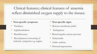
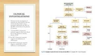
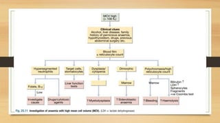
3. DEFINITION
• Anaemia refers to a state in which the level of hemoglobin in the blood is
below the reference range appropriate for age and sex.
• WHO criteria is Hb < 13 g/dL in men and Hb < 12 g/dL in women
• Hemoglobin is a protein found in red blood cells that carries oxygen from
the lungs to the body’s tissues and returns carbon dioxide from the tissues
back to the lungs .
4. ETIOLOGY
Decreased production of red blood cells
• Nutritional iron, and/or folic acid/vitamin B12 deficiency,
• Depressed bone marrow function (leukaemia, aplasia)
• Infections (HIV, TB)
Increased destruction of red blood cells (haemolysis)
• Malaria
• Drug side effects (dapsone, cotrimoxazole, Zidovudine)
• Congenital disorder, e.g. sickle cell anaemia
• Hypersplenism
Loss of red blood cells
• Acute and chronic blood loss (e.g. haemorrhage after trauma, hookworm infestation, pregnancy, abortion, heavy menstrual loss, schistosomiasis, massive or
chronic GI bleeding)
5. EPIDEMIOLOGY
• Anaemia affects about 30% of the total world population
• It is quite common in tropical settings, and often caused by multiple factors.
Children and young women are particularly at risk.
• In Uganda women of child bearing age are more susceptible to anaemia. Pregnant
women living in poverty are at greater risk of developing iron deficiency anaemia.
Iron deficiency is the leading cause of anaemia among pregnant women globally .
• In women of child-bearing age, menstrual blood loss, pregnancy and breastfeeding
contribute to iron deficiency by depleting iron stores.
6. General Clinical features
• Pallor of conjuctivae, mucous
membranes, palms, soles
• Fatigue, dizziness, palpitations,
headache, anorexia, sometimes
weight loss, low exercise tolerance
• Signs of heart failure if severe:
oedema in lower limbs, dyspnoea,
tachycardia, heart murmurs
• If due to acute blood loss: postural
hypotension, decreased cardiac out
put, tachycardia, sweating,
restlessness and thirst
• Look for signs of specific
pathology, e.g., splenomegaly,
malaria, nutrition deficiency,
haemolytic jaundice, etc.
7. Clinical features; clinical features of anaemia
reflect diminished oxygen supply to the tissues.
• Non-specific symptoms
• Tiredness
• Lightheadedness
• Breathlessness
• Development/worsening of
ischemic symptoms e.g. angina
• Non-specific signs
• Mucous membrane pallor
• Tachypnoea
• Raised jugular venous pressure
• Tachycardia
• Ankle oedema
• Postural hypotension
8. Compensatory mechanisms
Modulation of oxygen
affinity
Increased production of
erythropoietin
Redistribution of flow to
benefit the myocardium,
brain, and muscle
Increase in cardiac output
Reduction of mixed venous
oxygen tension to increase
the arterio-venous oxygen
difference
9.
10.
11. LABORATORY TESTS IN ANAEMIA
DIAGNOSIS
A.) Complete Blood Count(CBC)
1. Red Blood Cell Count
• i.) Haemoglobin
• ii.) Haematocrit
• iii.) Reticulocyte count.
2. Red Blood Cell Indices
• i.) Mean cell volume(MCV)
• ii.) Mean cell haemoglobin(MCH):
• iii.) Mean cell haemoglobin concentration(MCHC)
• iv.) Red Cell Distribution width(RDW)
13. Continuation
B) Iron supply studies
i.) Serum iron.
ii.) Total iron binding capacity.
iii.) Serum ferritin
C.) Marrow examination
1. Aspirate
i.) Myeloid/Erythroid precursor ratio
ii.) Cell morphology
iii.) Iron stain
14. Continuation
2. Biopsy
i.) Cellularity
ii.) Morphology
D.) Peripheral blood film
E.) Stool analysis for micro-bleeding, ova
and cysts.(very important)
■CBC and peripheral blood film are the
gold standard for anemia diagnosis.
15. CLINICAL
INVESTIGATIONS
• Schemes for the investigation of
anaemias are often based on the size
of the red cells, which is most
accurately indicated by the MCV in
the FBC. Commonly, in the presence
of anaemia:
• A normal MCV (normocytic
anaemia) suggests either acute blood
loss or the anaemia of chronic
disease (ACD)
• A low MCV (microcytic anaemia)
suggests iron deficiency or
thalassaemia.
• A high MCV (macrocytic anaemia)
suggests vitamin B12 or folate
deficiency or myelodysplasia
16.
17. Megaloblastic anemias
• This results from a deficiency of
vitamin B 12 or folic acid, or from
disturbances in folic acid
metabolism.
• It results in arrested nuclear
maturation but normal cytoplasmic
development: socalled
nucleocytoplasmic asynchrony.
19. Investigations in megaloblastic anaemia
Investigations
Blood smear: macrocytosis
Elevated MCH/MCV
Pancytopenia in severe cases
Full blood count: oval macrocytes, hyper-
segmentation of neutrophils,thrombocytopenia
Decreased serum Vitamin B12 or red cell folate
Management
Folic acid and vitamin B12 supplementation
Folic acid: 5 mg daily until haemoglobin levels
return to normal 1 mg IM daily for 5 days; then
weekly for a further 3 doses
Vitamin B12 – Follow with 1 mg every second
month for life in patients with pernicious anaemia
20. Note
• If vitamin B12 deficiency is suspected: (low leucocytes and platelets,
neuropsychiatric symptoms, vegan diet) DO NOT GIVE folic acid alone but
refer for further testing and treatment. Giving folic acid alone in patients
with vit.B 12 deficiency may precipitate permanent neurological deficit.
• Anaemia normally corrects within 1-2 months. White cell count and
thrombocytopenia normalise within 7-10 days
• DO NOT use ferrous-folate combination tablets to treat folic deficiency
because the quantity of folic acid is too low
21. VITAMIN B 12 DEFICIENCY
• The liver stores enough vitamin B 12 for 3 years and this, together with the
enterohepatic circulation, means that vitamin B12 deficiency takes years to
become manifest, even if all dietary intake is stopped or severe B12
malabsorption supervenes.
• The deficiency is also caused by fish tape worm infection (diphyllobothrium
latum), caused by eating undercooked or raw fish.
• The most common cause of Vit B 12 is pernicious anaemia.
22. Iron deficiency anaemia
This occurs when iron losses or physiological
requirements exceed absorption.
Causes
Poor nutritional intake
Chronic blood loss, e.g., infestation with
Ancylostoma, prolonged /excessive menstrual
bleeding, chronic gastrointestinal bleeding (e.g.,
chronic use of NSAIDS, large bowel tumors)
Clinical features
It usually develops slowly
As per general anaemia symptoms plus:
– Erosions of the corners of the mouth
– Brittle, fragile fingernails
23. Iron def.
Investigations.
Blood: CBC, Hb, (haematocrit (Hct) rarely <28%
unless iron deficiency is present)
Low MCV and Mean Corpuscular Hb (MCH)-
hypochromia
Hypochromic microcytic (small size) red cells
Investigate the cause of iron deficiency
treatment
Identify, and treat cause of iron deficiency
Adjust diet if poor diet is one of underlying
causes
Adult: Oral ferrous sulphate 200 mg (or ferrous
sulphate/folic acid 200/0.4 mg) every 8 hours
(equivalent to 180 mg elemental iron per day)
24. Iron deficiency anaemia
• Child: Oral ferrous sulphate 5 mg/kg (max 200 mg) every 8 hours (equivalent to
around 5 mg/kg elemental iron per day)
– Hb rises in 2-3 weeks and returns to normal after 2 months
– Treat for 6 months to 1 year to replenish stores
• Give an antihelminthic
– Albendazole 400 mg single dose
• Side effects of oral iron: diarrhoea, abdominal discomfort, constipation,
black stools.
25. Normocytic anaemia
Anaemia characterised by normal-sized red blood cells.
It is anaemia of chronic illness.
Causes
• Acute blood loss
• Haemolysis (destruction of red cells), e.g., auto-immune disorder,
hypersplenism, haemoglobin abnormalities (sickle cell disease, thalassemia),
drugs (sulphonamides,dapsone, primaquine)
• Decreased reticulocytosis (formation of new blood cells), e.g. chronic kidney
disease.
26. Normocytic anaemia
Investigations
Evidence of haemolysis
Full blood count smear: spherocytes
HIV serology
Treatment
Generally Identify and treat cause of anaemia
Medicine treatment
DO NOT treat with iron, folic acid or vitamin B12
unless there is clear documented deficiency
Treat all patients with folic acid 5 mg daily in
haemolytic anaemia
27.
28.
29. Anaemia of chronic disease
• It occurs in the setting of chronic infection, chronic inflammation or
neoplasia. The anaemia is not related to bleeding, haemolysis or marrow
infiltration, is mild, with haemoglobin in the range of 85–115 g/L, and is
usually associated with a normal MCV (normocytic, normochromic), though
this may be reduced in longstanding inflammation. The serum iron is low but
iron stores are normal or increased, as indicated by the ferritin or stainable
marrow iron.
• Treatment involves: Measures which reduce the severity of the underlying
disorder generally help to improve the ACD.
• Blood transfusion where necessary
31. cont
• Red blood cell haemolysis can be inherited or acquired
Inherited
Red cell membrane defects like hereditary spherocytosis and elliptocytosis
Hemoglobin abnormalities like SCD and thalassemia
Acquired
• Autoimmune
• Alloimmune
32. Aplastic Anaemia
• This is anaemia due to Bone marrow failure
• It leads to pancytopenia
• It is due to a reduction in pluripotent stem cells.
Clinical manifestations
Gum bleeding
Epistaxis
Investigation
• Pancytopenia
33. Management of anemia
The purpose of establishing the aetiology of the anaemia is to permit selection of a
specific and effective therapy. Below are some of the ways in which anaemia maybe
managed;
1. Corticosteroid therapy for the treatment of auto-immune haemolytic anaemia.
2. Splenectomy for the treatment of anaemia due to red cell defects(spherocytosis),
haemoglobinopathies.
3. Surgery so as to control bleeding in anaemic patients.
4. Cross matching and blood transfusion.
5. Oral administration of ferrous sulphate until the anaemia is corrected and for
several months afterwards to ensure that the body stores are replenished.
34. continuation
6. Administration of iron sucrose especially in patients with iron deficiency anaemia
associated with chronic kidney disease. Administered by mixing it in 0.9% NaCl
solution.
7. Increased dietary intake of iron through consumption of red meat.
8. Increased consumption of vegetables especially green leafy vegetables to improve
dietary intake of vitamin B12 and B9.
9. Bone marrow transplant for aplastic anaemia
10. Administration of oral folic acid 5mg daily for about 4 months to replenish body
stores.
11. Deworming.
35. Indications for blood transfusion
• The need for a blood transfusion depends on;
• i.) The degree of anaemia in terms of haemoglobin concentration.
• ii.) The clinical condition of the patient.
• iii.) Presence of active blood loss e.g. internal or external haemorrhage or red cell
destruction e.g. malaria, sepsis and haemolysis.
• Packed red blood cells are the recommended element for blood transfusion with a
reduced risk of transfusion associated circulatory overload.
• All blood products should be infused within 4 hours due to the risk of bacterial
proliferation in the blood component at room temperature.
36. Complications of anemia
• Tissue hypoxia
• Shock
• Coronary and pulmonary insufficiency
• Weakened immune system with increased susceptibility to infections.
• Lasting fatigue with decreased productivity.
• Obstetric complications e.g. preterm delivery.
37. Prevention of anemia
• Balanced diet rich in iron, vitamins and minerals with inclusion of foods such as
green leafy vegetables and fruits.
• Iron and folic acid supplementation during pregnancy.
• Prevention of malaria through sleeping under insecticide treated mosquito nets.
• Prevention of hook worm infestation especially in children through regular
deworming.
• Hygiene: avoid walking barefeet to avoid hook worm infestation, use of pit latrines
for faecal disposal, and practice good hand washing habits
• encourage periodic screening for children and pregnant mothers, and presumptive
iron therapy for either groups in cases of anaemia
• Early treatment of malaria, helminthic infections, etc.