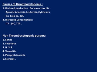
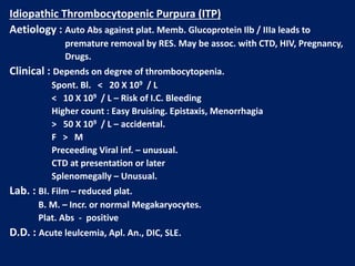
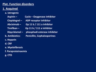
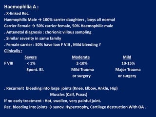
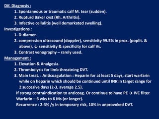
Haemostasis
Mechanism : 1. Immediate response : Vasoconst. + plat. Plug.
2. Secondary : Fibrin deposition .
Stage 1 : Pre-injury :
. Vascular endoth. Produce subs. Prevent adhesion of plat. & WBC to
vessel wall (NO2 , prostocyclin)
Stage 2 : Early Haemostasis :
. Plat. Adher. & coagulation activation → Thrombin
Stage 3 : Fibrin clot formation .
Stage 4 : Limiting clot formation by natural anticoagulants (antithrombin , TFPI
, protein C & S ).
Stage 5 : Fibrinolysis . Plasmin degrades fibrin → vessel recanalisation & tissue
repair .
Lecture (1)
Clotting Factors : Half life
I. Fibrinogen 4-5 days
II. Thrombin 3 days
III. Tissue F. -------
IV. Ca++ -------
V. Labile F. 12-13 h
VI. No longer used -------
VII. Proconvertin 4-7 h
VIII. AHF 8-10 h
IX. Christmas F 1 day
X. Stuart power F 2 days
XI. Plasma Thromboplastin antecedent 2-3 days
XII. Hagman F 1 day
XIII. Fibrin stabilisig F. 8 days
All produce by the liver , FV also by plat. & endoth. Cells .
VIT. K dependent factors II VII IX X (inhibited by WARFARIN)
Blood Coagulation :
1. Extrinsic pathway : initiated by F VII interacting with Tissue Factor.
2. Intrinsic pathway : initiated after exposure of subendoth. Collagen after vessel
damage . XII XI IX VIII
Extrinsic Pathway Intrinsic Pathway
TF XIa
VIIa IXa
VIIIa
Xa
Va
Prothrombin Thrombin
Fibrinogen Fibrin Monomer
XIIIa
Fibrin polymer clot
Platelets :
. Formed in Bone marrow from Megakaryocytes.
. Thrombopoiten (liver) stimulate formation & Maturation of plat.
. Circulate in Bl. For 8-10 days before destroyed in RES.
. 30% pooled in spleen.
. Discoid 2-4 Mm diameter.
. Invaginated surface Memb. to form Canaliculi.
Screening tests for bleeding disorders
1. Plat. Count (normal = 150-400 X 109 /L
2. Bleeding time (normal < 8 min.) prolonged in thrombocytopenia, abn. Plat. F, VWD,
vasc. & con. T dis.
3. Prothrombin time (PT) : assess extrinsic pathway (normal 9-12 s). F II, V, VII, X
4. Partial thromboplastin time (PTT) : assess intrinsic pathway (normal 26-36 s). F II, V,
VIII, IX, X, XI, XII, sever fibrinogen def. heparin, inhibitors
5. Prolonged PT & PTT = common pathway (X, V, II & I) , or > one factor def.
6. Fibrinogen conc. (normal 1.5-4 g/L) hypofibr. e.g. liver dis. DIC.
7. Mixing tests with normal plasma differentiate between coag. Factor def. &
presence of inhibitors (the prolonged time does not correct).
8. plat. Functions tests.
Bleeding Disorders : suspect if
1. Multiple sites
2. Spontaneous
3. Prolonged
1. Vascular purpura
a. Hereditary : Defective collagen .
b. Acquired : Immune, Inflammatory .
2. Platelets defects : Quantity, Quality
3. Coagulation defects :
Reduced synthesis of Fs
Abnormal Fs
Destruction (DIC)
Inhibitors
4. Fibrinolytic defects :
e.g. Excessive fibrinolysis following therepeutic thrombolysis
Clinical assessment
1. Site of bleeding
Ms, Joints, Retrop , indicate coagulation defect.
Purpura, Epistaxis, G.I.T., Menorrhagia, indicate low plat, plat. F. defect,
WWD.
Recurrent in single site – local abn.
2. Duration : Acq. Or Cong.
3. Precipitating Fs : Spont. , or traumatic.
4. Surgery, Exo, Tonsilectomy, Circumcision.
Immediate : Pri. Haemostasis
Delayed : Coag. def.
5. F.H. : Absence does not exclude hereditary dis.
6. Drugs .
Differentiation bet. Plat. dis & Coagul. dis
Plat dis. Coagul. dis.
Sex F > M M > F
Age 20-40 y childhood
Onset Immediate Delayed
Type Mucocut Deep Ts.
Local pr. Effective Not
Ex. ITP Haemophilia
Platelets Disorders
1. Thrombocytopenia < 150 X 109 / L
2. Thrombocytosis > 400 X 109 / L
3. Functional.
Thrmbocytopenia :
Spont. Bleeding – plat < 20 X 109 / L
< 10 X 109 /L – Risk of intracranial bleeding
Plat. Transfusion : indicated in
1. Bone marrow failure < 10 X 109 / L
2. Serious Haemorrhage.
Causes of thrombocytopenia :
1. Reduced production : Bone marrow dis.
Aplastic Anaemia, Leukemia, Cytotexics
B12 Folic ac. def.
2. Increased Consumption :
ITP , DIC, TTP .
Non Thrombocytopenic purpura
1. Senile
2. Factitious
3. H. S. P.
4. Vasculitis
5. Paraproteinaemia
6. Steroids .
Idiopathic Thrombocytopenic Purpura (ITP)
Aetiology : Auto Abs against plat. Memb. Glucoprotein Ilb / IIIa leads to
premature removal by RES. May be assoc. with CTD, HIV, Pregnancy,
Drugs.
Clinical : Depends on degree of thrombocytopenia.
Spont. Bl. < 20 X 109 / L
< 10 X 109 / L – Risk of I.C. Bleeding
Higher count : Easy Bruising. Epistaxis, Menorrhagia
> 50 X 109 / L – accidental.
F > M
Preceeding Viral inf. – unusual.
CTD at presentation or later
Splenomegally – Unusual.
Lab. : BI. Film – reduced plat.
B. M. – Incr. or normal Megakaryocytes.
Plat. Abs - positive
D.D. : Acute leulcemia, Apl. An., DIC, SLE.
Management of ITP :
. Stable, plat > 30 X 109 / L – No treatment, except with Surgery or Biopsy
. Spont. Bleeding – Prednisolone 1 mg / kg / day ( suppress Ab. Prod. and
inhibit phago. ) count will increase to normal or less in 2-4 weeks, then reduce
does gradually.
. Relapse – Reintroduce Steroids
. Immunoglobulin i.v. (Block Ab receptor on RES) + steroids in severe
haemostatic Failure
. Anti-D i.v. (Bind RBC and saturate Ab. Receptors in Rh + ve (+ spleen)
. Persistent or life threatening Bleeding needs plat. Transfusion
. 2 Relapses or Pri. Refractory needs splenectomy.
70% complete remission.
20-25% - improvement
5-10% require further treat. e.g. low dose steroids, Immunosup. (Rituximab,
Ciclosporin)
Thrombotic Thrombocytopenic Purpura (TTP) (HUS)
Aetiology : Autoimmune disorder, mediated by Abs. against ADAMTS – 13
enzyms .
(This enz. normally clears VWF multimers to produce normal functional units and
its deficiency results in large VWF multimers which cross – link platelets and leads
to microvascular occlusion by plat. Thrombi e.g. brain, kidney).
Idiopathic, associated with drugs (Ticlopidine, Ciclosporine), HIV, Malignancy.
Clinically : Sporadic, Fluctuating, Familial: reported, F > M , young – adolescent
1. Fever
2. Anemia : Microangiopathic haemolytic (Fragmented RBCs) ,
↑ Reticulocyte count, Jaundice, ↑ LDH, -ve coomb’s test.
3. Thrombocytopenia – Purpura, count usually < 20 X 109 /L
4. Neurological: Convulsion, Coma, Paralysis ( Transient or prolonged) ,
Psychological .
5. Renal : intermittent proteinuria, Renal Failure (more in HUS)
Lecture (2)
Lab. ↑ WBC, ↓ plat.
PT, PTT ------- Normal
B.M. ------- Normal or increase cellularity
Gingival Biosy – Helpful
DD : ITP
Prognosis : 10% Recurrent.
Untreated – 90% Mortality in 10 days
Treated – Mortality 20-30% at 6 months
Sudden death due to cardiac Microthrombi
Treatment : Plasmapharesis with FFP 12 bags/day for weeks – Months
Corticosteroids, Rituximab have a role.
Pregnant – May needs termination if no response
Thrombocytosis
1. Reactive (most Common) 2. Clonal
a. Chr. Infl. Dis. E.g. ulc. Collitis a. Pri. Thrombocythaemia (ET)
b. CTD e.g. Rh. Arthritis. b. PRV
c. Malignancy e.g. Hodgkin’s dis. c. Myelofibrosis.
d. Acute Haemolysis d. CML.
e. Fe. Def. An. e. MDS.
f. GI bleeding
g. Post – splenectomy
Clinically :
Those of the underlying dis.
Haemostasis rarely affected
PRV, ET, Myelofibrosis, may present with thrombosis and rarely bleeding
Reactive : small plat. No splenomegally.
Plat. Function disorders
1. Acquired
a. Iatrogenic
Aspirin – Cyclo – Oxygenase inhibitor
Clopidogrel – ADP receptor inhibitor
Abciximab – Gp 11 b / 111 a inhibitor
Tirofiban – Gp 11 b / 111 a inhibitor
Dipyridamol – phosphodi esterase inhibitor
b. Antibiotics: Penicillin, Cephalosporines
c. Heparin
d. CRF
e. Myelofibrosis
f. Paraproteinaemia
g. CTD
2. Congenital
a. Glanzmann’s thromboasthenia - Def. of GP 11b / 111a
AR, bleeding variable, often severe.
b. Bernard – Soulier dis. def. of GP 1b. AR
c. Defective plat. Granules – deficiency of dense granules
(b & c cause mild bleeding e.g. after trauma or surgery, rarely spont.)
Management :
Local mechanical pressure .
Antifibrinolytic e.g. Tranexamic acid may be useful .
Plat. Transf. in severe bleeding.
Recombinent FVIIa in resistant bleeding in Glanzmann.
VESSEL WALL ABNORMALITIES
1. Hereditary: HHT, EDD, Osteogenesis imperfecta
2. Inflammatory: Bacterial, Viral, Vasculitis.
3. Abn. Collage: Senile, Scurvy, Steroids.
4. Metaboli : Liver dis, Renal, Amyloidosis.
5. Others: Factitious, Paraproteinaemia, Drugs.
Hereditary Haemorrhagic Telengiectesia (HHT)
(Osler – Rendu – Weber synd.)
Clinically := A.D., Telengiectasia and small aneurysms on Fingers, lips, Face,
Tongue, Nasal passages, Lungs, G.I.T. / Usually appear at adulthood.
Signif. No. have Pul. A.V. malformation (PAVMS) → Arterial
Hypoxaemia due to Rt. To It. Shunt → paradoxical embolism →
stroke or cerebral abscess.
Recurrent Bleeding e.g. Epistaxis or Malaena → Fe def. An.
Treatment : Deficult bec. Of multiple Bleeding sites.
Regular Fe therapy
Local Cuatary or Laser, Oestrogens ?
Ehlers – Donalos Dis :
AD. , Collagen defect
Fragile Bl. Ves. And organ memb. → Bleeding and organ rupture
Joint Hypermobility
Scurvy
. Vit C def. → affect normal synthesis of collagen
. Perifollicular and patechial haemorrhage, sub- periosteal bleeding
. Diagnosis : Dietary History
Henoch – Schonlein Purpura (HSP)
. Children and young adults .
. Immune complex deposition in small ves. With vasculitis .
Often preceded by upper resp. inf.
Clinically :
1. Purpura: (raised) Buttocks and legs
2. Abdominal colicky pain and bleeding
3. Arthritis : Knees, Ankles .
4. Nephritis : 40% (May be later – 4 wks.)
Diagnosis : Clinical, Tissue biopsy ( IgA deposition in bl. Ves.)
Prognosis : good, Adverse if HTN, RF, Proteinuria > 1.5 g / day
Treatment : Nonspecific
Steroids for joint or GI involvment
Nephritis – pulse iv steroids ± immunosup.
COAGULATION DISORDERS
Congental :
X-linked : Haemoph. A and B
Autosomal : VWD, F II V VII X XI XII def.
Combined II VII IX X def.
Combined V and VIII
Hypofibrogenaemia
Dysfibrinogenaemia
Acquired
Under prod.: Liver failure.
Incr. Consumption: DIC
Immune – mediated: Acq. Haemophelia & VWD.
Others: Acq. F X def. (in amyloid)
Drug – induced
Inh. Of function : Heparin
Inh. Of synthesis : Warfarin
Lecture (3)
Haemophilia A :
. X-linked Rec.
Haemophilic Male → 100% carrier daughters , boys all normal
Carrier Female → 50% carrier female, 50% Haemophilic male
. Antenetal diagnosis : chorionic villous sampling
. Similar severity in same family
. Female carrier : 50% have low F VIII , Mild bleeding ?
Clinically :
Severe Moderate Mild
F VIII < 1% 2-10% 10-15%
Spont. Bl. Mild Trauma Major Trauma
or surgery or surgery
. Recurrent bleeding into large joints (Knee, Elbow, Ankle, Hip)
Muscles (Calf, Psoas)
If no early treatment : Hot, swollen, very painful joint.
Rec. bleeding into joints → synov. Hypertrophy, Cartilage destruction With OA .
Muscles Haematoma : depend on site
Psoas → compress fem. N
Calf → compartement synd. → Isch. , necrosis , fibrosis,
contracture and shortening of Achilles tendon.
Lab. ↑ PTT, Normal PT, plat. Count. & bleeding time , ↓ F VIII , -ve inhibitors.
Manag. : Avoid Trauma.
. Severe : 1- ↑ F VIII: (Donar Plasma or Recombinent) iv infusion F VIII
concentrate.
2- Rest of bleeding site
3- If bleeding settled → Mobilisation & physio.
4- Hepatitis A,B immunisation
5- Prophylactic: F VIII 2-3 X /wk – (at home) - expensive
. Mild : DDAVP (Desamino – des – aspartate – arginine vasopressin)
↑ F VIII X 4, it covers minor surgery e.g. Exo
Complications :
1- HIV , HBV , HCV (< 1986)
2- Anti F VIII Abs. ( inhibitors ) 20%
3- OA & deformities .
Haemophilia B (Christmas dis)
↓F IX
X – linked
Clincally similar to A
Treatment : F IX concentrate
Inhibitors < 1%
Von Willebrand Dis. (VWD)
AD , M > F / most common bleeding disorder , usually mild.
Function of VWF : 1- Carrier for F VIII (↓ VWF → ↓ F VIII).
2- Forms bridges bet. plat. & sub endoth. → plat. Function.
Classification : Type 1 : most common , ↓ quantity.
Type 2 : abn. Function (quality), 2A, 2B, 2M, 2N – Auto. Rec..
Type 3 : severe quantitative (AD).
Clinically : different severity in same family , bleeding similar to plat. Dysf.
Brusing , Epistaxis , Menorrhagia, G.I. bleeding.
severe bleeding: ± after trauma or surgery.
Lab. : ↑ BT, ↑PTT, normal PT , ↓ VWF, secondary ↓ F VIII.
Manag. : Mild bleeding : local means or DDAVP , Tranexamic ac. may be useful
in mucosal bleeding.
Severe or persistent bl. : selected F VIII concentrate (contain F VIII +
VWF ).
Aquired Bleeding Disorders :
Disseminated Intravascular Coagulation (DIC)
Definition : Acq. Thrombohemorrhagic disorder, characterise by systemic
activation of coagulation (Thrombosis) , with simultaneous coagulation Fs.
(Notably F V & VIII) , plat. & fibrinogin consumption causing bleeding.
Lysis of fibrin →fibrin degredation products ( FDP ) including D – dimers.
Causes :
1- Infection ( sepsis ).
2- Trauma.
3- Obstetric (Amniotic fluid embolism, placental abruption, pre-eclampsia).
4- Severe liver failure.
5- Malignancy.
6- Tissue destruction (Pancreatitis, burn).
7- Toxic / immulog. (ABO incompatab. , snake bite).
Clinically :
, 1- Signs of Thrombosis: Jaundice, blue toes, fingers gangrene, delerium,
coma, respiratory failure, oliguria.
2- Signs of bleeding: S.C. bleeding, I.C. bleeding, Epistaxis, Gingival,
vaginal, renal .
Lab.
Anaemia ( Microangiopathic Haemolytic ).
↑ PT & PTT
↓ Fibrinogen
↑ Bilirubin, ↓ plat. , FDP +ve
Manag.: Treat. Of underlying cause.
Deal with acidosis, dehyd., RF, Hypoxia.
FFP, cryoppt., plat. – if bleeding, or cover intervention (not roteinly)
Heparin – prophylactic doses (unless contraindicated) to treat
established thrombosis.
Liver diseases : ↓ synthesis of coagulation Fs.
↓ clearance of plasminogin activators.
↓ plat. (Hypersplenism)
↓ vit K absorp. (cholestatic jaundice) → ↓ F II VII IX X
Lab. ↑ PT , ↑ PTT (in severe cases).
Treatment FFP , Plat. Vit. K
Renal Failure : Bleeding propotional to Urea concentration.
Similar to Plat. Dysf. Type (GIT is common).
Causes : An. , ↓ plat. , waste products.
Treatment Dialysis, plat. transf., Bl. , cryo. , DDAVP.
Vit K deficiency :
Sources : Green vegetables , intestinal flora.
Causes : ↓ intake, malabsorp., biliary obst. , antibiotics.
Clinical : plat. dysf. type of bleeding.
Lab : ↑ PT, ↑ PTT (in severe cases).
Treatment : Vit. K , im or iv for 3 days + treat. Of underlying cause.
Thrombotic Disorders
Deep Vein Thrombosis: (DVT)
Mortality: 1-3%
Predisposing Factors:
1. Patients Factors: ↑Age, Obesity, Varicose veins, Previouse DVT, F.H.
Pregnancy, CCP, Puerperium.
2. Surgical: Major surgury, ( > 30 min ) , abdom., pelvic or lower limb ortho..
3. Medical: M.I., H.F., Inflm. bowel dis., Neph. Synd., Pneumonia, Stroke.
4. Haematology :PRV, ET, Deficiency of anticoagulants (antithrombin, prot. C,
prot. S), PNH, FV Leiden, Prothrombin gene G20210A .
5. Antiphospholipid synd.
Clinically :
Lower limb DVT : Typically Unilat. (may be bilat.).
Pain, swelling, high temp. , tender, dilated superficial Vs..
Examine thigh for DVT (proximal Involv.) → ↑ risk of PE.
Lecture (4)
Dif. Diagnosis :
1. Spontaneous or traumatic calf M. tear (sudden).
2. Rupturd Baker cyst (Rh. Arthritis).
3. Infective cellulitis (well demarkated swelling).
Investigations :
1. D-diamer.
2. compression ultrasound (doppler), sensitivity 99.5% in prox. (poplit. &
above), ↓ sensitivity & specificity for calf Vs.
3. Contrast venography – rarely used.
Management :
1. Elevation & Analgesia.
2. Thrombolysis for limb threatning DVT.
3. Main treat. : Anticoagulation : Heparin for at least 5 days, start warfarin
while on heparin which should be continued until INR in target range for
2 succesive days (2-3, average 2.5).
If strong contraindication to anticoag. Or continue to have PE → IVC filter.
Warfarin – 6 wks to 6 Ms (or longer).
Recurrence : 2-3% /y in temporary risk, 10% in unprovoked DVT.
Post – thrombotic synd.: Damage to ven. valves by thrombous → persistant
leg swelling, heaviness & discolouration, sometimes ulceration.
Patient PT
X international sensitivity index‫ـــــــــــــــــــــــــــــــــــ‬INR =
Normal control
Indications for Anticoagulation
Heparin Warfarin
1. Prev. & treat. of VTE. 1. Prev. & treat. of VTE.
2. Percut. Coronary interv. 2. Art. Embolism.
3. Post thrombolysis MI . 3. AF with stroke risk.
4. Unstable Angina pectoris. 4. Mobile mural thrombus.
5. Non Q wave MI. 5. Extensive Ant. MI.
6. Acute periph. Art. Occlusion. 6. Dilated cardiomyopathy.
7. Cardio pul. By pass. 7. Cardioversion, Isch. Stroke .
APS
8. MR & MS + AF, Rec. ven. Throm.
mechanical prosth. H. valves
Heparin : potentiate antithrombin anticoagul. activity .
1.Unfractioned( UFH) .
2.Low molecular wt.(LMWH).
Advantage of LMWH :
1. 100% bioavailability =reliable dose dependent anticoagulation,no need for
monitoring,except in very thin pt. & R.F.
2. Once S.C./d .(UFH continuous iv infusion In therapeutic doses, X2 / S.C. in
prophylactic doses) .
3. ↓ Osteoporosis & Thrombocytopenia.
Dose :
1.UFH :
Therapeutic : 5000 u iv followed by 20u/Kg/h by contin. Iv inf.
Assess by PTT(Target 1.5-2.5 the control).
Prophylactic : 5000 u X 2 / S.C.
2. LMWH :
a. Enoxoparin :
Therapeutic : 150 u / Kg /S.C. daily.
Prophylactic : 2000 – 4000 u /S.C. daily.
b. Tinzaparin :
Therapeutic : 175 u / Kg/ S.C. daily.
Prophylactic 3500 - 4500 u/ S.C. daily.
Complications :
1. Bleeding : Treatment : Stop Heparin is usually enough (short half life)
If necessary protamine sulphate (Anti-dote).
2. Thrombocytopenia :
a. Early : First 24 hours (benign, due to plat. Clumping).
b. Late : 5 – 14 days after treatment (anti-heparin/PF4 Abs. , bind to &
activate plat. & prothrombotic state causing thrombocytopenia.
Clinically : Asymtomatic – thrombosis in 50% , poor prognosis.
Manag. : Stop Heparin give lepirudin.
3. Osteoporosis (Heparin > 6 Ms ) .
Warfarin : inhibit vit K depend. Fs (II VII IX X).
Take 3 days for anticoagulation.
Drug interaction
1. Potentiate : Sulphonamides, Cimetidine, Allopurinol, Metronidazole,
Thyroxin, Aspirin, Antibiotics.
2. Inhibition : Barbiturates, Rifampicin, CCP, Pregnancy .
Dose : 10 mg on 1st & 2nd day , 5 mg 3rd day, & then according to INR.
Complications :
Bleeding : 1% fatal (intracranial)
Manag.: Look for the cause,e.g.drug interaction or↑dose,stop
warfarin temporarilly.
Minor : 1-2.5mg vit. K iv.
Major : 5-10 mg vit. K iv. Slowly + FFP ( 15-30 ml / kg)
Prolonged INR with no bleeding : Stop or reduce the next Dose, small
Dose vit. K orally or iv. 1-2.5 mg.
especially if INR > 8
Prophylaxis of ven. Thrombosis
Moderate risk High risk
1. Major surgery ( > 40 y + risk F) 1. Major abd. Or pelvic surgery for malig.
or with history of DVT or known
thrombophilia.
2. Major medical illness e.g. 2. Major Hip or Knee surgery.
HF, MI, Sepsis, Active malig. , 3. Neurosurgery
Neph. Synd. , Stroke + Paralysis
Methods
1. Mechanical : intermit. Pneumatic compr. , /feet pump, compression stoking
2. Pharm. : LMWH, UFH, Warfarin, Aspirin.
Contraindications
1. Recent surgury
2. Pre-Existing Haemorrhagic state ( liver dis. , Haemophilia )
3. Peptic ulcer.
4. Recent cerebral Hemorrhage.
5. Uncontrolled HTN
6. Frequent falls in old age.
Antiphospholipid Syndrome (AS)
Acquired prethrombotic tendency, in which arterial & or 0r venous thrombosis
assosciated with finding in the plasma of lupus anticoagulant (LA) & or
seropositivity for antiphosphoslipid Ab.,most commonly Anticardiolipin Ab. (ACA)
Clinically
1. Primary APS
2.Secondary APS is associated with : SLE, Rh.arthritis, Systemic sclerosis,
Behcets dis,Temporal arteritis.
Criteria A : Clinical:
1.Vascular thrombosis,one or more episode of arterial or venous or small
vessel thrombosis in any tissue/ organ.
2.Pregnancy morbidity:
a.One or more unexplained death >10 wk gest.
b.3 or more unexplained spont. Abortion,<10 wk gest.
c.One or more premature birth <34 wk gest.
B :Lab:
1.Anticardiolipin Ab. On 2 or more occasions, 6 Wks apart.
2.Lupus anticoagulant Ab. On 2 or more occasions, 6Wks apart.
3.Prolonged PTT.
Diagnosis : At least ONE clinical & ONE lab. Criteria
Treatment:
Arterial thrombosis( stroke): Warfarin.
Recurrent VTE :Long term anticoagulation
Pregnancy :Heparin + Aspirin (Increase chance of succesful pregnancy).
Blood Products & transfusin
Donation :
1.Whole Blood .
2. Apheresis e.g. platelets.
Blood components :
1.Red cell concentrate: (most of plasma removed ), ABO &Rh compatab. required.
Indication: Acute blood loss replac.& severe An.
2.Plat. Concentrate: ABO compatab.preferable.
Indication: Thrombocytopenia, Plat. Dysfunction.
3.FFP (150- 300 ml): ABO compat. recommended. Dose: 15 ml / Kg.
Indic.:Replac. Of coagul. Fs def. , TTP .
4.Cryoprecipitate: (10-12ml / pack): Contain fibrinogen(150-300mg) & coagulation
Fs (80- 120u FvIII & VWF )
Indication: hypo- Fibrinogenaemia , VWD ,Haemophelia.
Lecture (5)
Plasma Derivatives :
1.Coaguiation Fs : F VIII , F IX , recently: recobbinent DNA technology.
2.Immunoglobulins : IV IgG (Ab.def., ITP, Gullian Bar. Synd.)
Prophylaxis of Varicella zooster.
Anti Rhesus D Ig
3.Human albumin :
5% :colloid fluid ( Crystalloid sol. Cheaper)
20% :Hypoprot. Odema (Nephrotic synd.), Ascitis in chr.
liver dis.
ABO Blood Groups :
Group Red cell A or B Ag Abs. in plasma % UK
O None Anti A & B 46
A A Anti B 42
B B Anti A 9
AB A &B None
3
Donar Blood
Test for HIV ,HBV ,SYPHILIS ,ABO &Rh
Red cells FFP Pooled plat. --
Storage 4 C - 30 C 22
35 d 1 yr 5 d (agitated)
Adverse effects
Death: o.5/ 100 000
1%: Symptoms e.g. fever , itch ,urticaria (repeated transfusions) .
Any symptoms or signs _take it seriously__WARNING !
1. ABO incompatability :
Recep.IgM (Anti-A ,Anti-B or Anti AB + Transfused red cells
Activate complement system
intravascular haemolysis, hypoten. RF ,peribronchial odema &
smooth M.cont 2. Other
immunological complic. e.g. Transf. assosiated lung injury.
, 3. Infections: HBV, HCV, HIV, less than 1/1000 000,(bact. Contam. of plat.packs)
Conclusion: avoid unnecesary transfusion.
Signs & Symptoms of acute transfusion reaction :
(Fever, Chills, Tachycardia, Hyper or Hypotension, Collapse , Rigors, Flushing,
Urticaria, bone,M.,Chest & abd. Pain, SOB, Nausea , Resp.distress )
Stop Transfusion
Temp, PR, BP, pt.identity, Bl.pack, compatability form
1. Reaction only Fever or urticaria
↓ ↓
FEVER URTICARIA
*Febrile Non Haemolytic Transf. Reaction * Mild Allergic Reaction
Treatment: Paracetamol Chlorpheramine 10mg iv
Restart Transf. after 15-30min.at slower rate
under observation if no more
complications
Suspect ABO incomoatability
! Wrong Bl. Transfusion !
YES NO
ABO incompat. Severe Allergic Reaction
Pack & giving set Blood Bank Bronch. Spasm,Angiodema,Abd.pain
i.v.Saline, Urine Output(catheter) Hypoten.
U.O.<100ml/h: give Frusemide
Treat DIC NO YES
Bact. Contam. Stop Transf.
Pack & Giving Set back Chlorphen. 10 mg i.v.
to Bl.Bank, Bl.Culture, O2,Salbutamol Neb.
Repeat Bl.group & comp. Adrenaline 0.5 mg i.m. in
FBC, Coagul.screen ,Bioch. Severe Hypot.or Bronchosp.
GUE. Send pack &giving set to Bl.
Broad spectrum antibiotics. Bank.
O2, Fluid.
.
No Bact. Contam.
If acute Dyspnoea. Hypot. Bl. Gases, CXR, CVP, Pul.cap. Pr.
↓ ↓
High CVP Normal CVP
Fluid Overload Transf. related Acute Lung Injury
O2, Frusemide 40-80 mg (Typically withen 6h )
SOB, cough, CXR: bilat. nodular infilt.
Stop Transf. 100% O2 ,
Treat as RDS, Ventilation if necessary.
Bone Marrow & peripheral stem cell Transplant (BMT)
Allogenic :Stem cell from Donar, given i.v. to patient( previously conditioned
by chemo ± radioth. → engraft into pt.BM (function 3-4 wks).
Sources :
1. Related (HLA identical subling ).
2. Unrelated ( closely HLA matched Volunteer ) .
Indications :
1.Neoplastic disorders (Leukemia, AML, ALL, CML, MDS, Myelofibrosis )
2. Failure of haemostasis e.g.( Aplastic an.)
3.Inherited Defects e.g.( Thalassemia ) .
Complications : Mucositis, infection, bleeding, pneumonitis, infertility, Chr.GVHD.
secondary malignant dis.
20 yr. +HLA identical subling.>Best results : pt. with minimal residual dis. .Age
25 % die from proceedure ,GVHD,
Long survival :50% for leukemia.
GVHD :
Aetiology: cytotoxic activity of donor T lymphocytes ,which become
sensitised to their new host,regarding it as foreign.This may cause
acute or chr. GVHD .
Acute: Ist 100 d, one third of pt. , mild – lethal, → Skin rash, jaundice,
diarrhoea.
Prevention: HLA matching + Immunosupp.drugs
Chronic :May follow acute or independent, later than acute,resemble CTD.
Reduced intensity BMT :
↓ mortality
↓ Intensive conditioning
↓ Toxic , old pt.
↑ relapse ?
Autologous :
Pt. own stem cell,from Bl. Or BM ,harvested & frozen → conditioning
→reinfuse .
Indications : Dis. do not primarilly involving haematological Tissues, or
Pt. who had achieved good Remission.
e.g. AML CR2, Myeloma, poor –risk Hodgkin,High- grade Non-Hodgkin.
Marrow recovery : 2-3 wks .
No risk of GVHD.
↓ mortality .
↑ Recurrence .
Stem cells treated to remove any residual leukemic cells is still invesigated.
Polycythaemia
0.52<180 gr / L in M PCV<Hb
0.48<165gr/ L in F PCV
Classification : RBC Mass Plasma Vol
True Increased Normal
Relative Normal Decreased
Relative : e.g. Dehydration, Diuretics, Alcohol.
True : Primary Secondar
Myeloprplif. Disorders ↑EPO (Hypoxia) Inappropriate ↑
( PRV) ↑ Altitude Renal dis.
Lung dis. CHD, Hepatoma
↑ Affinity Hb Bronchogen. Ca.
Uterine fibroid.
Phaeochromocyt.
Cerebellar haemangioblas-
stoma.
Polycythemia Rubra Vera
Age: more than 40y.
Clinically :
1. Incidental.
2. Symtomatic :Hyperviscocity : Lassitude, loss of conc., headache, diziness,
blackouts, pruritis (worse after hot bath) , epistaxis, peripheral art. dis.
e.g. CVA (thrombosis) , venous thromboembolism,.
Peptic ulcer is common (sometimes bleeding ) .
Plethoric face , majority : palpable spleen .
Diagnosis: ↑ RC mass , ↑PCV , ↑ Plat.& Neutrophil (often) , JAK -2 Mut.(97%)
Hypercellular B.M. + Absence of secondary causes .
Treatment : ↓ risk of thrombosis : Venesection 400-500 ml (elderly less) every
5-7 wks → PCV 45%- & MAINTAIN AT THIS LEVEL .
Hydroxycarbamide ( Hydroxyurea), or ᾱ interferon → supress marrow prolif.
& ↓ transform. Into myelofibrosis , control spleen size .
RA P 32 In old pt.
Prognosis : Survival 10- 20 y with Treatment. CVA & IHD 60%
Convertion to other myeloprolif. Dis. 15% , Acute leukemia ( RA P)
WBC DISORDERS
⁄ L* Normal Count : 4 –10 X 10
9 * Lecoerythroblastic picture : presence of immature WBC & ERYTHROBLAST
in peripheral Bl.
* Peripheral bl. Contain Neutrophils (N) ,Lymphocytes (L),Eosinophils (E),
Basophils (B) ,Monocytes(M) .
* Myelocytes & Metamyelocytes may appear in peripheral Bl.,e.g. in infection.
* Leucocytosis : ↑ Total WBC or only one type of cell (differential) .
* Leucopenia : ↓ Total WBC or only one type of cell .
NEUTROPHILS : 2---7 X 10 9 ∕ L
Recognise ,ingest & destroy foreign particles,spend 6-10 h in circul.→ removed
by spleen .
* Neutropenia: < 1.5 x 10 9 ∕ L ( Asymptomatic –Sepsis )
If < o.5 x 10 9 / L – Critical , Fever, sore throat ,perianal pain,skin
inflam. → SHOCK if no antibiotics .
Lecture (6)
Neutrophelia Neutropenia,
Infection: Bacterial ,Fungal Viral ,Bacterial ( TF) , Protozoal(
malaria)
Trauma: Surgery ,Burn . CTD, Alcohol.
Infarction: MI, PE ,Sickle C.Crises. BM infilt.: Leukemia,MDS,
Inflam.: Gout,R.arth.,U.C. Drugs
Malignant: Solid Tumours,Hodgkins.
Myeloprolif.: PRV,CML.
Physiological: Exercise, Pregnancy.
Drugs causing Neutropenia
Analgesia,Anti-infl.: Gold,Penicillamine. Anti-Thyroid : Carbimazole.
Anti-Arrhythmics: Quinidine . Anti-Hypert.: Captopril, Nifedipine.
Anti-Depres.: Amitryptylline. Anti-Malarial: Chloroquine.
Anti-Convulsants: Phenytoin, Carbim.
Antibiotics: Sulphonamide,Penicillin, Chloramphenicol.
Miscellaneous:
Cimetidine,Ranitidine,Chlorpropamide,Zidovudine
Lymphocytes : 1.5- 4 x 10 9 / L
T Lymphocytes : Mediate cell immunity.
B Lymphocytes : Mediate humeral
immunity.
Lymphocytosis Lymphopenia
Infection :Viral, Bacterial (Pertussis) Inflam.: CTD.
Lymphoprplif.: CLL, Lymphoma. Lymphoma ,
Post- splenectomy R.F., Steroids, Severe comb.im.def.
Cytotoxics.
Eosinophils : 0.04 -0.4 X10 9 / L
1. Intracellular killing of Protozoa &Hellminthics., 2.Allergic reaction.
Eosinophilia :
1. Allergy : Hay fever,asthma,eczema.
2.Infection ;Parasitic.
3.Drug hypersensitivity: gold,sulphonamide.
4:Skin dis.
5:CTD. Polyarteritis nodosa,Hypersensitivity.
Basophils : 0.01 -0.1 x 10 9/ L
Involved in hypersensitivity reaction.
Basophilia : 1. Myeloprolif.dis.: PRV, CML.
2. Inflam.: Acute hypersensitivity ,Ulcer. Collitis(UC).
3.Fe def. An.
Monocytes : 0.2 – 0.8 x 10 9 / L
Migrate into tissue → Macrophage .
Monocytosis : 1. Infection (TB).
2.Inflam. : CTD, UC.
3.Malig.: Solid tumours.
Lymphadenopathy
Causes : Infection : 1.Bacterial : strept. ,TB, brucellosis.Typhoid fever.
2.Viral ;EBV, HIV.
3.Protoz. :Toxoplasmosis.
4. :Fungal :Histoplasmosis.
Neoplastic : 1. Primary : Lymphoma, Leukemia
2.Secondary : lung, breast, thyroid, stomach.
CTD :Rh. Arthritis,SLE.
Sarcoidosis,Amyloidosis.
Drugs :Phenytoin.
History :Speed of onset,Pain,Associated symptoms,Wt. loss,Night sweat.,Itching.
Exam.: Site,Localised or Generalised.
Consistency: Hard,Soft,Rubbery.
Area of drainage, + General exam.
Investigations : CBP ( ↑ N eutrophils = Inflam. ,Haematological dis.), ESR, CXR,
Biopsy.
Splenomegally
Causes : 1.Congestive: 4.Haematological:
a.Intrahep. Portal HT. a.Red cell disorders
Cirrhosis,Hepatic V. Throm. Megalob. An.,Haemoglobinopat.
b.Extrahep.Portal HT. b.Autoimmune haemolytic An.
c.Cardiac:CHF, Const.peric. c.Myeloprolif.disorders :CML,PRV,
2.Infective: Myelofibrosis,ET.
a.Bact.:Endocarditis,Septicemia, d.Neoplastic:Leukemia, Lymphoma.
b.Viral :Hepatitis,EBV,Cytomegal. 5.Other Malignancies:Metast.(rare)
c.Protoz.:Malaria,Kala-azar. 6.Gauchers dis.
d.Fungal:Histoplasmosis.
7.Miscellan.:Cysts,Amyl.Thyrotoxico.
3.Inflammatory/Granulomatous:
Feltys syndrome,SLE,Sarcoidosis.
Clinically: + Liver → Lympho. Or Myeloprolif.dis.,Liver dis.,Amyloid.
+ LN → LymphoProlif.
Abdom. discomfort+ backache, splen. Infarc.→ severe abd. Pain.,rupture ?
Investig.: FBC (Pancytopenia =Hypersplenism ?) ,Abd. US & CT ,CXR (Mediast. LN )
BM ,Splenectomy if all negative.
Haematological Malignancies
Corruption of the processes controlling proliferation & apoptosis of bl. Cells.
If mature cells involved → Chronic Leukemia or Low grade Lymphoma
Primitive cells involved → Acute Leukemia or High grade Lymphoma
Leukemias : 100/ 100 000 /y .Acute Leukemia ˂ 50 % M ˃ F ,
Acute: All ages , ALL ↑ in children, AML ↑ in Old.
Risk Factors : 1. Ionising radiation
2. Cytotoxic drugs
3.Retroviruses.
4.Genetics
5.Immunological.
Lecture (7)
Acute Leukemia : ALL & AML
Failure of cell maturation,accumulation Of useless immature cells in B.M. on the
expense of normal haemopoeitic elements,with spilling of immature cells into Bl.
Clinical Featurea are: marrow failure ( Anemia, Bleeding, or Infection)
Investigations :
CBP : Anaemia ,WBC : ↓ 1 X 10 9/ L --- ↑ 500 X 10 9/ L (Majority ˂ 100 X 10 9/L.
Platelets : usually very low.
BLAST in Peripheral Bl.
BM : Confirm the diagnosis, replacement of normal elements by leukaemic
blast cells ( ˃ 20 % of the cells ),presence of Auer rodes in the cytoplasm
of the blast cells = AML.
Management :
1. Decide whether to give specific treatment or not,(It is aggressive with ↑S.E.)
which may not be appropriate for elderly or pts.with other serious dis.→ supp-
-ortive treatment.
2.Specific treatment :
Drugs commonly used in treat. Of Acute Leukaemia
phase ALL AML
Induction Vivcristine (iv) Daunorubicine(iv)
Prednisolone(po) Cytarabine(iv)
L-asparaginase(im) Etoposide(iv&po)
Daunorubicine(iv)
Methotrexate(IT)
Consolidation Daunorubicine(iv) Cytarabine(iv)
Cytarabine(iv) Amsacrine(iv)
Etoposide(iv) Mitoxantrone(iv)
Methotrexate(iv)
Maintenance Prednisolone(po)
Vincristine(iv)
Mercaotopurine(po)
Methotrexate(po)
Supportivt treatment:
Anaemia : Red Cell Concentrate Transfusion.
Bleeding :Thrombocytopenic bleeding require platelets.Prphylactic plat.transf.to
maintain plat.c.˃ 10 X 10 9/L.Treat any Coagulation abnormalities.
Infection : Fever ˃ 38oC , ˃ 1h in Neutropenic pt.indicate possible septicemia.Start
Broadspectrum antibiotic(Gentomycin + piperacillin± Vancomycin),to
continue for at least 3ds after the fever resolved.
Flucanazole for oral & pharangeal moniliasis.
Amphotericin iv for systemic fungal inf.(candida or aspergillosis) for 3wks.
Herpes simplex and zoster : antiviral(aciclovir).
Isolation is debatable (psychological).
Metabolic problems :Monitor fluid, hepatic, renal function,anorexia & deficult drinking
→ iv fluid & electrolytes.Cellular breakdown during induction(tumour lysis
syndrome) → ↑K, ↑Uric acid, ↑phosphate,& ↑Ca. → RF ?
Allopurinol + iv Hydration .
Psychological : Keep pt. informed ,answer question,allay fear as possible.
Prognosis : Median survival without treat.is 5wks….Few Ms with supportive treatment.
80% ˂ 60y age avhieve remission with specific treat.
Survival depends on risk factors (chromosomal analysis)
5y survival for AML 21%-76%, ALL 20-37% .
Chronic Myeloid Leukemia ( CML )
* Myeloproliferative stem cell disorder resulting in prolif. Of all haematopoeitic
Lineage but predominatly in the granulocytes series.chiefly 30-80y, 20% of all
leukemia.
* 95% philadelphia (ph ch) = shortened ch. 22 resulting from translocation of
material with ch 9.
* BCR(breapoint cluster region on chr.22 + Fragment from chr. 9 carries ABL
oncogen → BCR ABL Gene codes for protein with Tyrosine Kinase activity→
play a role in the dis. As an oncogene.
CML has 3 phases:
1.Chronic phase:.The dis. is responsive to treat., lasting 3-5y.
With the introduction of imatinib → ˃ 5Y.
2.Accelerated phase :(not always seen), dis. control more difficult.
3.Blast crises:dis transforms into acute leukemia (AML%70 ,ALL30%),Refractory to
treat.,It is the major cause of death.Prior to imatinib therapy, 10% CML→
AML/y, now only 0.4-2.5% after up to 5y treat.
ph. Ch. –ve CML: 0ld pt., ↑Male, ↓Plat. ↑ Monocyte,respond poorly to treat.
Median survival 1y.
Lecture (8)
Clinically: 25% - asymptomatic at diagnosis.
Tiredness 37% Anorexia 12%
SOB 21% Abdom.fullness 10%
Abdom. Pain 21% Bruising 7 %
Lethargy 13% Vague ill health 7%
*Splenomegally 90% (10% massive) ± friction rub(splenic infarction)
*Hepatomegally 50%
*LN Unusual.
Investigations:1.FBC: An.(normochromic normocytic).WBC 10-600X10 9/L(full range
of granulocytes precursors from myeloblast to mature neutrophils,
myeloblast ˂ 10%, ↑Eosinoph.&Basophils.
Platelets: ↑ in ⅓ (up to 2000x109/L.Nucleated RBC are common.
Accelerated phase: ↑ % of more primitive cells.
Blast transformation:Dramatic ↑ Blast in circul.,↓plat.
Basophils ↑ with dis. Progress.
2. Bone marrow:to confirm diagnosis &phase of the dis.(morphology &
chromosome analysis .
3. Bl. LDH & Uricacid ↑ ( ↑ cell breakdown)
Management:
Chronic phase:1.Imatinib inhibit BCR ABL Tyrosine Kinase(TK) activity & reduce
the uncontrolled prolif.of WBC.. It is Ist line therapy in chronic phase
complete cytogenic response (disappearance of ph chr.) in 76% at 18 Ms
of therapy.
Failure of response or progress on imatinib → 2nd geeration TK Inh. e.g.
Dasatinib,or Nilotinib
: 2. Allogenic BMT
3. Hyroxycarbamide (Hydroxyurea):still used for initial control of the dis.or
palliative treat.(no effect on ph chr. & on onset of blast transformation).
4. Interferon-ἀ : was Ist line of treat.± Ara-C: control CML in 70% of cases.
Accelerated phase: Pt. presented with this phase ,imatinib is indicated if not
already
received. Hydroxyurea is also effective. Low dose cytarabine can be tried.
Blast Transform.: ALL resppnse better than AML.(supportive treat.)
Pts. Progressing to advanced phase on imatinib may respond to 2nd gener. TK inh.or
BMT.
Chronic Lymphocytic Leukemia (CLL)
* Most common leukemia(30%).M:F is 2:1, median age at presentation 65-70y.
* B lymphocytes fail to transform & produce Abs →Increasing mass of immuno-
incompetent cells → ↓immune function & norrmal BM haematopoeisis.
Clinically:
*insidious onset. 70% incidental diagnosis (routine FBC).
*Anaemia, Infection, Painless Lymphadenopathy & systemic symptoms e.g.Night
sweat,or wt. loss.
Investigations:
1.FBC: Mature lymphocytes ˃ 5x 10 9/L.characteristic morphology & markers, CD19
& CD23.
2.↑ Reticulocyte count & +ve coombs test.= haemolytic An.(may occur)
3.S. Igs: to assess the degree of immunosuppression(common & progressive.
4.B.M. exam. Is not essential for diagnosis,but helpful for prpgnosis(diffuse
involv.= poor prognosis),& to monitor response to therapy.
Main prognostic factor is stage of the dis.,CD38 ,Mutation of IgVH Genes
suggest poor prognosis.
Staging of CLL:
Stage A: 60%,no Anaemia,Normal Plat.count, ˂ 3 areas of LN enlargment.
Stage B: 30%,no Anaemia,N Plat., 3 or more areas of LN enlarg.
Stage C : 10%,Anaemia & or ↓ Plat.,regardless of the No. of areas of LN enlarg .
Management:
* No specific treat.for most stage A,unleaa progression,life expectancy is normal.
*Treatment:
1. BM failure.
2. Massive or progressive LN PATHY or Splenomegally.
3. Systemic symptoms e.g. wt. loss or night sweat.
4. Rapidly ↑ lymphocyte count.
5. Autoimmune haemolytic An.,or ↓ Plat.
Stage B & C :Chlorambucil
Recently Fludarabine + Cyclophosphamide → ↑ rem.rates & dis. Free
survival ( ↑ risk of infection )
B.M. Failure or autoimmune cytopenias → corticosteroid.
Supportive care: Symptomatic, Anaemia or thrombocytopenia → Transfusion.
Treat. Of infec., Ig for hypogammaglobulinaemia.
Radiotherapy: LN causing discomfort or obstruction.& for symptomatic splenomegaly
Splenectomy : may be , to improve low Bl. Count. due to autoimmune destruction
or due to hypersplenism & can relieve massive splenomegally.
* Prognosis: Overall survival is 6y., stageC 2-3y, ,50% die from infection.
Rarely → Aggressive high grade Lymphoma(Richters syndrome).
Prolymphocytic Leukemia
* Variant of CLL, mainly in Male ˃ 60y. 25% T cell variety
* Massive splenomegaly, + Little Lymphadenopathy.
* WBC often ˃ 400 x 10 9/ L (Characteristic cell is large lymphocyte with
prominent nucleolus.
* Treat. : Is generally unsuccessful & prognosis very poor.
Leukapharesis, Splenectomy, & chemo. May be tried.
Hairy cell Leukemia :
* Rare,chronic lymphoproliferative B cell disorder, M 6:F 1 , median age 50y.
* General ill health, & recurrent infection.
* Splenomegaly 90%, LN unusual.
* Lab.:Severe Neutropenia,Monocytosis,& characteristic HAIRY CELL in Bl. &
B.M..
→ CD25 & CD103.
* Treat.: Cladribine & Deoxycoformycin.
Lymphoma
* Neoplasm arise from lymphoid tissue.majority are of B cell origion.diagnosed
from pathological changes on biopsy as Hodgkin or Non-Hodgkin lymphoma.
* Non-Hodgkin lymphomas are classified as:
1. High - grade: rapidly dividing, present for Wks.before diagnosis, life threatining.
2. Low - grade: slowly dividing, present for Mns.before diagnosis,indolent.
Hodgkin lymphoma (HL)
* Histological hallmark is Reed –Sternberg cells.
* 4/ 100 000/y, slight M. excess,median age 31y
Aetiology unknown,↑in educated background & small families, X3 ↑ likely with
PMH of inf. Mononucleosis ,(no assosciation with EBV).
* Pathological classification:
1.Nodular lymphocyte predominant,(5%),slow growing,localised,rarely fatal.
2.Classical HL:
a.Nodular sclerosing (70%), ↑in young F.
b.Mixed Cellularity (20%), ↑elderly.
c. lymphocyte-rich: (5%) ↑M.
d.Lymphocyte –depleted : (rare)
Lecture (9)
Clinically :
* PAINLESS,RUBBERY LN-pathy, ↑ neck,& supraclavicular. Young with Nod. Sclerosis
may have large mediastinal masses (asymptomatic or dry cough & SOB).
Isolated subdiaphragmatic nodes ˂ 10% at diagnosis.
* Hepatosplenomegaly may be present but not always indicate dis. In them.
* Extranodal spread to bone brain or skin --- rare.
Clinical Stages: (Ann Arbor)
1.Stage I : Single LN region(I)or extralymphatic site(IE).
2.Stage II: 2 or more LN regions(II),or an extralymph.site & LN regions on the same
side of (above or below) the diaphgram(IIE).
3. Stage III: LN regions on both sides of the diaph.with(IIIE) or without (III) localised
extralymph. Involv. Or involv. Of spleen(IIIs) or both (IIISE).
4. Stage IV: Diffuse involv. Of one or more extralymph. Tissue e.g. liver or BM.
Note: 1. Each stsge is subclassified into :
A. No systemic symtoms.
B. Systemic symptoms ( Weight loss,drenching sweat)
2. Lymphatic structure = LN, Spleen, Thymus,Waldeyers ring, Appendix &
peyers patches.
Investigations:
* FBC: may be Normal. Anaemia or Lymphopenia :poor prognostic F.. ± ↑ E , N &
ESR.
* Renal f. tests: (to insure normal function before treat.).
* Liver f. tests: Abnormal may reflect hepatic infilt.,LN in porta hapatis → obs.j.
* LDH: ↑ = Poor prognostic F.
* CXR: Medast mass ?
* CT: chest, abd., pelvis → staging. Bulky dis.= ˃ 1o cm single node mass(poor
prog.F) * LN biopsy:surgical or percut. Needle biopsy.
Management:
* Historically: stage IA & IIA : Local radiotherapy(Radio.T)
*.Recently: Early-stage dis.has better outcome if chemotherapy(Chemo.T)
included.
Majority are now treated with Chemo,T.+ Radio.T.
ABVD regimen (doxorubicin,vinblastine,bleomycin & dacarbazine).
Early –stage: 4 courses ABVD,followed by radio.T. to involved LN.
Response by CT & positron emision tomography(PET).
S.E.of ABVD: doxorubicin → cardiac,bleomycin → pulmonary.
↓ MDS /AML & infertility.
Advanced stage: Chemo.T. alone. 6-8 cycles ABVD., Therapty resistant → BMT.
Prognosis:
Early- stage: 90% CR, majority cured.
Advanced –stage: 45-70% cured.
Relapse within a year → good salvage rate with autologous BMT.
Relapse after 1y → Chemo.T
Non- Hodgkin lymphoma ( NHL )
* Monoclonal prolif. Of lymphoid cells of B cell (70%) or T cell (30%) origion.
* 12 new cases / 100 000/y. slight ↑ M ,median age 65-70y.
Aetiology:
* Late manifest. Of HIV.
* EBV,HTLV.(Certain lymphoma).
* H pylori –Gastric lymphoma.
*Chrom. Lesion: t(14:18) –Follicular lymphoma.
*Immunosuppressed pts.after organ transplant,congen. immunodef states.
Clinically,the most important factor is grade:
* High grade: Hige prolif. rate,rapidly producing symptoms,fatal if untreated,
potentially curable.
* Low grade: Low prolif. Rate,may be asymptomatic for many Ms before
presentation.
indolent course, not curable.
*85% are either high grade diffuse large Bcell , or low grade follicular
lymphoma.
Other forms include : mantle cellL.&MALT lymphoma (less common).
Clinically:
* Often widely spread at presentation,including extranodal sites.
* LN-pathy + systemic upset: Weight loss,sweats,fever & itching. ±
H.splenomegally.
* Extranodal involvolves BM,gut, thyroid,lung,skin,testis,brain & bone.
BM involv. is more common in low grade (50-60%) than high grade (10%).
Compression syndrome: gut obst.,ascitis,SVC obst. & SC compression.
* Staging is same as HL,but NHL is more likely to be stage III & IV at presentation.
Investigations: as in HL + the followings:
1.Routine BM aspirate & trephine biopsy.
2.Immunophenotyping of surface antigens to distiguish T & B cell tumours.
3. Igs: some lymphomas are associated with IgG or IgM paraproteins( serve as
markers for treatment response.
4. Uric acid ↑ with treat..
5. HIV.
Management:
Low grade lymphoma:
* Aymptomatic: may not require treat.
Indications:marked systemic symptoms, LN causing discomfort or disfigurement.
* Options:
1. Radiotherapy: localised stage I (rare)
2. Chemotherapy: Chlorambucil (po), well tolerated but not curative.
More intensive iv chemo. In young → better quality of life
but no survival benefit,
3.Monoclonal Ab. Therapy: target surface Ag.on tumour cells.→ cell apoptosis.
Rituximab(R)(anti-CD20) → durable reaponse in 60%,when given alone.
Ist line therapy = R-CVP ( Cyclophosphamide,Vincristine,Prednisolone)
4.Relapse: ↑ dose Chemo. + BMT (Study is going on).
High grade lymphoma: needs treat. At initial presentation.
1. Chemotherapy: > 90%: CHOP(Cyclophosphamide,Doxorubicin,Vincristine,Predn)
2. Radiotherapy:Few stage I,residual bulky dis. After chemo,compression(e.g. SC.)
3.Monoclonal Ab. Therapy: R-CHOP → ↑ complete response rate & survival.
4.Autologous BMT: relapsed chemosensitive pt.
Prognosis:
Low grade: Indolent, remitting & relapsing,median survival- 10y.
High grade: 80% respond initially to treat. But only 35% have dis.-free survival at 5y.
Paraproteinaemia:
Gammopathy: Overproduction of 1 or more Ig.( detected by electrophoresis).
Polyclonal: Acute or chr. Inflam,e.g. Infection,Sarcoidosis,Autoimmu
disorder, Malignancy.
Monoclonal:(=M Protein) occure with N or ↓ other I gs.
Myeloma,Lymphoma,Amyloidosis,CTD e.g. Rh. arthritis ,HIV,Solid tumours,
or no underlying dis.
gammapathy of uncertain origion (MGUS):
Common condition with increasing age, 1% > 50y, 5% > 80y.
Paraprotein is present in Bl. but with no other features of MM or WM ,Lymphoma
or related dis.
Clinical: Usually asymptomatic, normal FBC & Biochemistry.Ig : small amount with
no immune paresis. BM → Plasma cells < 10% of nucleated cells.
Prognosis: After 20y → 25% MM or related disorder.
Annual monitoring.
Lecture (10)
Waldenstrom macroglobulinaemia
Low- grade lymphoplasmacytoid lymphoma associated with IgM paraprotein,
causing clinical features of hyperviscocity syndrome. Rare, ↑ elderly, ↑ M.
Clinically: Nose bleed, bruising,confusion & visual disturbance.
May present with An.,systemic symptoms, splenomegaly,or LN-pathy.
Lab.: ↑ IgM + ↑ plasma viscocity.
BM: Infiltration with lymphoid cells & prominent mast cells.
Manag.:
Severe hyperviscocity & An. → plasmapheresis.
Chlorambucil: effective but slow.
Fludarabine: more effective.
Rituximab: also effective
Multiple Myeloma (MM)
* Malignant prolif. Of plasma cells.
Normally: Plasma cell produce polyclonal Ig = variety of heavy chains are produced,
& each may be of kappa or lambda light chain type.
Myeloma: Plasma cells produce Ig of a single heavy & light chain (monoclonal prot-
ein = paraprotein), in some cases only light chain is produced →appear
in urine (Bence Jones proteinuria).
Classification:
Type of paraprotein %
IgG 55
IgA 21
Light chain only 22
Others(D,E non-secretory) 2
*Majority of malignant plasma cells are present in BM ,small No. in circulation.
*Malignant plasma cells produce cytokines → stimulate osteoclast →bone reabso-
rption. → lytic lesions →pain,fracture,& ↑ Ca++.
*BM involv. → An. or pancytopenia.
Clinically:
4/100 000/y, M2:FI,Median age 60-70y,
PATHOLOGY EFFECTS SYMPTOMS
Marrow involv. Bone erosion by osteoclast Pain
path. Fracture Severe local pain
↑Ca++ Lethargy,thirst.
BM failure → An. Tiredness
↑ paraprot.& L chain Renal damage Renal failure
↑ viscocity Blurred vission, headach
Amyloidosis Nephrotic syndrome
↓ Normal plasma cells ↓ immunity ↑ infection(UTI,Resp.)
Diagnosis: Requires 2 of the followings:
1. BM malignant plasma cells.(BM aspiration)
2. S & or urinary paraprotein.(Bl & urine protein electrophoresis)
3. Skeletal lytic lesions.(X-rays/skeletal survey)
other investigations:
1. FBC: (Degree of BM failure),↑ ESR (not specific), urea & electrolytes, creatinine,
uric ac., S.Ca++ & albumin.
coagulaton screen?, B2 microglobulin, MRI( SC compr.).
2.Bl. & urine immunoelectrophoresis(type of paraprotein).
3.Quantification of paraprotein,& other Igs(immune paresis).
Management:
Asymptomatic: treat. may not be required.
Immediate support:
1. High fluid intake to treat ranal impairment & hyper Ca++.
2. Analgesia for bone pain.
3. Biphosphanates for hyperCa++(it also reduce bone pain & risk of fracture,may
cause apoptosis of malignant plasma cells)
4.Allopurinol:prevent urate nephropathy.
5. Plasmapheresis for hyperviscocity.
Chemotherapy:
1. Old pts.: Ist line chemotherapy: Thalidomide + Melphalan + Prednisolone →
median survival to 51 Ms. 2.
Young Pts.: Ist line chemo. To max. response(plateau phase),then autologous BMT
(prolong survival but no cure).
3. Relapse: Bortezomib.
Radiotherapy:
For localised pain not responding to analgesia,pathological fracture & for
emergency treat. Of SC compr. Complicating extradural plasmacytoma.
Prognosis: Poor prognostic features: ↑ B2-microglobulin, ↓ albumin, ↓Hb or
↑Ca++ at presentation. 5% survive 5y.
Myeloproliferative disorders:
Group of diseases characterised by clonal prolif. Of marrow erythroid precursors(PRV),
Megakaryocytes( Essential thrombocythaemia & Myelofibrosis) or myeloid cell CML), some
having overlaping features,& often prpgression from one to another e.g. PRV to
myelofibrosis.
MYELOFIBROSIS:
BM is initially hypercellular,with excess abnormal megakaryocytes which release
growth factors e.g. platelet-derived growth factor,to the marrow microenviroment
→ reactive prolif. Of fibroblasts →marrow fibrosis.
Clinically: Age ↑ ˃ 50y, Lassitude, wt. loss, night sweat. SPLEEN MASSIVELY ENLARGED
(extramed-ullary haematopoiesis).
Lab.: 1.FBC: Leukoerythroblastic An. ↑ reticulocyte count,tear drop RBC. WBC: ↓ - ↑ .
Plateles: ↑ , N, ↑, Giant form may be seen. 2. ↑uric ac., folate def. is common.
3. BM aspirate: difficult, Biopsy: Excess megakaryocytes,↑ reticulin & fibrous T. replac.
4. JAK -2 mutation 50%.
Management & prognosis:
Median survival: 4y.( range 1-20y).
Treat.: Control symptoms:
RBC transf. for An.
Folic ac. To prevent def.
Hydroxycarbamide to control splenic size, WBC count, systemic
symptoms.
Splenectomy: grossly enlarged,or pancytopenia(hypersplenism)
BMT: may be considered for young pt.
Essential thrombocythaemia;
Malignant prolif. Of megakaryocytes result in raised level of circulating platelets,
that are often dysfunctional.
Reactive causes of ↑plat. must be excluded.
Clinically: Median age: 60y, may be asymptomatic with ↑ plat. Count.
Vascular occlusion or Bleeding.
Small % → Acute leukemia or Myelofibrosis.
Lab.: ↑ plat.,JAK-2mutation 50%
Treat.:
* ↓ risk pt. ( age < 40y, plat.c. < 1000 x 10 9/L & no bleeding or thrombosis) →
may not require treat.
* Plat, c. > 1000 x 10 9/L, with symptoms, or with risk factors for thrombosis(DM,
HTN),→ Treat. to control plat.c../ e.g. Hydroxycarbamide or Anagrelide(inh. Of
megakaryocytes maturation). Radioactive phosphorus( P32) for old age.
* Aspirin for all pts. To reduce risk of thrombosis.
Infectious mononucleosis (IM)
*Acute viral illness,most often caused by EBV(CMV, HIV-1 & Toxoplasmosis may
cause similar clinical syndrome).acquired from asymtomatic excreters via saliva
by droplet infection or kissing.
Clinically: I.P.:Prolonged, undetermined.
Fever, Headache,& malaise,followed by severe pharyngitis (± tonsillar exudate, Ant. &
Post. cervical LN-pathy), palatal petechiae, periorbital oedema,splenomegaly,
inguinal & axillary LN. & macular, petechial,or erythema multiforme rashes may
occure.Fever resolved in 2 wks & fatigue in another 2 wks.
Death is rare : resp.obst., splenic rupture, thrombocytopenia, or encephalitis.
Investigations:
1. FBC: Atyical Lymphocytes in peripheral Bl.
2. Paul-Bunnel or Monospot test: detect heterophil Ab.present during acute illness
& convalescence (to be repeated if initially –ve).
3.Specific EBV serology (immunofluorescence) can be used to confirm the diagnosis
Complications:
Common Uncommon Rare
Severe pharyngeal oedema Neurological Ruptured spleen
Antibiotic-induced rash Cranial nerve palsies Resp. obstruction
(80-90% with Ampicillin) Polyneuritis Agranulocytosis
Prolonged post-viral fatigue Transverse myelitis Lymphoprplif. Dis.
Hepatitis(80%) Meningoencephalitis
Jaundice( <10%) Haematological
Haemolytic An.
Thrombocytopnia
Renal Abnormal GUE
Interstitial nephritis
Cardiac
Myocarditis,ECG abn.
Pericarditis
Management:
1.Symptomatic:
2.If throat culture revealed B-haemolytic streptococci → penicillin.(avoid
ampicillin
& amoxicillin → macular rash .
3.Severe pharyng. Oedema → prednisolone 30 mg 5d.
4.Antiviral: not effective.
5.Avoid contact sports (splenic rupture) until splenomegay has completely
resolved .

Hematology

  • 1.
    Haemostasis Mechanism : 1.Immediate response : Vasoconst. + plat. Plug. 2. Secondary : Fibrin deposition . Stage 1 : Pre-injury : . Vascular endoth. Produce subs. Prevent adhesion of plat. & WBC to vessel wall (NO2 , prostocyclin) Stage 2 : Early Haemostasis : . Plat. Adher. & coagulation activation → Thrombin Stage 3 : Fibrin clot formation . Stage 4 : Limiting clot formation by natural anticoagulants (antithrombin , TFPI , protein C & S ). Stage 5 : Fibrinolysis . Plasmin degrades fibrin → vessel recanalisation & tissue repair . Lecture (1)
  • 2.
    Clotting Factors :Half life I. Fibrinogen 4-5 days II. Thrombin 3 days III. Tissue F. ------- IV. Ca++ ------- V. Labile F. 12-13 h VI. No longer used ------- VII. Proconvertin 4-7 h VIII. AHF 8-10 h IX. Christmas F 1 day X. Stuart power F 2 days XI. Plasma Thromboplastin antecedent 2-3 days XII. Hagman F 1 day XIII. Fibrin stabilisig F. 8 days All produce by the liver , FV also by plat. & endoth. Cells . VIT. K dependent factors II VII IX X (inhibited by WARFARIN)
  • 3.
    Blood Coagulation : 1.Extrinsic pathway : initiated by F VII interacting with Tissue Factor. 2. Intrinsic pathway : initiated after exposure of subendoth. Collagen after vessel damage . XII XI IX VIII Extrinsic Pathway Intrinsic Pathway TF XIa VIIa IXa VIIIa Xa Va Prothrombin Thrombin Fibrinogen Fibrin Monomer XIIIa Fibrin polymer clot
  • 4.
    Platelets : . Formedin Bone marrow from Megakaryocytes. . Thrombopoiten (liver) stimulate formation & Maturation of plat. . Circulate in Bl. For 8-10 days before destroyed in RES. . 30% pooled in spleen. . Discoid 2-4 Mm diameter. . Invaginated surface Memb. to form Canaliculi. Screening tests for bleeding disorders 1. Plat. Count (normal = 150-400 X 109 /L 2. Bleeding time (normal < 8 min.) prolonged in thrombocytopenia, abn. Plat. F, VWD, vasc. & con. T dis. 3. Prothrombin time (PT) : assess extrinsic pathway (normal 9-12 s). F II, V, VII, X 4. Partial thromboplastin time (PTT) : assess intrinsic pathway (normal 26-36 s). F II, V, VIII, IX, X, XI, XII, sever fibrinogen def. heparin, inhibitors 5. Prolonged PT & PTT = common pathway (X, V, II & I) , or > one factor def. 6. Fibrinogen conc. (normal 1.5-4 g/L) hypofibr. e.g. liver dis. DIC. 7. Mixing tests with normal plasma differentiate between coag. Factor def. & presence of inhibitors (the prolonged time does not correct). 8. plat. Functions tests.
  • 5.
    Bleeding Disorders :suspect if 1. Multiple sites 2. Spontaneous 3. Prolonged 1. Vascular purpura a. Hereditary : Defective collagen . b. Acquired : Immune, Inflammatory . 2. Platelets defects : Quantity, Quality 3. Coagulation defects : Reduced synthesis of Fs Abnormal Fs Destruction (DIC) Inhibitors 4. Fibrinolytic defects : e.g. Excessive fibrinolysis following therepeutic thrombolysis
  • 6.
    Clinical assessment 1. Siteof bleeding Ms, Joints, Retrop , indicate coagulation defect. Purpura, Epistaxis, G.I.T., Menorrhagia, indicate low plat, plat. F. defect, WWD. Recurrent in single site – local abn. 2. Duration : Acq. Or Cong. 3. Precipitating Fs : Spont. , or traumatic. 4. Surgery, Exo, Tonsilectomy, Circumcision. Immediate : Pri. Haemostasis Delayed : Coag. def. 5. F.H. : Absence does not exclude hereditary dis. 6. Drugs .
  • 7.
    Differentiation bet. Plat.dis & Coagul. dis Plat dis. Coagul. dis. Sex F > M M > F Age 20-40 y childhood Onset Immediate Delayed Type Mucocut Deep Ts. Local pr. Effective Not Ex. ITP Haemophilia
  • 8.
    Platelets Disorders 1. Thrombocytopenia< 150 X 109 / L 2. Thrombocytosis > 400 X 109 / L 3. Functional. Thrmbocytopenia : Spont. Bleeding – plat < 20 X 109 / L < 10 X 109 /L – Risk of intracranial bleeding Plat. Transfusion : indicated in 1. Bone marrow failure < 10 X 109 / L 2. Serious Haemorrhage.
  • 9.
    Causes of thrombocytopenia: 1. Reduced production : Bone marrow dis. Aplastic Anaemia, Leukemia, Cytotexics B12 Folic ac. def. 2. Increased Consumption : ITP , DIC, TTP . Non Thrombocytopenic purpura 1. Senile 2. Factitious 3. H. S. P. 4. Vasculitis 5. Paraproteinaemia 6. Steroids .
  • 12.
    Idiopathic Thrombocytopenic Purpura(ITP) Aetiology : Auto Abs against plat. Memb. Glucoprotein Ilb / IIIa leads to premature removal by RES. May be assoc. with CTD, HIV, Pregnancy, Drugs. Clinical : Depends on degree of thrombocytopenia. Spont. Bl. < 20 X 109 / L < 10 X 109 / L – Risk of I.C. Bleeding Higher count : Easy Bruising. Epistaxis, Menorrhagia > 50 X 109 / L – accidental. F > M Preceeding Viral inf. – unusual. CTD at presentation or later Splenomegally – Unusual. Lab. : BI. Film – reduced plat. B. M. – Incr. or normal Megakaryocytes. Plat. Abs - positive D.D. : Acute leulcemia, Apl. An., DIC, SLE.
  • 15.
    Management of ITP: . Stable, plat > 30 X 109 / L – No treatment, except with Surgery or Biopsy . Spont. Bleeding – Prednisolone 1 mg / kg / day ( suppress Ab. Prod. and inhibit phago. ) count will increase to normal or less in 2-4 weeks, then reduce does gradually. . Relapse – Reintroduce Steroids . Immunoglobulin i.v. (Block Ab receptor on RES) + steroids in severe haemostatic Failure . Anti-D i.v. (Bind RBC and saturate Ab. Receptors in Rh + ve (+ spleen) . Persistent or life threatening Bleeding needs plat. Transfusion . 2 Relapses or Pri. Refractory needs splenectomy. 70% complete remission. 20-25% - improvement 5-10% require further treat. e.g. low dose steroids, Immunosup. (Rituximab, Ciclosporin)
  • 16.
    Thrombotic Thrombocytopenic Purpura(TTP) (HUS) Aetiology : Autoimmune disorder, mediated by Abs. against ADAMTS – 13 enzyms . (This enz. normally clears VWF multimers to produce normal functional units and its deficiency results in large VWF multimers which cross – link platelets and leads to microvascular occlusion by plat. Thrombi e.g. brain, kidney). Idiopathic, associated with drugs (Ticlopidine, Ciclosporine), HIV, Malignancy. Clinically : Sporadic, Fluctuating, Familial: reported, F > M , young – adolescent 1. Fever 2. Anemia : Microangiopathic haemolytic (Fragmented RBCs) , ↑ Reticulocyte count, Jaundice, ↑ LDH, -ve coomb’s test. 3. Thrombocytopenia – Purpura, count usually < 20 X 109 /L 4. Neurological: Convulsion, Coma, Paralysis ( Transient or prolonged) , Psychological . 5. Renal : intermittent proteinuria, Renal Failure (more in HUS) Lecture (2)
  • 18.
    Lab. ↑ WBC,↓ plat. PT, PTT ------- Normal B.M. ------- Normal or increase cellularity Gingival Biosy – Helpful DD : ITP Prognosis : 10% Recurrent. Untreated – 90% Mortality in 10 days Treated – Mortality 20-30% at 6 months Sudden death due to cardiac Microthrombi Treatment : Plasmapharesis with FFP 12 bags/day for weeks – Months Corticosteroids, Rituximab have a role. Pregnant – May needs termination if no response
  • 19.
    Thrombocytosis 1. Reactive (mostCommon) 2. Clonal a. Chr. Infl. Dis. E.g. ulc. Collitis a. Pri. Thrombocythaemia (ET) b. CTD e.g. Rh. Arthritis. b. PRV c. Malignancy e.g. Hodgkin’s dis. c. Myelofibrosis. d. Acute Haemolysis d. CML. e. Fe. Def. An. e. MDS. f. GI bleeding g. Post – splenectomy Clinically : Those of the underlying dis. Haemostasis rarely affected PRV, ET, Myelofibrosis, may present with thrombosis and rarely bleeding Reactive : small plat. No splenomegally.
  • 20.
    Plat. Function disorders 1.Acquired a. Iatrogenic Aspirin – Cyclo – Oxygenase inhibitor Clopidogrel – ADP receptor inhibitor Abciximab – Gp 11 b / 111 a inhibitor Tirofiban – Gp 11 b / 111 a inhibitor Dipyridamol – phosphodi esterase inhibitor b. Antibiotics: Penicillin, Cephalosporines c. Heparin d. CRF e. Myelofibrosis f. Paraproteinaemia g. CTD
  • 21.
    2. Congenital a. Glanzmann’sthromboasthenia - Def. of GP 11b / 111a AR, bleeding variable, often severe. b. Bernard – Soulier dis. def. of GP 1b. AR c. Defective plat. Granules – deficiency of dense granules (b & c cause mild bleeding e.g. after trauma or surgery, rarely spont.) Management : Local mechanical pressure . Antifibrinolytic e.g. Tranexamic acid may be useful . Plat. Transf. in severe bleeding. Recombinent FVIIa in resistant bleeding in Glanzmann.
  • 22.
    VESSEL WALL ABNORMALITIES 1.Hereditary: HHT, EDD, Osteogenesis imperfecta 2. Inflammatory: Bacterial, Viral, Vasculitis. 3. Abn. Collage: Senile, Scurvy, Steroids. 4. Metaboli : Liver dis, Renal, Amyloidosis. 5. Others: Factitious, Paraproteinaemia, Drugs. Hereditary Haemorrhagic Telengiectesia (HHT) (Osler – Rendu – Weber synd.) Clinically := A.D., Telengiectasia and small aneurysms on Fingers, lips, Face, Tongue, Nasal passages, Lungs, G.I.T. / Usually appear at adulthood. Signif. No. have Pul. A.V. malformation (PAVMS) → Arterial Hypoxaemia due to Rt. To It. Shunt → paradoxical embolism → stroke or cerebral abscess. Recurrent Bleeding e.g. Epistaxis or Malaena → Fe def. An. Treatment : Deficult bec. Of multiple Bleeding sites. Regular Fe therapy Local Cuatary or Laser, Oestrogens ?
  • 26.
    Ehlers – DonalosDis : AD. , Collagen defect Fragile Bl. Ves. And organ memb. → Bleeding and organ rupture Joint Hypermobility Scurvy . Vit C def. → affect normal synthesis of collagen . Perifollicular and patechial haemorrhage, sub- periosteal bleeding . Diagnosis : Dietary History
  • 27.
    Henoch – SchonleinPurpura (HSP) . Children and young adults . . Immune complex deposition in small ves. With vasculitis . Often preceded by upper resp. inf. Clinically : 1. Purpura: (raised) Buttocks and legs 2. Abdominal colicky pain and bleeding 3. Arthritis : Knees, Ankles . 4. Nephritis : 40% (May be later – 4 wks.) Diagnosis : Clinical, Tissue biopsy ( IgA deposition in bl. Ves.) Prognosis : good, Adverse if HTN, RF, Proteinuria > 1.5 g / day Treatment : Nonspecific Steroids for joint or GI involvment Nephritis – pulse iv steroids ± immunosup.
  • 30.
    COAGULATION DISORDERS Congental : X-linked: Haemoph. A and B Autosomal : VWD, F II V VII X XI XII def. Combined II VII IX X def. Combined V and VIII Hypofibrogenaemia Dysfibrinogenaemia Acquired Under prod.: Liver failure. Incr. Consumption: DIC Immune – mediated: Acq. Haemophelia & VWD. Others: Acq. F X def. (in amyloid) Drug – induced Inh. Of function : Heparin Inh. Of synthesis : Warfarin Lecture (3)
  • 31.
    Haemophilia A : .X-linked Rec. Haemophilic Male → 100% carrier daughters , boys all normal Carrier Female → 50% carrier female, 50% Haemophilic male . Antenetal diagnosis : chorionic villous sampling . Similar severity in same family . Female carrier : 50% have low F VIII , Mild bleeding ? Clinically : Severe Moderate Mild F VIII < 1% 2-10% 10-15% Spont. Bl. Mild Trauma Major Trauma or surgery or surgery . Recurrent bleeding into large joints (Knee, Elbow, Ankle, Hip) Muscles (Calf, Psoas) If no early treatment : Hot, swollen, very painful joint. Rec. bleeding into joints → synov. Hypertrophy, Cartilage destruction With OA .
  • 33.
    Muscles Haematoma :depend on site Psoas → compress fem. N Calf → compartement synd. → Isch. , necrosis , fibrosis, contracture and shortening of Achilles tendon. Lab. ↑ PTT, Normal PT, plat. Count. & bleeding time , ↓ F VIII , -ve inhibitors. Manag. : Avoid Trauma. . Severe : 1- ↑ F VIII: (Donar Plasma or Recombinent) iv infusion F VIII concentrate. 2- Rest of bleeding site 3- If bleeding settled → Mobilisation & physio. 4- Hepatitis A,B immunisation 5- Prophylactic: F VIII 2-3 X /wk – (at home) - expensive . Mild : DDAVP (Desamino – des – aspartate – arginine vasopressin) ↑ F VIII X 4, it covers minor surgery e.g. Exo Complications : 1- HIV , HBV , HCV (< 1986) 2- Anti F VIII Abs. ( inhibitors ) 20% 3- OA & deformities .
  • 34.
    Haemophilia B (Christmasdis) ↓F IX X – linked Clincally similar to A Treatment : F IX concentrate Inhibitors < 1%
  • 36.
    Von Willebrand Dis.(VWD) AD , M > F / most common bleeding disorder , usually mild. Function of VWF : 1- Carrier for F VIII (↓ VWF → ↓ F VIII). 2- Forms bridges bet. plat. & sub endoth. → plat. Function. Classification : Type 1 : most common , ↓ quantity. Type 2 : abn. Function (quality), 2A, 2B, 2M, 2N – Auto. Rec.. Type 3 : severe quantitative (AD). Clinically : different severity in same family , bleeding similar to plat. Dysf. Brusing , Epistaxis , Menorrhagia, G.I. bleeding. severe bleeding: ± after trauma or surgery. Lab. : ↑ BT, ↑PTT, normal PT , ↓ VWF, secondary ↓ F VIII. Manag. : Mild bleeding : local means or DDAVP , Tranexamic ac. may be useful in mucosal bleeding. Severe or persistent bl. : selected F VIII concentrate (contain F VIII + VWF ).
  • 37.
    Aquired Bleeding Disorders: Disseminated Intravascular Coagulation (DIC) Definition : Acq. Thrombohemorrhagic disorder, characterise by systemic activation of coagulation (Thrombosis) , with simultaneous coagulation Fs. (Notably F V & VIII) , plat. & fibrinogin consumption causing bleeding. Lysis of fibrin →fibrin degredation products ( FDP ) including D – dimers. Causes : 1- Infection ( sepsis ). 2- Trauma. 3- Obstetric (Amniotic fluid embolism, placental abruption, pre-eclampsia). 4- Severe liver failure. 5- Malignancy. 6- Tissue destruction (Pancreatitis, burn). 7- Toxic / immulog. (ABO incompatab. , snake bite).
  • 38.
    Clinically : , 1-Signs of Thrombosis: Jaundice, blue toes, fingers gangrene, delerium, coma, respiratory failure, oliguria. 2- Signs of bleeding: S.C. bleeding, I.C. bleeding, Epistaxis, Gingival, vaginal, renal . Lab. Anaemia ( Microangiopathic Haemolytic ). ↑ PT & PTT ↓ Fibrinogen ↑ Bilirubin, ↓ plat. , FDP +ve Manag.: Treat. Of underlying cause. Deal with acidosis, dehyd., RF, Hypoxia. FFP, cryoppt., plat. – if bleeding, or cover intervention (not roteinly) Heparin – prophylactic doses (unless contraindicated) to treat established thrombosis.
  • 40.
    Liver diseases :↓ synthesis of coagulation Fs. ↓ clearance of plasminogin activators. ↓ plat. (Hypersplenism) ↓ vit K absorp. (cholestatic jaundice) → ↓ F II VII IX X Lab. ↑ PT , ↑ PTT (in severe cases). Treatment FFP , Plat. Vit. K Renal Failure : Bleeding propotional to Urea concentration. Similar to Plat. Dysf. Type (GIT is common). Causes : An. , ↓ plat. , waste products. Treatment Dialysis, plat. transf., Bl. , cryo. , DDAVP. Vit K deficiency : Sources : Green vegetables , intestinal flora. Causes : ↓ intake, malabsorp., biliary obst. , antibiotics. Clinical : plat. dysf. type of bleeding. Lab : ↑ PT, ↑ PTT (in severe cases). Treatment : Vit. K , im or iv for 3 days + treat. Of underlying cause.
  • 41.
    Thrombotic Disorders Deep VeinThrombosis: (DVT) Mortality: 1-3% Predisposing Factors: 1. Patients Factors: ↑Age, Obesity, Varicose veins, Previouse DVT, F.H. Pregnancy, CCP, Puerperium. 2. Surgical: Major surgury, ( > 30 min ) , abdom., pelvic or lower limb ortho.. 3. Medical: M.I., H.F., Inflm. bowel dis., Neph. Synd., Pneumonia, Stroke. 4. Haematology :PRV, ET, Deficiency of anticoagulants (antithrombin, prot. C, prot. S), PNH, FV Leiden, Prothrombin gene G20210A . 5. Antiphospholipid synd. Clinically : Lower limb DVT : Typically Unilat. (may be bilat.). Pain, swelling, high temp. , tender, dilated superficial Vs.. Examine thigh for DVT (proximal Involv.) → ↑ risk of PE. Lecture (4)
  • 42.
    Dif. Diagnosis : 1.Spontaneous or traumatic calf M. tear (sudden). 2. Rupturd Baker cyst (Rh. Arthritis). 3. Infective cellulitis (well demarkated swelling). Investigations : 1. D-diamer. 2. compression ultrasound (doppler), sensitivity 99.5% in prox. (poplit. & above), ↓ sensitivity & specificity for calf Vs. 3. Contrast venography – rarely used. Management : 1. Elevation & Analgesia. 2. Thrombolysis for limb threatning DVT. 3. Main treat. : Anticoagulation : Heparin for at least 5 days, start warfarin while on heparin which should be continued until INR in target range for 2 succesive days (2-3, average 2.5). If strong contraindication to anticoag. Or continue to have PE → IVC filter. Warfarin – 6 wks to 6 Ms (or longer). Recurrence : 2-3% /y in temporary risk, 10% in unprovoked DVT.
  • 44.
    Post – thromboticsynd.: Damage to ven. valves by thrombous → persistant leg swelling, heaviness & discolouration, sometimes ulceration. Patient PT X international sensitivity index‫ـــــــــــــــــــــــــــــــــــ‬INR = Normal control Indications for Anticoagulation Heparin Warfarin 1. Prev. & treat. of VTE. 1. Prev. & treat. of VTE. 2. Percut. Coronary interv. 2. Art. Embolism. 3. Post thrombolysis MI . 3. AF with stroke risk. 4. Unstable Angina pectoris. 4. Mobile mural thrombus. 5. Non Q wave MI. 5. Extensive Ant. MI. 6. Acute periph. Art. Occlusion. 6. Dilated cardiomyopathy. 7. Cardio pul. By pass. 7. Cardioversion, Isch. Stroke . APS 8. MR & MS + AF, Rec. ven. Throm. mechanical prosth. H. valves
  • 45.
    Heparin : potentiateantithrombin anticoagul. activity . 1.Unfractioned( UFH) . 2.Low molecular wt.(LMWH). Advantage of LMWH : 1. 100% bioavailability =reliable dose dependent anticoagulation,no need for monitoring,except in very thin pt. & R.F. 2. Once S.C./d .(UFH continuous iv infusion In therapeutic doses, X2 / S.C. in prophylactic doses) . 3. ↓ Osteoporosis & Thrombocytopenia. Dose : 1.UFH : Therapeutic : 5000 u iv followed by 20u/Kg/h by contin. Iv inf. Assess by PTT(Target 1.5-2.5 the control). Prophylactic : 5000 u X 2 / S.C. 2. LMWH : a. Enoxoparin : Therapeutic : 150 u / Kg /S.C. daily. Prophylactic : 2000 – 4000 u /S.C. daily. b. Tinzaparin : Therapeutic : 175 u / Kg/ S.C. daily. Prophylactic 3500 - 4500 u/ S.C. daily.
  • 46.
    Complications : 1. Bleeding: Treatment : Stop Heparin is usually enough (short half life) If necessary protamine sulphate (Anti-dote). 2. Thrombocytopenia : a. Early : First 24 hours (benign, due to plat. Clumping). b. Late : 5 – 14 days after treatment (anti-heparin/PF4 Abs. , bind to & activate plat. & prothrombotic state causing thrombocytopenia. Clinically : Asymtomatic – thrombosis in 50% , poor prognosis. Manag. : Stop Heparin give lepirudin. 3. Osteoporosis (Heparin > 6 Ms ) .
  • 47.
    Warfarin : inhibitvit K depend. Fs (II VII IX X). Take 3 days for anticoagulation. Drug interaction 1. Potentiate : Sulphonamides, Cimetidine, Allopurinol, Metronidazole, Thyroxin, Aspirin, Antibiotics. 2. Inhibition : Barbiturates, Rifampicin, CCP, Pregnancy . Dose : 10 mg on 1st & 2nd day , 5 mg 3rd day, & then according to INR. Complications : Bleeding : 1% fatal (intracranial) Manag.: Look for the cause,e.g.drug interaction or↑dose,stop warfarin temporarilly. Minor : 1-2.5mg vit. K iv. Major : 5-10 mg vit. K iv. Slowly + FFP ( 15-30 ml / kg) Prolonged INR with no bleeding : Stop or reduce the next Dose, small Dose vit. K orally or iv. 1-2.5 mg. especially if INR > 8
  • 48.
    Prophylaxis of ven.Thrombosis Moderate risk High risk 1. Major surgery ( > 40 y + risk F) 1. Major abd. Or pelvic surgery for malig. or with history of DVT or known thrombophilia. 2. Major medical illness e.g. 2. Major Hip or Knee surgery. HF, MI, Sepsis, Active malig. , 3. Neurosurgery Neph. Synd. , Stroke + Paralysis Methods 1. Mechanical : intermit. Pneumatic compr. , /feet pump, compression stoking 2. Pharm. : LMWH, UFH, Warfarin, Aspirin. Contraindications 1. Recent surgury 2. Pre-Existing Haemorrhagic state ( liver dis. , Haemophilia ) 3. Peptic ulcer. 4. Recent cerebral Hemorrhage. 5. Uncontrolled HTN 6. Frequent falls in old age.
  • 49.
    Antiphospholipid Syndrome (AS) Acquiredprethrombotic tendency, in which arterial & or 0r venous thrombosis assosciated with finding in the plasma of lupus anticoagulant (LA) & or seropositivity for antiphosphoslipid Ab.,most commonly Anticardiolipin Ab. (ACA) Clinically 1. Primary APS 2.Secondary APS is associated with : SLE, Rh.arthritis, Systemic sclerosis, Behcets dis,Temporal arteritis. Criteria A : Clinical: 1.Vascular thrombosis,one or more episode of arterial or venous or small vessel thrombosis in any tissue/ organ. 2.Pregnancy morbidity: a.One or more unexplained death >10 wk gest. b.3 or more unexplained spont. Abortion,<10 wk gest. c.One or more premature birth <34 wk gest. B :Lab: 1.Anticardiolipin Ab. On 2 or more occasions, 6 Wks apart. 2.Lupus anticoagulant Ab. On 2 or more occasions, 6Wks apart. 3.Prolonged PTT. Diagnosis : At least ONE clinical & ONE lab. Criteria
  • 50.
    Treatment: Arterial thrombosis( stroke):Warfarin. Recurrent VTE :Long term anticoagulation Pregnancy :Heparin + Aspirin (Increase chance of succesful pregnancy).
  • 51.
    Blood Products &transfusin Donation : 1.Whole Blood . 2. Apheresis e.g. platelets. Blood components : 1.Red cell concentrate: (most of plasma removed ), ABO &Rh compatab. required. Indication: Acute blood loss replac.& severe An. 2.Plat. Concentrate: ABO compatab.preferable. Indication: Thrombocytopenia, Plat. Dysfunction. 3.FFP (150- 300 ml): ABO compat. recommended. Dose: 15 ml / Kg. Indic.:Replac. Of coagul. Fs def. , TTP . 4.Cryoprecipitate: (10-12ml / pack): Contain fibrinogen(150-300mg) & coagulation Fs (80- 120u FvIII & VWF ) Indication: hypo- Fibrinogenaemia , VWD ,Haemophelia. Lecture (5)
  • 52.
    Plasma Derivatives : 1.CoaguiationFs : F VIII , F IX , recently: recobbinent DNA technology. 2.Immunoglobulins : IV IgG (Ab.def., ITP, Gullian Bar. Synd.) Prophylaxis of Varicella zooster. Anti Rhesus D Ig 3.Human albumin : 5% :colloid fluid ( Crystalloid sol. Cheaper) 20% :Hypoprot. Odema (Nephrotic synd.), Ascitis in chr. liver dis.
  • 53.
    ABO Blood Groups: Group Red cell A or B Ag Abs. in plasma % UK O None Anti A & B 46 A A Anti B 42 B B Anti A 9 AB A &B None 3
  • 54.
    Donar Blood Test forHIV ,HBV ,SYPHILIS ,ABO &Rh Red cells FFP Pooled plat. -- Storage 4 C - 30 C 22 35 d 1 yr 5 d (agitated)
  • 55.
    Adverse effects Death: o.5/100 000 1%: Symptoms e.g. fever , itch ,urticaria (repeated transfusions) . Any symptoms or signs _take it seriously__WARNING ! 1. ABO incompatability : Recep.IgM (Anti-A ,Anti-B or Anti AB + Transfused red cells Activate complement system intravascular haemolysis, hypoten. RF ,peribronchial odema & smooth M.cont 2. Other immunological complic. e.g. Transf. assosiated lung injury. , 3. Infections: HBV, HCV, HIV, less than 1/1000 000,(bact. Contam. of plat.packs) Conclusion: avoid unnecesary transfusion.
  • 56.
    Signs & Symptomsof acute transfusion reaction : (Fever, Chills, Tachycardia, Hyper or Hypotension, Collapse , Rigors, Flushing, Urticaria, bone,M.,Chest & abd. Pain, SOB, Nausea , Resp.distress ) Stop Transfusion Temp, PR, BP, pt.identity, Bl.pack, compatability form 1. Reaction only Fever or urticaria ↓ ↓ FEVER URTICARIA *Febrile Non Haemolytic Transf. Reaction * Mild Allergic Reaction Treatment: Paracetamol Chlorpheramine 10mg iv Restart Transf. after 15-30min.at slower rate under observation if no more complications
  • 57.
    Suspect ABO incomoatability !Wrong Bl. Transfusion ! YES NO ABO incompat. Severe Allergic Reaction Pack & giving set Blood Bank Bronch. Spasm,Angiodema,Abd.pain i.v.Saline, Urine Output(catheter) Hypoten. U.O.<100ml/h: give Frusemide Treat DIC NO YES Bact. Contam. Stop Transf. Pack & Giving Set back Chlorphen. 10 mg i.v. to Bl.Bank, Bl.Culture, O2,Salbutamol Neb. Repeat Bl.group & comp. Adrenaline 0.5 mg i.m. in FBC, Coagul.screen ,Bioch. Severe Hypot.or Bronchosp. GUE. Send pack &giving set to Bl. Broad spectrum antibiotics. Bank. O2, Fluid. .
  • 58.
    No Bact. Contam. Ifacute Dyspnoea. Hypot. Bl. Gases, CXR, CVP, Pul.cap. Pr. ↓ ↓ High CVP Normal CVP Fluid Overload Transf. related Acute Lung Injury O2, Frusemide 40-80 mg (Typically withen 6h ) SOB, cough, CXR: bilat. nodular infilt. Stop Transf. 100% O2 , Treat as RDS, Ventilation if necessary.
  • 59.
    Bone Marrow &peripheral stem cell Transplant (BMT) Allogenic :Stem cell from Donar, given i.v. to patient( previously conditioned by chemo ± radioth. → engraft into pt.BM (function 3-4 wks). Sources : 1. Related (HLA identical subling ). 2. Unrelated ( closely HLA matched Volunteer ) . Indications : 1.Neoplastic disorders (Leukemia, AML, ALL, CML, MDS, Myelofibrosis ) 2. Failure of haemostasis e.g.( Aplastic an.) 3.Inherited Defects e.g.( Thalassemia ) . Complications : Mucositis, infection, bleeding, pneumonitis, infertility, Chr.GVHD. secondary malignant dis. 20 yr. +HLA identical subling.>Best results : pt. with minimal residual dis. .Age 25 % die from proceedure ,GVHD, Long survival :50% for leukemia.
  • 60.
    GVHD : Aetiology: cytotoxicactivity of donor T lymphocytes ,which become sensitised to their new host,regarding it as foreign.This may cause acute or chr. GVHD . Acute: Ist 100 d, one third of pt. , mild – lethal, → Skin rash, jaundice, diarrhoea. Prevention: HLA matching + Immunosupp.drugs Chronic :May follow acute or independent, later than acute,resemble CTD. Reduced intensity BMT : ↓ mortality ↓ Intensive conditioning ↓ Toxic , old pt. ↑ relapse ?
  • 61.
    Autologous : Pt. ownstem cell,from Bl. Or BM ,harvested & frozen → conditioning →reinfuse . Indications : Dis. do not primarilly involving haematological Tissues, or Pt. who had achieved good Remission. e.g. AML CR2, Myeloma, poor –risk Hodgkin,High- grade Non-Hodgkin. Marrow recovery : 2-3 wks . No risk of GVHD. ↓ mortality . ↑ Recurrence . Stem cells treated to remove any residual leukemic cells is still invesigated.
  • 62.
    Polycythaemia 0.52<180 gr /L in M PCV<Hb 0.48<165gr/ L in F PCV Classification : RBC Mass Plasma Vol True Increased Normal Relative Normal Decreased Relative : e.g. Dehydration, Diuretics, Alcohol. True : Primary Secondar Myeloprplif. Disorders ↑EPO (Hypoxia) Inappropriate ↑ ( PRV) ↑ Altitude Renal dis. Lung dis. CHD, Hepatoma ↑ Affinity Hb Bronchogen. Ca. Uterine fibroid. Phaeochromocyt. Cerebellar haemangioblas- stoma.
  • 63.
    Polycythemia Rubra Vera Age:more than 40y. Clinically : 1. Incidental. 2. Symtomatic :Hyperviscocity : Lassitude, loss of conc., headache, diziness, blackouts, pruritis (worse after hot bath) , epistaxis, peripheral art. dis. e.g. CVA (thrombosis) , venous thromboembolism,. Peptic ulcer is common (sometimes bleeding ) . Plethoric face , majority : palpable spleen . Diagnosis: ↑ RC mass , ↑PCV , ↑ Plat.& Neutrophil (often) , JAK -2 Mut.(97%) Hypercellular B.M. + Absence of secondary causes . Treatment : ↓ risk of thrombosis : Venesection 400-500 ml (elderly less) every 5-7 wks → PCV 45%- & MAINTAIN AT THIS LEVEL . Hydroxycarbamide ( Hydroxyurea), or ᾱ interferon → supress marrow prolif. & ↓ transform. Into myelofibrosis , control spleen size . RA P 32 In old pt. Prognosis : Survival 10- 20 y with Treatment. CVA & IHD 60% Convertion to other myeloprolif. Dis. 15% , Acute leukemia ( RA P)
  • 64.
    WBC DISORDERS ⁄ L*Normal Count : 4 –10 X 10 9 * Lecoerythroblastic picture : presence of immature WBC & ERYTHROBLAST in peripheral Bl. * Peripheral bl. Contain Neutrophils (N) ,Lymphocytes (L),Eosinophils (E), Basophils (B) ,Monocytes(M) . * Myelocytes & Metamyelocytes may appear in peripheral Bl.,e.g. in infection. * Leucocytosis : ↑ Total WBC or only one type of cell (differential) . * Leucopenia : ↓ Total WBC or only one type of cell . NEUTROPHILS : 2---7 X 10 9 ∕ L Recognise ,ingest & destroy foreign particles,spend 6-10 h in circul.→ removed by spleen . * Neutropenia: < 1.5 x 10 9 ∕ L ( Asymptomatic –Sepsis ) If < o.5 x 10 9 / L – Critical , Fever, sore throat ,perianal pain,skin inflam. → SHOCK if no antibiotics . Lecture (6)
  • 67.
    Neutrophelia Neutropenia, Infection: Bacterial,Fungal Viral ,Bacterial ( TF) , Protozoal( malaria) Trauma: Surgery ,Burn . CTD, Alcohol. Infarction: MI, PE ,Sickle C.Crises. BM infilt.: Leukemia,MDS, Inflam.: Gout,R.arth.,U.C. Drugs Malignant: Solid Tumours,Hodgkins. Myeloprolif.: PRV,CML. Physiological: Exercise, Pregnancy. Drugs causing Neutropenia Analgesia,Anti-infl.: Gold,Penicillamine. Anti-Thyroid : Carbimazole. Anti-Arrhythmics: Quinidine . Anti-Hypert.: Captopril, Nifedipine. Anti-Depres.: Amitryptylline. Anti-Malarial: Chloroquine. Anti-Convulsants: Phenytoin, Carbim. Antibiotics: Sulphonamide,Penicillin, Chloramphenicol. Miscellaneous: Cimetidine,Ranitidine,Chlorpropamide,Zidovudine
  • 68.
    Lymphocytes : 1.5-4 x 10 9 / L T Lymphocytes : Mediate cell immunity. B Lymphocytes : Mediate humeral immunity. Lymphocytosis Lymphopenia Infection :Viral, Bacterial (Pertussis) Inflam.: CTD. Lymphoprplif.: CLL, Lymphoma. Lymphoma , Post- splenectomy R.F., Steroids, Severe comb.im.def. Cytotoxics. Eosinophils : 0.04 -0.4 X10 9 / L 1. Intracellular killing of Protozoa &Hellminthics., 2.Allergic reaction. Eosinophilia : 1. Allergy : Hay fever,asthma,eczema. 2.Infection ;Parasitic. 3.Drug hypersensitivity: gold,sulphonamide. 4:Skin dis. 5:CTD. Polyarteritis nodosa,Hypersensitivity.
  • 70.
    Basophils : 0.01-0.1 x 10 9/ L Involved in hypersensitivity reaction. Basophilia : 1. Myeloprolif.dis.: PRV, CML. 2. Inflam.: Acute hypersensitivity ,Ulcer. Collitis(UC). 3.Fe def. An. Monocytes : 0.2 – 0.8 x 10 9 / L Migrate into tissue → Macrophage . Monocytosis : 1. Infection (TB). 2.Inflam. : CTD, UC. 3.Malig.: Solid tumours.
  • 73.
    Lymphadenopathy Causes : Infection: 1.Bacterial : strept. ,TB, brucellosis.Typhoid fever. 2.Viral ;EBV, HIV. 3.Protoz. :Toxoplasmosis. 4. :Fungal :Histoplasmosis. Neoplastic : 1. Primary : Lymphoma, Leukemia 2.Secondary : lung, breast, thyroid, stomach. CTD :Rh. Arthritis,SLE. Sarcoidosis,Amyloidosis. Drugs :Phenytoin. History :Speed of onset,Pain,Associated symptoms,Wt. loss,Night sweat.,Itching. Exam.: Site,Localised or Generalised. Consistency: Hard,Soft,Rubbery. Area of drainage, + General exam. Investigations : CBP ( ↑ N eutrophils = Inflam. ,Haematological dis.), ESR, CXR, Biopsy.
  • 74.
    Splenomegally Causes : 1.Congestive:4.Haematological: a.Intrahep. Portal HT. a.Red cell disorders Cirrhosis,Hepatic V. Throm. Megalob. An.,Haemoglobinopat. b.Extrahep.Portal HT. b.Autoimmune haemolytic An. c.Cardiac:CHF, Const.peric. c.Myeloprolif.disorders :CML,PRV, 2.Infective: Myelofibrosis,ET. a.Bact.:Endocarditis,Septicemia, d.Neoplastic:Leukemia, Lymphoma. b.Viral :Hepatitis,EBV,Cytomegal. 5.Other Malignancies:Metast.(rare) c.Protoz.:Malaria,Kala-azar. 6.Gauchers dis. d.Fungal:Histoplasmosis. 7.Miscellan.:Cysts,Amyl.Thyrotoxico. 3.Inflammatory/Granulomatous: Feltys syndrome,SLE,Sarcoidosis. Clinically: + Liver → Lympho. Or Myeloprolif.dis.,Liver dis.,Amyloid. + LN → LymphoProlif. Abdom. discomfort+ backache, splen. Infarc.→ severe abd. Pain.,rupture ? Investig.: FBC (Pancytopenia =Hypersplenism ?) ,Abd. US & CT ,CXR (Mediast. LN ) BM ,Splenectomy if all negative.
  • 75.
    Haematological Malignancies Corruption ofthe processes controlling proliferation & apoptosis of bl. Cells. If mature cells involved → Chronic Leukemia or Low grade Lymphoma Primitive cells involved → Acute Leukemia or High grade Lymphoma Leukemias : 100/ 100 000 /y .Acute Leukemia ˂ 50 % M ˃ F , Acute: All ages , ALL ↑ in children, AML ↑ in Old. Risk Factors : 1. Ionising radiation 2. Cytotoxic drugs 3.Retroviruses. 4.Genetics 5.Immunological. Lecture (7)
  • 76.
    Acute Leukemia :ALL & AML Failure of cell maturation,accumulation Of useless immature cells in B.M. on the expense of normal haemopoeitic elements,with spilling of immature cells into Bl. Clinical Featurea are: marrow failure ( Anemia, Bleeding, or Infection) Investigations : CBP : Anaemia ,WBC : ↓ 1 X 10 9/ L --- ↑ 500 X 10 9/ L (Majority ˂ 100 X 10 9/L. Platelets : usually very low. BLAST in Peripheral Bl. BM : Confirm the diagnosis, replacement of normal elements by leukaemic blast cells ( ˃ 20 % of the cells ),presence of Auer rodes in the cytoplasm of the blast cells = AML. Management : 1. Decide whether to give specific treatment or not,(It is aggressive with ↑S.E.) which may not be appropriate for elderly or pts.with other serious dis.→ supp- -ortive treatment. 2.Specific treatment :
  • 81.
    Drugs commonly usedin treat. Of Acute Leukaemia phase ALL AML Induction Vivcristine (iv) Daunorubicine(iv) Prednisolone(po) Cytarabine(iv) L-asparaginase(im) Etoposide(iv&po) Daunorubicine(iv) Methotrexate(IT) Consolidation Daunorubicine(iv) Cytarabine(iv) Cytarabine(iv) Amsacrine(iv) Etoposide(iv) Mitoxantrone(iv) Methotrexate(iv) Maintenance Prednisolone(po) Vincristine(iv) Mercaotopurine(po) Methotrexate(po)
  • 82.
    Supportivt treatment: Anaemia :Red Cell Concentrate Transfusion. Bleeding :Thrombocytopenic bleeding require platelets.Prphylactic plat.transf.to maintain plat.c.˃ 10 X 10 9/L.Treat any Coagulation abnormalities. Infection : Fever ˃ 38oC , ˃ 1h in Neutropenic pt.indicate possible septicemia.Start Broadspectrum antibiotic(Gentomycin + piperacillin± Vancomycin),to continue for at least 3ds after the fever resolved. Flucanazole for oral & pharangeal moniliasis. Amphotericin iv for systemic fungal inf.(candida or aspergillosis) for 3wks. Herpes simplex and zoster : antiviral(aciclovir). Isolation is debatable (psychological). Metabolic problems :Monitor fluid, hepatic, renal function,anorexia & deficult drinking → iv fluid & electrolytes.Cellular breakdown during induction(tumour lysis syndrome) → ↑K, ↑Uric acid, ↑phosphate,& ↑Ca. → RF ? Allopurinol + iv Hydration . Psychological : Keep pt. informed ,answer question,allay fear as possible. Prognosis : Median survival without treat.is 5wks….Few Ms with supportive treatment. 80% ˂ 60y age avhieve remission with specific treat. Survival depends on risk factors (chromosomal analysis) 5y survival for AML 21%-76%, ALL 20-37% .
  • 83.
    Chronic Myeloid Leukemia( CML ) * Myeloproliferative stem cell disorder resulting in prolif. Of all haematopoeitic Lineage but predominatly in the granulocytes series.chiefly 30-80y, 20% of all leukemia. * 95% philadelphia (ph ch) = shortened ch. 22 resulting from translocation of material with ch 9. * BCR(breapoint cluster region on chr.22 + Fragment from chr. 9 carries ABL oncogen → BCR ABL Gene codes for protein with Tyrosine Kinase activity→ play a role in the dis. As an oncogene. CML has 3 phases: 1.Chronic phase:.The dis. is responsive to treat., lasting 3-5y. With the introduction of imatinib → ˃ 5Y. 2.Accelerated phase :(not always seen), dis. control more difficult. 3.Blast crises:dis transforms into acute leukemia (AML%70 ,ALL30%),Refractory to treat.,It is the major cause of death.Prior to imatinib therapy, 10% CML→ AML/y, now only 0.4-2.5% after up to 5y treat. ph. Ch. –ve CML: 0ld pt., ↑Male, ↓Plat. ↑ Monocyte,respond poorly to treat. Median survival 1y. Lecture (8)
  • 85.
    Clinically: 25% -asymptomatic at diagnosis. Tiredness 37% Anorexia 12% SOB 21% Abdom.fullness 10% Abdom. Pain 21% Bruising 7 % Lethargy 13% Vague ill health 7% *Splenomegally 90% (10% massive) ± friction rub(splenic infarction) *Hepatomegally 50% *LN Unusual. Investigations:1.FBC: An.(normochromic normocytic).WBC 10-600X10 9/L(full range of granulocytes precursors from myeloblast to mature neutrophils, myeloblast ˂ 10%, ↑Eosinoph.&Basophils. Platelets: ↑ in ⅓ (up to 2000x109/L.Nucleated RBC are common. Accelerated phase: ↑ % of more primitive cells. Blast transformation:Dramatic ↑ Blast in circul.,↓plat. Basophils ↑ with dis. Progress. 2. Bone marrow:to confirm diagnosis &phase of the dis.(morphology & chromosome analysis . 3. Bl. LDH & Uricacid ↑ ( ↑ cell breakdown)
  • 86.
    Management: Chronic phase:1.Imatinib inhibitBCR ABL Tyrosine Kinase(TK) activity & reduce the uncontrolled prolif.of WBC.. It is Ist line therapy in chronic phase complete cytogenic response (disappearance of ph chr.) in 76% at 18 Ms of therapy. Failure of response or progress on imatinib → 2nd geeration TK Inh. e.g. Dasatinib,or Nilotinib : 2. Allogenic BMT 3. Hyroxycarbamide (Hydroxyurea):still used for initial control of the dis.or palliative treat.(no effect on ph chr. & on onset of blast transformation). 4. Interferon-ἀ : was Ist line of treat.± Ara-C: control CML in 70% of cases. Accelerated phase: Pt. presented with this phase ,imatinib is indicated if not already received. Hydroxyurea is also effective. Low dose cytarabine can be tried. Blast Transform.: ALL resppnse better than AML.(supportive treat.) Pts. Progressing to advanced phase on imatinib may respond to 2nd gener. TK inh.or BMT.
  • 87.
    Chronic Lymphocytic Leukemia(CLL) * Most common leukemia(30%).M:F is 2:1, median age at presentation 65-70y. * B lymphocytes fail to transform & produce Abs →Increasing mass of immuno- incompetent cells → ↓immune function & norrmal BM haematopoeisis. Clinically: *insidious onset. 70% incidental diagnosis (routine FBC). *Anaemia, Infection, Painless Lymphadenopathy & systemic symptoms e.g.Night sweat,or wt. loss. Investigations: 1.FBC: Mature lymphocytes ˃ 5x 10 9/L.characteristic morphology & markers, CD19 & CD23. 2.↑ Reticulocyte count & +ve coombs test.= haemolytic An.(may occur) 3.S. Igs: to assess the degree of immunosuppression(common & progressive. 4.B.M. exam. Is not essential for diagnosis,but helpful for prpgnosis(diffuse involv.= poor prognosis),& to monitor response to therapy. Main prognostic factor is stage of the dis.,CD38 ,Mutation of IgVH Genes suggest poor prognosis. Staging of CLL: Stage A: 60%,no Anaemia,Normal Plat.count, ˂ 3 areas of LN enlargment. Stage B: 30%,no Anaemia,N Plat., 3 or more areas of LN enlarg. Stage C : 10%,Anaemia & or ↓ Plat.,regardless of the No. of areas of LN enlarg .
  • 90.
    Management: * No specifictreat.for most stage A,unleaa progression,life expectancy is normal. *Treatment: 1. BM failure. 2. Massive or progressive LN PATHY or Splenomegally. 3. Systemic symptoms e.g. wt. loss or night sweat. 4. Rapidly ↑ lymphocyte count. 5. Autoimmune haemolytic An.,or ↓ Plat. Stage B & C :Chlorambucil Recently Fludarabine + Cyclophosphamide → ↑ rem.rates & dis. Free survival ( ↑ risk of infection ) B.M. Failure or autoimmune cytopenias → corticosteroid. Supportive care: Symptomatic, Anaemia or thrombocytopenia → Transfusion. Treat. Of infec., Ig for hypogammaglobulinaemia. Radiotherapy: LN causing discomfort or obstruction.& for symptomatic splenomegaly Splenectomy : may be , to improve low Bl. Count. due to autoimmune destruction or due to hypersplenism & can relieve massive splenomegally. * Prognosis: Overall survival is 6y., stageC 2-3y, ,50% die from infection. Rarely → Aggressive high grade Lymphoma(Richters syndrome).
  • 91.
    Prolymphocytic Leukemia * Variantof CLL, mainly in Male ˃ 60y. 25% T cell variety * Massive splenomegaly, + Little Lymphadenopathy. * WBC often ˃ 400 x 10 9/ L (Characteristic cell is large lymphocyte with prominent nucleolus. * Treat. : Is generally unsuccessful & prognosis very poor. Leukapharesis, Splenectomy, & chemo. May be tried. Hairy cell Leukemia : * Rare,chronic lymphoproliferative B cell disorder, M 6:F 1 , median age 50y. * General ill health, & recurrent infection. * Splenomegaly 90%, LN unusual. * Lab.:Severe Neutropenia,Monocytosis,& characteristic HAIRY CELL in Bl. & B.M.. → CD25 & CD103. * Treat.: Cladribine & Deoxycoformycin.
  • 94.
    Lymphoma * Neoplasm arisefrom lymphoid tissue.majority are of B cell origion.diagnosed from pathological changes on biopsy as Hodgkin or Non-Hodgkin lymphoma. * Non-Hodgkin lymphomas are classified as: 1. High - grade: rapidly dividing, present for Wks.before diagnosis, life threatining. 2. Low - grade: slowly dividing, present for Mns.before diagnosis,indolent. Hodgkin lymphoma (HL) * Histological hallmark is Reed –Sternberg cells. * 4/ 100 000/y, slight M. excess,median age 31y Aetiology unknown,↑in educated background & small families, X3 ↑ likely with PMH of inf. Mononucleosis ,(no assosciation with EBV). * Pathological classification: 1.Nodular lymphocyte predominant,(5%),slow growing,localised,rarely fatal. 2.Classical HL: a.Nodular sclerosing (70%), ↑in young F. b.Mixed Cellularity (20%), ↑elderly. c. lymphocyte-rich: (5%) ↑M. d.Lymphocyte –depleted : (rare) Lecture (9)
  • 97.
    Clinically : * PAINLESS,RUBBERYLN-pathy, ↑ neck,& supraclavicular. Young with Nod. Sclerosis may have large mediastinal masses (asymptomatic or dry cough & SOB). Isolated subdiaphragmatic nodes ˂ 10% at diagnosis. * Hepatosplenomegaly may be present but not always indicate dis. In them. * Extranodal spread to bone brain or skin --- rare. Clinical Stages: (Ann Arbor) 1.Stage I : Single LN region(I)or extralymphatic site(IE). 2.Stage II: 2 or more LN regions(II),or an extralymph.site & LN regions on the same side of (above or below) the diaphgram(IIE). 3. Stage III: LN regions on both sides of the diaph.with(IIIE) or without (III) localised extralymph. Involv. Or involv. Of spleen(IIIs) or both (IIISE). 4. Stage IV: Diffuse involv. Of one or more extralymph. Tissue e.g. liver or BM. Note: 1. Each stsge is subclassified into : A. No systemic symtoms. B. Systemic symptoms ( Weight loss,drenching sweat) 2. Lymphatic structure = LN, Spleen, Thymus,Waldeyers ring, Appendix & peyers patches.
  • 98.
    Investigations: * FBC: maybe Normal. Anaemia or Lymphopenia :poor prognostic F.. ± ↑ E , N & ESR. * Renal f. tests: (to insure normal function before treat.). * Liver f. tests: Abnormal may reflect hepatic infilt.,LN in porta hapatis → obs.j. * LDH: ↑ = Poor prognostic F. * CXR: Medast mass ? * CT: chest, abd., pelvis → staging. Bulky dis.= ˃ 1o cm single node mass(poor prog.F) * LN biopsy:surgical or percut. Needle biopsy. Management: * Historically: stage IA & IIA : Local radiotherapy(Radio.T) *.Recently: Early-stage dis.has better outcome if chemotherapy(Chemo.T) included. Majority are now treated with Chemo,T.+ Radio.T. ABVD regimen (doxorubicin,vinblastine,bleomycin & dacarbazine). Early –stage: 4 courses ABVD,followed by radio.T. to involved LN. Response by CT & positron emision tomography(PET). S.E.of ABVD: doxorubicin → cardiac,bleomycin → pulmonary. ↓ MDS /AML & infertility. Advanced stage: Chemo.T. alone. 6-8 cycles ABVD., Therapty resistant → BMT.
  • 99.
    Prognosis: Early- stage: 90%CR, majority cured. Advanced –stage: 45-70% cured. Relapse within a year → good salvage rate with autologous BMT. Relapse after 1y → Chemo.T
  • 100.
    Non- Hodgkin lymphoma( NHL ) * Monoclonal prolif. Of lymphoid cells of B cell (70%) or T cell (30%) origion. * 12 new cases / 100 000/y. slight ↑ M ,median age 65-70y. Aetiology: * Late manifest. Of HIV. * EBV,HTLV.(Certain lymphoma). * H pylori –Gastric lymphoma. *Chrom. Lesion: t(14:18) –Follicular lymphoma. *Immunosuppressed pts.after organ transplant,congen. immunodef states. Clinically,the most important factor is grade: * High grade: Hige prolif. rate,rapidly producing symptoms,fatal if untreated, potentially curable. * Low grade: Low prolif. Rate,may be asymptomatic for many Ms before presentation. indolent course, not curable. *85% are either high grade diffuse large Bcell , or low grade follicular lymphoma. Other forms include : mantle cellL.&MALT lymphoma (less common).
  • 101.
    Clinically: * Often widelyspread at presentation,including extranodal sites. * LN-pathy + systemic upset: Weight loss,sweats,fever & itching. ± H.splenomegally. * Extranodal involvolves BM,gut, thyroid,lung,skin,testis,brain & bone. BM involv. is more common in low grade (50-60%) than high grade (10%). Compression syndrome: gut obst.,ascitis,SVC obst. & SC compression. * Staging is same as HL,but NHL is more likely to be stage III & IV at presentation. Investigations: as in HL + the followings: 1.Routine BM aspirate & trephine biopsy. 2.Immunophenotyping of surface antigens to distiguish T & B cell tumours. 3. Igs: some lymphomas are associated with IgG or IgM paraproteins( serve as markers for treatment response. 4. Uric acid ↑ with treat.. 5. HIV.
  • 102.
    Management: Low grade lymphoma: *Aymptomatic: may not require treat. Indications:marked systemic symptoms, LN causing discomfort or disfigurement. * Options: 1. Radiotherapy: localised stage I (rare) 2. Chemotherapy: Chlorambucil (po), well tolerated but not curative. More intensive iv chemo. In young → better quality of life but no survival benefit, 3.Monoclonal Ab. Therapy: target surface Ag.on tumour cells.→ cell apoptosis. Rituximab(R)(anti-CD20) → durable reaponse in 60%,when given alone. Ist line therapy = R-CVP ( Cyclophosphamide,Vincristine,Prednisolone) 4.Relapse: ↑ dose Chemo. + BMT (Study is going on). High grade lymphoma: needs treat. At initial presentation. 1. Chemotherapy: > 90%: CHOP(Cyclophosphamide,Doxorubicin,Vincristine,Predn) 2. Radiotherapy:Few stage I,residual bulky dis. After chemo,compression(e.g. SC.) 3.Monoclonal Ab. Therapy: R-CHOP → ↑ complete response rate & survival. 4.Autologous BMT: relapsed chemosensitive pt.
  • 103.
    Prognosis: Low grade: Indolent,remitting & relapsing,median survival- 10y. High grade: 80% respond initially to treat. But only 35% have dis.-free survival at 5y.
  • 104.
    Paraproteinaemia: Gammopathy: Overproduction of1 or more Ig.( detected by electrophoresis). Polyclonal: Acute or chr. Inflam,e.g. Infection,Sarcoidosis,Autoimmu disorder, Malignancy. Monoclonal:(=M Protein) occure with N or ↓ other I gs. Myeloma,Lymphoma,Amyloidosis,CTD e.g. Rh. arthritis ,HIV,Solid tumours, or no underlying dis. gammapathy of uncertain origion (MGUS): Common condition with increasing age, 1% > 50y, 5% > 80y. Paraprotein is present in Bl. but with no other features of MM or WM ,Lymphoma or related dis. Clinical: Usually asymptomatic, normal FBC & Biochemistry.Ig : small amount with no immune paresis. BM → Plasma cells < 10% of nucleated cells. Prognosis: After 20y → 25% MM or related disorder. Annual monitoring. Lecture (10)
  • 105.
    Waldenstrom macroglobulinaemia Low- gradelymphoplasmacytoid lymphoma associated with IgM paraprotein, causing clinical features of hyperviscocity syndrome. Rare, ↑ elderly, ↑ M. Clinically: Nose bleed, bruising,confusion & visual disturbance. May present with An.,systemic symptoms, splenomegaly,or LN-pathy. Lab.: ↑ IgM + ↑ plasma viscocity. BM: Infiltration with lymphoid cells & prominent mast cells. Manag.: Severe hyperviscocity & An. → plasmapheresis. Chlorambucil: effective but slow. Fludarabine: more effective. Rituximab: also effective
  • 106.
    Multiple Myeloma (MM) *Malignant prolif. Of plasma cells. Normally: Plasma cell produce polyclonal Ig = variety of heavy chains are produced, & each may be of kappa or lambda light chain type. Myeloma: Plasma cells produce Ig of a single heavy & light chain (monoclonal prot- ein = paraprotein), in some cases only light chain is produced →appear in urine (Bence Jones proteinuria). Classification: Type of paraprotein % IgG 55 IgA 21 Light chain only 22 Others(D,E non-secretory) 2 *Majority of malignant plasma cells are present in BM ,small No. in circulation. *Malignant plasma cells produce cytokines → stimulate osteoclast →bone reabso- rption. → lytic lesions →pain,fracture,& ↑ Ca++. *BM involv. → An. or pancytopenia.
  • 107.
    Clinically: 4/100 000/y, M2:FI,Medianage 60-70y, PATHOLOGY EFFECTS SYMPTOMS Marrow involv. Bone erosion by osteoclast Pain path. Fracture Severe local pain ↑Ca++ Lethargy,thirst. BM failure → An. Tiredness ↑ paraprot.& L chain Renal damage Renal failure ↑ viscocity Blurred vission, headach Amyloidosis Nephrotic syndrome ↓ Normal plasma cells ↓ immunity ↑ infection(UTI,Resp.) Diagnosis: Requires 2 of the followings: 1. BM malignant plasma cells.(BM aspiration) 2. S & or urinary paraprotein.(Bl & urine protein electrophoresis) 3. Skeletal lytic lesions.(X-rays/skeletal survey) other investigations: 1. FBC: (Degree of BM failure),↑ ESR (not specific), urea & electrolytes, creatinine, uric ac., S.Ca++ & albumin. coagulaton screen?, B2 microglobulin, MRI( SC compr.). 2.Bl. & urine immunoelectrophoresis(type of paraprotein). 3.Quantification of paraprotein,& other Igs(immune paresis).
  • 110.
    Management: Asymptomatic: treat. maynot be required. Immediate support: 1. High fluid intake to treat ranal impairment & hyper Ca++. 2. Analgesia for bone pain. 3. Biphosphanates for hyperCa++(it also reduce bone pain & risk of fracture,may cause apoptosis of malignant plasma cells) 4.Allopurinol:prevent urate nephropathy. 5. Plasmapheresis for hyperviscocity. Chemotherapy: 1. Old pts.: Ist line chemotherapy: Thalidomide + Melphalan + Prednisolone → median survival to 51 Ms. 2. Young Pts.: Ist line chemo. To max. response(plateau phase),then autologous BMT (prolong survival but no cure). 3. Relapse: Bortezomib. Radiotherapy: For localised pain not responding to analgesia,pathological fracture & for emergency treat. Of SC compr. Complicating extradural plasmacytoma. Prognosis: Poor prognostic features: ↑ B2-microglobulin, ↓ albumin, ↓Hb or ↑Ca++ at presentation. 5% survive 5y.
  • 111.
    Myeloproliferative disorders: Group ofdiseases characterised by clonal prolif. Of marrow erythroid precursors(PRV), Megakaryocytes( Essential thrombocythaemia & Myelofibrosis) or myeloid cell CML), some having overlaping features,& often prpgression from one to another e.g. PRV to myelofibrosis. MYELOFIBROSIS: BM is initially hypercellular,with excess abnormal megakaryocytes which release growth factors e.g. platelet-derived growth factor,to the marrow microenviroment → reactive prolif. Of fibroblasts →marrow fibrosis. Clinically: Age ↑ ˃ 50y, Lassitude, wt. loss, night sweat. SPLEEN MASSIVELY ENLARGED (extramed-ullary haematopoiesis). Lab.: 1.FBC: Leukoerythroblastic An. ↑ reticulocyte count,tear drop RBC. WBC: ↓ - ↑ . Plateles: ↑ , N, ↑, Giant form may be seen. 2. ↑uric ac., folate def. is common. 3. BM aspirate: difficult, Biopsy: Excess megakaryocytes,↑ reticulin & fibrous T. replac. 4. JAK -2 mutation 50%.
  • 114.
    Management & prognosis: Mediansurvival: 4y.( range 1-20y). Treat.: Control symptoms: RBC transf. for An. Folic ac. To prevent def. Hydroxycarbamide to control splenic size, WBC count, systemic symptoms. Splenectomy: grossly enlarged,or pancytopenia(hypersplenism) BMT: may be considered for young pt.
  • 115.
    Essential thrombocythaemia; Malignant prolif.Of megakaryocytes result in raised level of circulating platelets, that are often dysfunctional. Reactive causes of ↑plat. must be excluded. Clinically: Median age: 60y, may be asymptomatic with ↑ plat. Count. Vascular occlusion or Bleeding. Small % → Acute leukemia or Myelofibrosis. Lab.: ↑ plat.,JAK-2mutation 50% Treat.: * ↓ risk pt. ( age < 40y, plat.c. < 1000 x 10 9/L & no bleeding or thrombosis) → may not require treat. * Plat, c. > 1000 x 10 9/L, with symptoms, or with risk factors for thrombosis(DM, HTN),→ Treat. to control plat.c../ e.g. Hydroxycarbamide or Anagrelide(inh. Of megakaryocytes maturation). Radioactive phosphorus( P32) for old age. * Aspirin for all pts. To reduce risk of thrombosis.
  • 116.
    Infectious mononucleosis (IM) *Acuteviral illness,most often caused by EBV(CMV, HIV-1 & Toxoplasmosis may cause similar clinical syndrome).acquired from asymtomatic excreters via saliva by droplet infection or kissing. Clinically: I.P.:Prolonged, undetermined. Fever, Headache,& malaise,followed by severe pharyngitis (± tonsillar exudate, Ant. & Post. cervical LN-pathy), palatal petechiae, periorbital oedema,splenomegaly, inguinal & axillary LN. & macular, petechial,or erythema multiforme rashes may occure.Fever resolved in 2 wks & fatigue in another 2 wks. Death is rare : resp.obst., splenic rupture, thrombocytopenia, or encephalitis. Investigations: 1. FBC: Atyical Lymphocytes in peripheral Bl. 2. Paul-Bunnel or Monospot test: detect heterophil Ab.present during acute illness & convalescence (to be repeated if initially –ve). 3.Specific EBV serology (immunofluorescence) can be used to confirm the diagnosis
  • 117.
    Complications: Common Uncommon Rare Severepharyngeal oedema Neurological Ruptured spleen Antibiotic-induced rash Cranial nerve palsies Resp. obstruction (80-90% with Ampicillin) Polyneuritis Agranulocytosis Prolonged post-viral fatigue Transverse myelitis Lymphoprplif. Dis. Hepatitis(80%) Meningoencephalitis Jaundice( <10%) Haematological Haemolytic An. Thrombocytopnia Renal Abnormal GUE Interstitial nephritis Cardiac Myocarditis,ECG abn. Pericarditis
  • 118.
    Management: 1.Symptomatic: 2.If throat culturerevealed B-haemolytic streptococci → penicillin.(avoid ampicillin & amoxicillin → macular rash . 3.Severe pharyng. Oedema → prednisolone 30 mg 5d. 4.Antiviral: not effective. 5.Avoid contact sports (splenic rupture) until splenomegay has completely resolved .