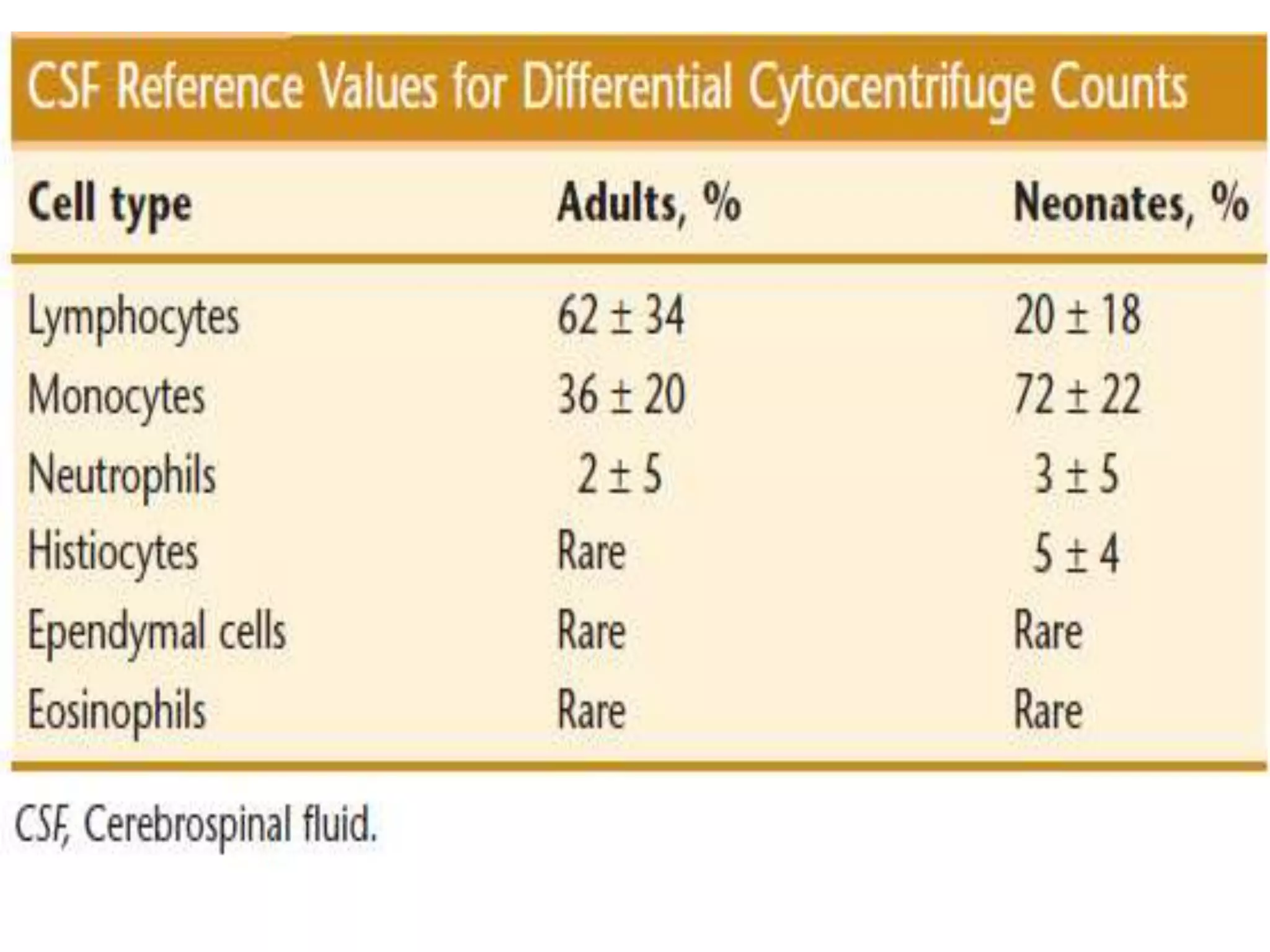
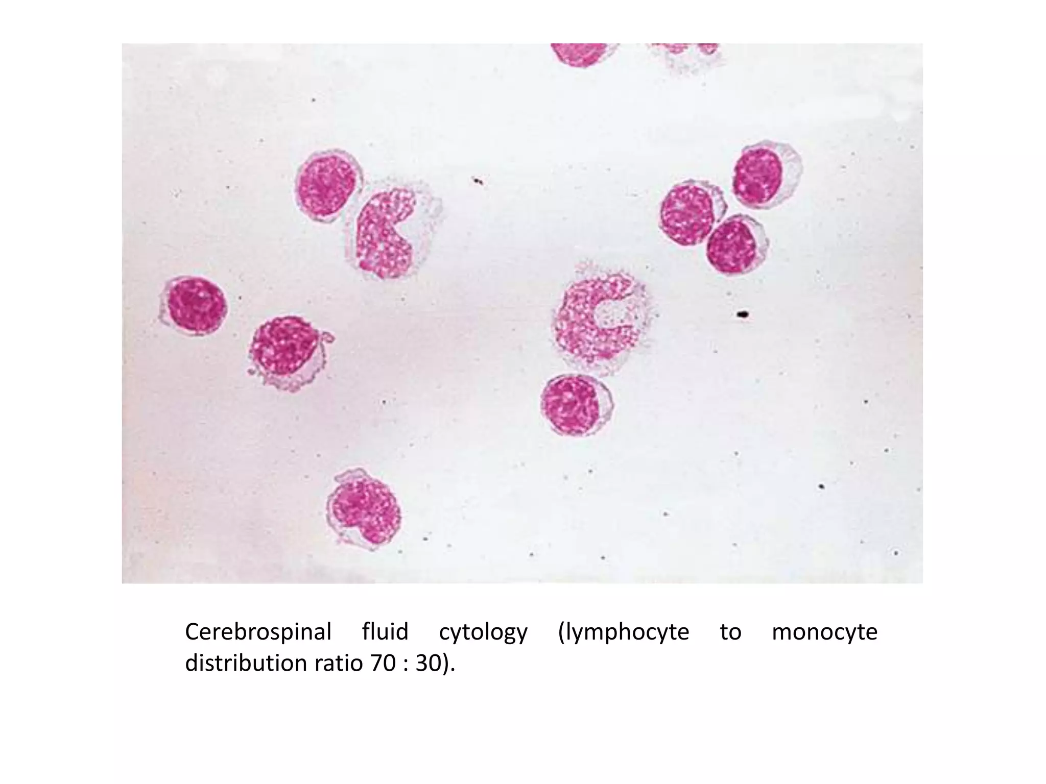
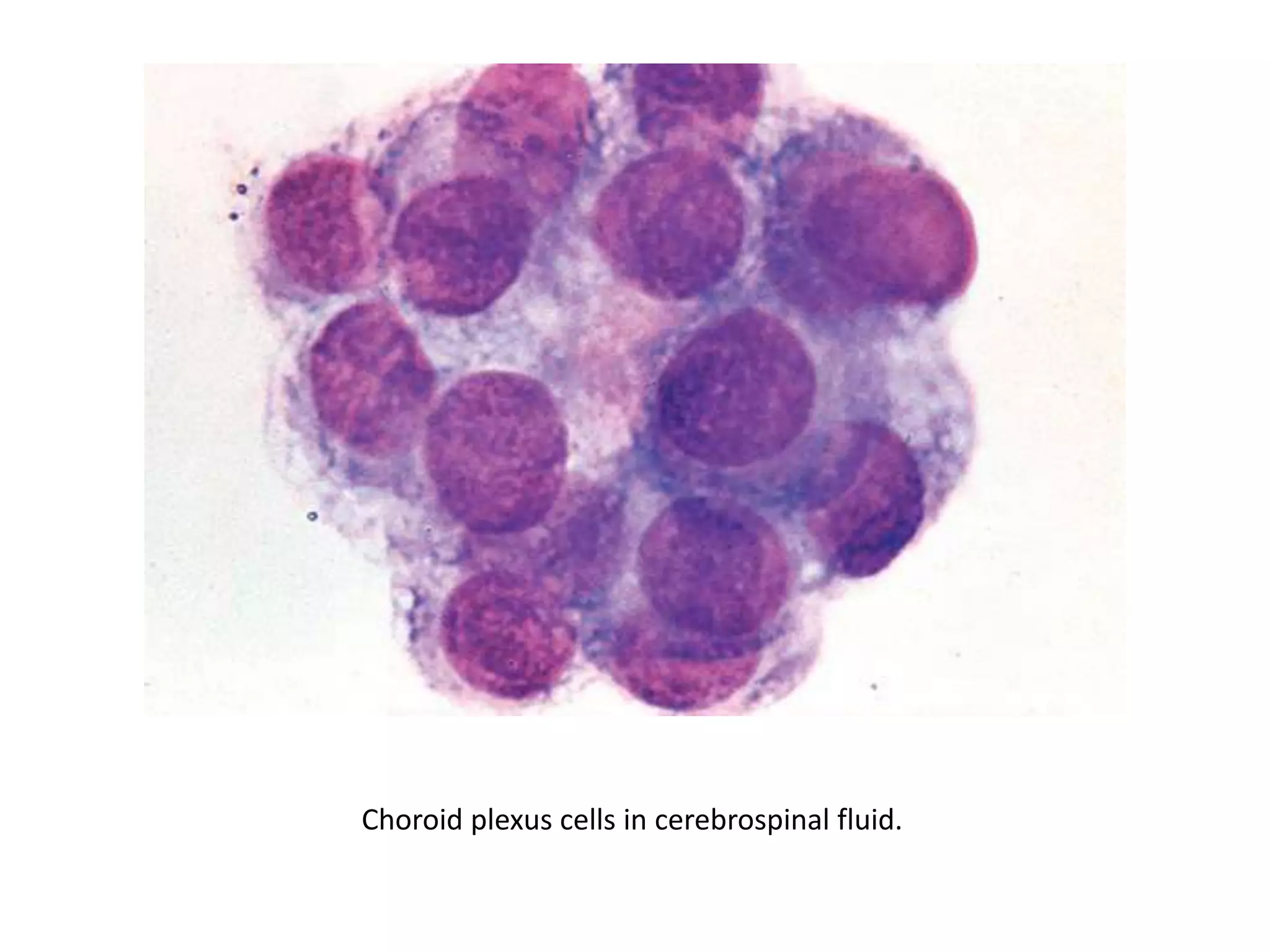
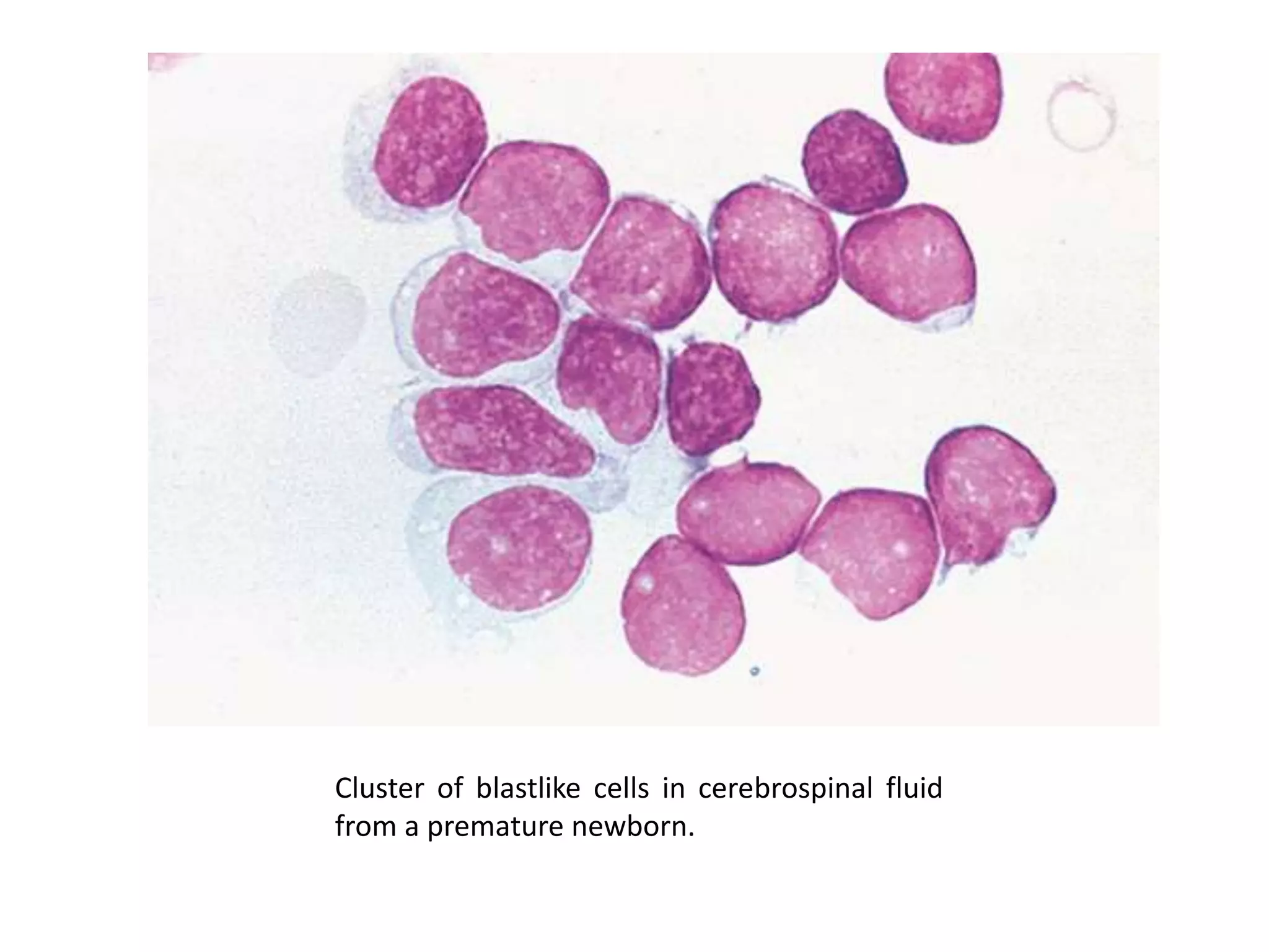
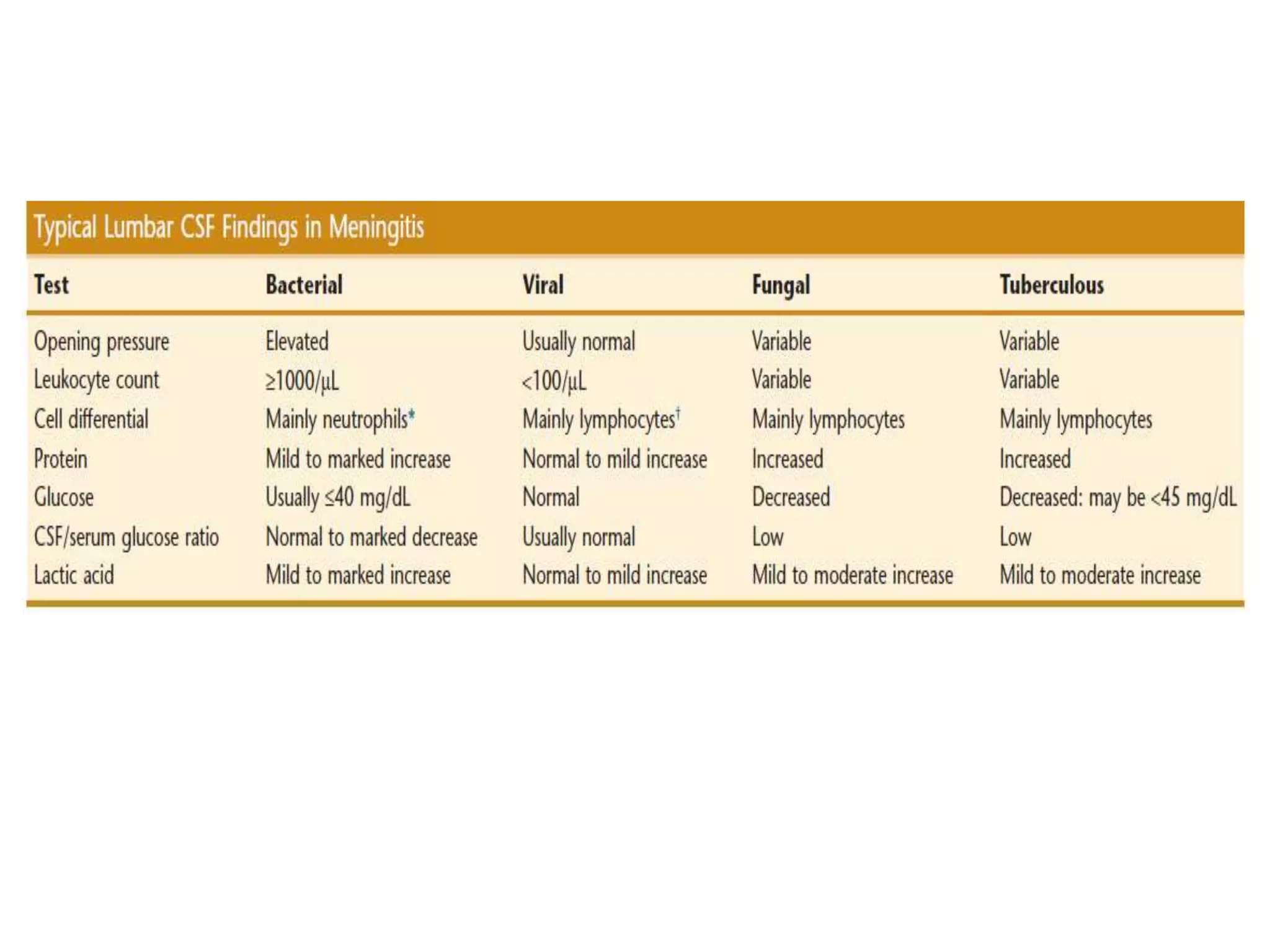
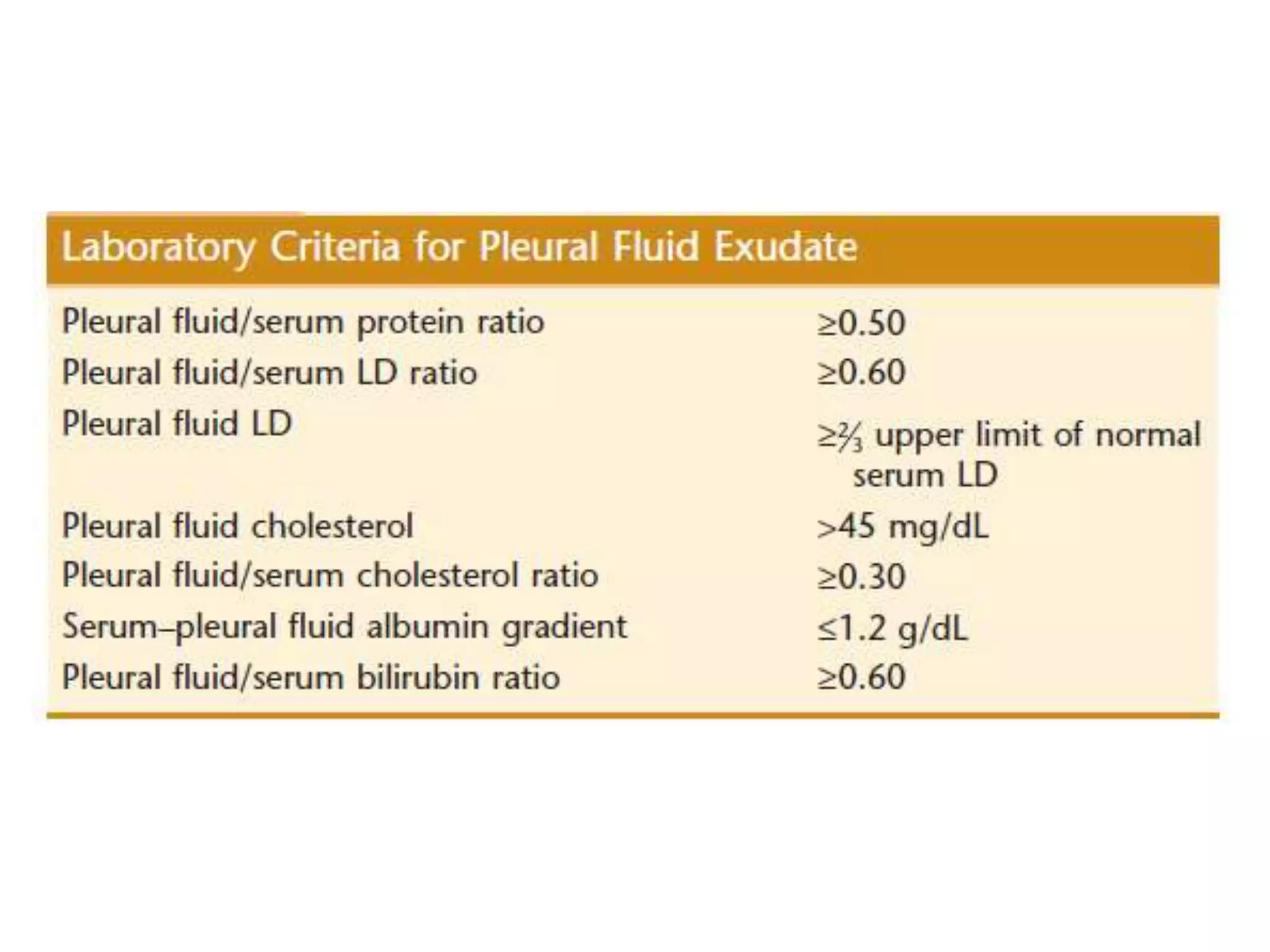
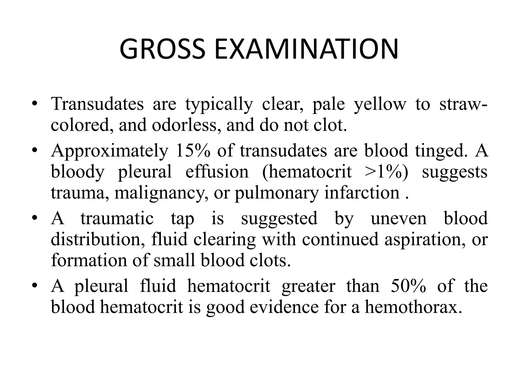
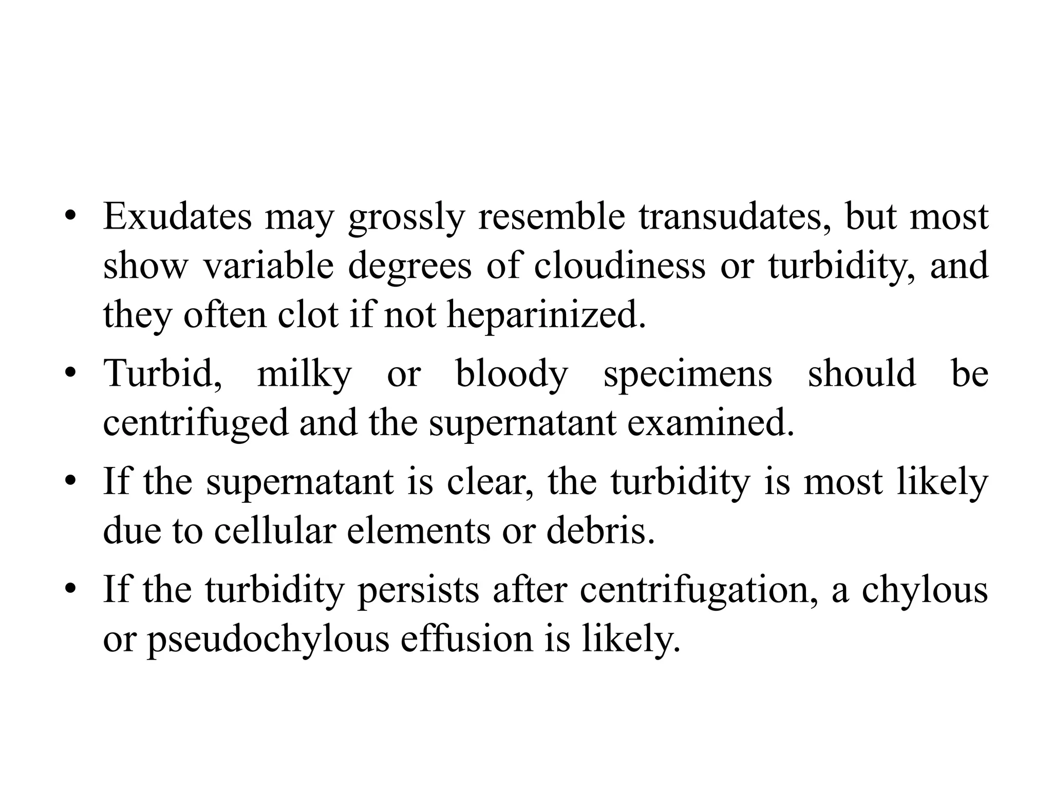
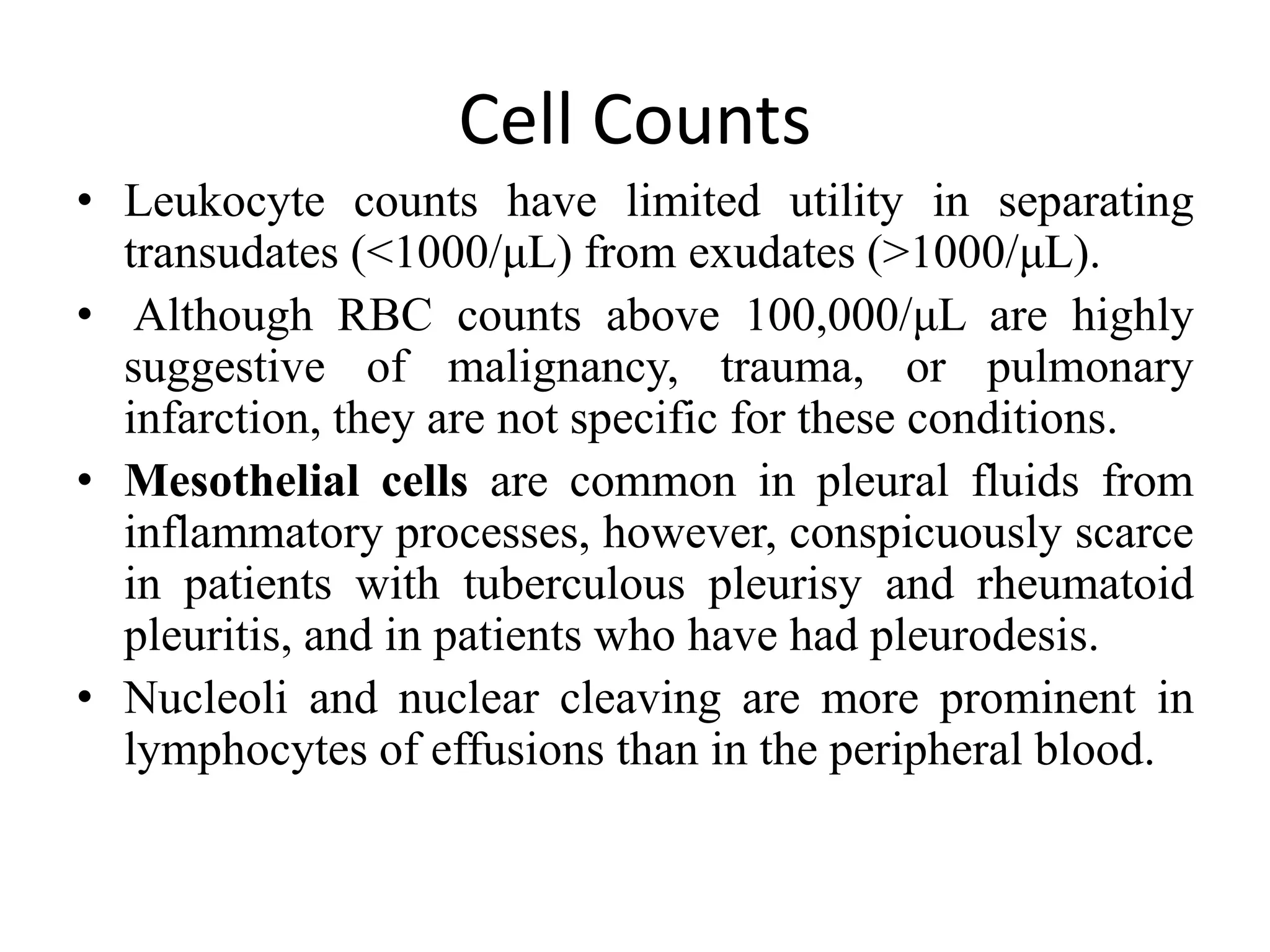
SlideShare a Scribd company logo
EXAMINATION OF BODY FLUIDS
Dr Utkarsh Sharma
Moderated by: Dr Navita Gupta
EXAMINATION OF CSF
• In adults, approximately 500 mL of
cerebrospinal fluid (CSF) is produced each day
(0.3 to 0.4 mL/min). The total adult volume
varies from 90 to 150 mL.
• In neonates, the volume varies from 10 to 60
mL.
• the total CSF volume is replaced every 5 to 7
hours
• The normal opening adult pressure is 90 to 180
mm of water in the lateral decubitus position
with the legs and neck in a neutral position.
• Opening pressures above 250 mm H2O are
diagnostic of intracranial hypertension.
• If the opening pressure is greater than 200 mm
H2O in a relaxed patient, no more than 2.0 mL
should be withdrawn.
• The CSF specimen is usually divided into three
serially collected sterile tubes:
Tube 1 for chemistry and immunology studies;
Tube 2 for microbiological examination
Tube 3 for cell count and differential.
 An additional tube may be inserted in the No. 3
position for cytology if a malignancy is suspected.
• Note: Tube 1 should never be used for
microbiology because it may be contaminated
with skin bacteria.
• Glass tubes should be avoided because cell
adhesion to glass affects the cell count and
differential.
• Specimens should be delivered to the laboratory
and processed quickly to minimize cellular
degradation, which begins within 1 hour of
collection.
• Refrigeration is contraindicated for samples in
which flow cytometry is likely to be needed for
detection of leukemia or lymphoma cells.
GROSS EXAMINATION
• Normal CSF is crystal clear and colorless and has
a viscosity similar to that of water.
• Clot formation may be present in patients with
traumatic taps, complete spinal block (Froin’s
syndrome), or suppurative or tuberculous
meningitis.
• It is not usually seen in patients with
subarachnoid hemorrhage.
• Clots may interfere with cell count accuracy by
entrapping inflammatory cells and/or by
interfering with automated instrument counting
• Viscous CSF may be encountered in patients
with
 Metastatic mucin producing adenocarcinomas.
 Cryptococcal meningitis due to capsular
polysaccharide
Liquid nucleus pulposus resulting from needle
injury to the annulus fibrosus.
• Pink-red CSF usually indicates the presence of
blood and is grossly bloody when the RBC
count exceeds 6000/μL. It may originate from
a subarachnoid hemorrhage, intracerebral
hemorrhage, or cerebral infarct or from a
traumatic spinal tap.
• Xanthochromia commonly refers to a pale
pink to yellow color in the supernatant of
centrifuged CSF.
Visible CSF xanthochromia may also be due to the
following:
• (1) oxyhemoglobin resulting from artifactual red blood
cell lysis caused by detergent contamination of the needle
or collecting tube, or a delay of longer than 1 hour
without refrigeration before examination;
• (2) bilirubin (bilirhachia) in jaundiced patients;
• (3) CSF protein levels over 150 mg/dL,
• (4) disinfectant contamination;
• (5) carotenoids (orange) in people with dietary
hypercarotenemia (i.e., hypervitaminosis A);
• (6) melanin (brownish) from meningeal metastatic
melanoma; and
• (7) rifampin therapy (red-orange).
Differential Diagnosis of Bloody CSF
• A traumatic tap occurs in about 20% of lumbar
punctures. Distinction of a traumatic puncture from a
pathologic hemorrhage is, therefore, of vital importance.
• In a traumatic tap, the hemorrhagic fluid usually clears
between the first and third collected tubes but remains
relatively uniform in subarachnoid hemorrhage.
• Xanthochromia, microscopic evidence of
erythrophagocytosis, or hemosiderin-laden macrophages
indicate a subarachnoid bleed in the absence of a prior
traumatic tap. RBC lysis begins as early as 1 to 2 hours
after a traumatic tap. Thus, rapid evaluation is necessary
to avoid false-positive results.
MICROSCOPIC EXAMINATION
Total Cell Count
• The normal leukocyte cell count in adults is 0 to 5
cells/μL.
• It is higher in neonates, ranging from 0 to 30
cells/μL, with the upper limit of normal
decreasing to adult values by adolescence.
• No RBCs should be present in normal CSF. If
numerous (except with a traumatic tap), a
pathologic process is probable (e.g., trauma,
malignancy, infarct, hemorrhage).
Differential Cell Count
• A differential performed in a counting chamber is
unsatisfactory because the low cell numbers give
rise to poor precision, and identifying the cell
type beyond granulocytes and “mononuclears” is
difficult in a wet preparation.
• Direct smears of the centrifuged CSF sediment
are also subject to significant error from cellular
distortion and fragmentation.
• The cytocentrifuge method is rapid, requires
minimal training, and allows Wright’s staining of
air-dried cytospins. It is the recommended method
for differential cell counts in all body fluids.
• In adults, normal CSF contains small numbers of
lymphocytes and monocytes in an approximate 70 : 30
ratio.
• A higher proportion of monocytes is present in young
children, in whom up to 80% may be normal.
• No general consensus regarding an upper limit of normal
for PMNs has been established. Many laboratories
accept up to 7% neutrophils with a normal WBC count.
• The number of PMNs may be decreased by as much as
68% within the first 2 hours after lumbar puncture owing
to cell lysis .
• A total PMN count of over 1180 cells/μL (or more than
2000 WBCs/μL) has a 99% predictive value for bacterial
meningitis.
Cerebrospinal fluid cytology (lymphocyte to monocyte
distribution ratio 70 : 30).
Choroid plexus cells in cerebrospinal fluid.
Cluster of blastlike cells in cerebrospinal fluid
from a premature newborn.
Hemosiderin-laden macrophages (siderophages) from the
cerebrospinal fluid of a patient with subarachnoid
hemorrhage. Hemosiderin crystals (golden-yellow) are also
present.
Acute lymphoblastic leukemia in cerebrospinal fluid. Note
uniformity of the blast cells.
Burkitt’s lymphoma in cerebrospinal fluid. The cells
are characterized by blue cytoplasm with vacuoles
and a slightly clumped chromatin pattern.
CSF protein levels of 15 to 45 mg/
dL have long been accepted as the “normal”
reference range.
EXAMINATION OF PLEURAL FLUID
• The pleural cavity is a potential space lined by
mesothelium of the visceral and parietal pleurae.
• The pleural cavity normally contains a small amount of
fluid that facilitates movement of the two membranes
against each other.
• An accumulation of fluid, called an effusion, results from
an imbalance of fluid production and reabsorption.
• Except for an EDTA tube for total and differential cell
counts, the specimen should be collected in heparinized
tubes to avoid clotting.
• Because serous effusions are more forgiving than
CSF in maintaining cellular integrity, fresh specimens
for cytology may be stored for up to 48 hours in the
refrigerator with satisfactory results
• Initial classification of a pleural fluid as a transudate
or an exudate greatly simplifies the process of
arriving at a correct final diagnosis.
• Transudates generally require no further workup.
GROSS EXAMINATION
• Transudates are typically clear, pale yellow to straw-
colored, and odorless, and do not clot.
• Approximately 15% of transudates are blood tinged. A
bloody pleural effusion (hematocrit >1%) suggests
trauma, malignancy, or pulmonary infarction .
• A traumatic tap is suggested by uneven blood
distribution, fluid clearing with continued aspiration, or
formation of small blood clots.
• A pleural fluid hematocrit greater than 50% of the
blood hematocrit is good evidence for a hemothorax.
• Exudates may grossly resemble transudates, but most
show variable degrees of cloudiness or turbidity, and
they often clot if not heparinized.
• Turbid, milky or bloody specimens should be
centrifuged and the supernatant examined.
• If the supernatant is clear, the turbidity is most likely
due to cellular elements or debris.
• If the turbidity persists after centrifugation, a chylous
or pseudochylous effusion is likely.
Cell Counts
• Leukocyte counts have limited utility in separating
transudates (<1000/μL) from exudates (>1000/μL).
• Although RBC counts above 100,000/μL are highly
suggestive of malignancy, trauma, or pulmonary
infarction, they are not specific for these conditions.
• Mesothelial cells are common in pleural fluids from
inflammatory processes, however, conspicuously scarce
in patients with tuberculous pleurisy and rheumatoid
pleuritis, and in patients who have had pleurodesis.
• Nucleoli and nuclear cleaving are more prominent in
lymphocytes of effusions than in the peripheral blood.
Mesothelial cells in pleural fluid
Small cell carcinoma of the lung showing typical
molding of nuclei.
EXAMINATION OF PERITONEAL FLUID
• Ascites is the pathologic accumulation of excess fluid
in the peritoneal cavity.
• Up to 50 mL of fluid is normally present in this
mesothelial-lined space.
• Diagnostic paracentesis is performed in most patients
with new ascites, or if there is a change in the clinical
picture of a patient with ascites, such as rapid fluid
accumulation or fever development.
• A minimum of 30 mL is needed for complete
evaluation.
• If possible, at least 100 mL should be provided for
cytologic examination.
GROSS EXAMINATION
• Transudates are generally pale yellow and clear.
• Exudates are cloudy or turbid because of the presence of
leukocytes, tumor cells, or increased protein levels.
• The presence of food particles, foreign material, or green-
yellow bile staining in a DPL specimen suggests
perforation of the gastrointestinal or biliary tract.
• Acute pancreatitis and cholecystitis may also cause
greenish discoloration.
• As little as 15 mL of blood per liter of fluid produces a
bright red opaque color.
• Bloody ascites is also seen in malignancy and
tuberculosis.
MICROSCOPIC EXAMINATION
• The total leukocyte count is useful in distinguishing
ascites due to uncomplicated cirrhosis from
spontaneous bacterial peritonitis (SBP), which is
caused by migration of bacteria from the intestine
into the ascitic fluid.
• Approximately 90% of patients with SBP will have
leukocyte counts greater than 500/μL, more than 50%
of which are neutrophils.
• Eosinophilia (>10%) is most commonly associated
with the chronic inflammatory process associated
with chronic peritoneal dialysis, lymphoma, and
ruptured hydatid cyst.
Semen Analysis
• In addition to its use in the evaluation of reproductive
dysfunction, particularly infertility, semen analysis is used
to select donors for therapeutic insemination and to
monitor the success of surgical procedures, such as
varicocelectomy and vasectomy.
• The patient should be instructed to collect semen after 2
to 5 days of sexual abstinence.
• Longer periods of abstinence usually result in a higher
semen volume but reduced sperm motility.
• In such a case, a second semen specimen may be
collected 2 hours after the first sample is collected.
• The semen specimen should be delivered to the
laboratory within 1 hour of collection and kept warm
during transportation.
• Two specimens collected within 2- to 3-week intervals
should be used for evaluation, and if they are markedly
different, additional specimens should be collected.
• Semen should be thoroughly mixed before
examination, and its viscosity recorded.
• The appearance of a yellow hue in a semen specimen is
associated with pyospermia, and a rust color is due to
small bleedings in the seminal vesicle.
• A hemocytometer or microchamber should be used for the
sperm count.
• Total sperm count is then calculated by multiplying the
dilution factor (normal concentration range, 15-50
million/mL) by its volume (normal range, 2-5 mL).
• Progressive motility (normal range 32% or above) is
expressed as the percentage of sperm that move. In addition,
forward movement is graded.
• Sperm that move rapidly in a straight line with little yaw
and lateral movement are grade 4
• if they move more slowly, grade 3.
• Grade 2 sperm move even more slowly and with substantial
yaw.
• Grade 1 sperm have no forward progression.
• Zero progression denotes absence of any motility.
• If motility is less than 50%, a viability stain of eosin
Y with nigrosin as a counterstain is done. In bright-
field microscopy, dead sperm will stain red, whereas
live sperm will exclude the dye and appear unstained.
• In samples with no visible sperm, such as
postvasectomy semen, the entire sample should be
centrifuged and the pellet examined for intact or
damaged sperm fragments. The analysis should be
repeated in 4 to 6 months.
• Agglutination occurs when motile sperm stick to one
another in an orientation that is reproducible within a
given specimen, such as head to head, tail to tail,
midpiece to midpiece, or mixed ways, depending on the
specificity of sperm antibodies.
• Agglutination suggests an immunologic cause of
infertility, and a description of the type of agglutination
should be recorded.
• This can usually be distinguished from clumping due to
bacterial infection or tissue debris, which typically
involves nonspecific orientation of the sperm.
• Round cells should be differentiated into two classes:
immature germ cells with a single or double highly
condensed nucleus with a relatively large area of
cytoplasm, and polymorphonuclear leukocytes, which
are smaller than the germ cells and have a lower
nuclear/cytoplasmic ratio.
• Peroxidase staining specifically identifies the
polymorphonuclear leukocytes in the presence of
lymphocytes and other cells that normally occur in
semen.
• Bacterial contamination should be noted, as should the
presence or absence of epithelial cells.
• Typically, more than 4% of the sperm in a semen
specimen should exhibit normal morphology.
• A strict morphology score of greater than 4% of normal
indicates excellent fertilizing capacity. Scores between
0% and 3% predict probable inability to fertilize.
• Wide variability in the size of the acrosomal cap is the
most obvious characteristic of abnormal sperm. An
acrosomal cap less than one third of the head surface is
considered abnormal, as are retention of a cytoplasmic
droplet greater than one-half of the head size and a tail
less than 45 μm long.
• Of particular note is the direct relationship between
acrosome size and the frequency of fertilization or
pregnancy.

More Related Content

What's hot

Examination of cerebrospinal fluid presentation mode
Examination of cerebrospinal fluid presentation modeExamination of cerebrospinal fluid presentation mode
Examination of cerebrospinal fluid presentation mode
Pavulraj Selvaraj
 
Reticulocyte count
Reticulocyte countReticulocyte count
Reticulocyte count
Prbn Shah
 
Blood collection and preservation
Blood collection  and preservationBlood collection  and preservation
Blood collection and preservation
globalsoin
 
Peritoneal examination
Peritoneal examinationPeritoneal examination
Peritoneal examination
Nasir Nazeer
 

What's hot (20)

Bleeding and clotting time
Bleeding and clotting time  Bleeding and clotting time
Bleeding and clotting time
 
Gastric analysis
Gastric analysisGastric analysis
Gastric analysis
 
Stool Examinations
Stool ExaminationsStool Examinations
Stool Examinations
 
PLATELET COUNT by Dr. Pandian M .pptx
PLATELET COUNT by Dr. Pandian M .pptxPLATELET COUNT by Dr. Pandian M .pptx
PLATELET COUNT by Dr. Pandian M .pptx
 
Examination of cerebrospinal fluid presentation mode
Examination of cerebrospinal fluid presentation modeExamination of cerebrospinal fluid presentation mode
Examination of cerebrospinal fluid presentation mode
 
Cerebrospinal fluid sample collection
Cerebrospinal fluid  sample collectionCerebrospinal fluid  sample collection
Cerebrospinal fluid sample collection
 
Chemical examination of urine
Chemical examination of urineChemical examination of urine
Chemical examination of urine
 
AEC COUNT
AEC COUNTAEC COUNT
AEC COUNT
 
Pcv
PcvPcv
Pcv
 
Stool examination
Stool examinationStool examination
Stool examination
 
Crossmatching
CrossmatchingCrossmatching
Crossmatching
 
Reticulocyte count
Reticulocyte countReticulocyte count
Reticulocyte count
 
Blood specimen collection
Blood specimen collectionBlood specimen collection
Blood specimen collection
 
Blood collection and its types
Blood collection and its typesBlood collection and its types
Blood collection and its types
 
Introduction to hematology
Introduction to hematologyIntroduction to hematology
Introduction to hematology
 
Bm examination
Bm examinationBm examination
Bm examination
 
Erythrocyte Sedimentation Rate (ESR)
Erythrocyte Sedimentation Rate (ESR)Erythrocyte Sedimentation Rate (ESR)
Erythrocyte Sedimentation Rate (ESR)
 
Blood collection and preservation
Blood collection  and preservationBlood collection  and preservation
Blood collection and preservation
 
Complete blood count test
Complete blood count testComplete blood count test
Complete blood count test
 
Peritoneal examination
Peritoneal examinationPeritoneal examination
Peritoneal examination
 

Similar to Pathological examination of body fluids

CEREBROSPINAL FLUID IN HEALTH AND DISEASE.pptx
CEREBROSPINAL FLUID IN HEALTH AND  DISEASE.pptxCEREBROSPINAL FLUID IN HEALTH AND  DISEASE.pptx
CEREBROSPINAL FLUID IN HEALTH AND DISEASE.pptx
Emmanuellaodia
 
4_2018_02_21!02_23_11_PM.ppt
4_2018_02_21!02_23_11_PM.ppt4_2018_02_21!02_23_11_PM.ppt
4_2018_02_21!02_23_11_PM.ppt
ssuser75fd45
 

Similar to Pathological examination of body fluids (20)

Asiatic Fluid
Asiatic  FluidAsiatic  Fluid
Asiatic Fluid
 
body fluid analysis ppt.pptx
body fluid analysis ppt.pptxbody fluid analysis ppt.pptx
body fluid analysis ppt.pptx
 
CSF-cerebrospinal fluid
CSF-cerebrospinal fluidCSF-cerebrospinal fluid
CSF-cerebrospinal fluid
 
Peripheral smear
Peripheral smear Peripheral smear
Peripheral smear
 
Fluid cytology in CSF
Fluid cytology in CSFFluid cytology in CSF
Fluid cytology in CSF
 
Blood count
Blood countBlood count
Blood count
 
Csf cytological studies and its interpretation1
Csf cytological studies and its interpretation1Csf cytological studies and its interpretation1
Csf cytological studies and its interpretation1
 
PCV.pdf
PCV.pdfPCV.pdf
PCV.pdf
 
Laboratory analysis of csf
Laboratory analysis of csfLaboratory analysis of csf
Laboratory analysis of csf
 
CSF Examination
CSF ExaminationCSF Examination
CSF Examination
 
Instruments and procedures for undergraduates.pptx
Instruments and procedures for undergraduates.pptxInstruments and procedures for undergraduates.pptx
Instruments and procedures for undergraduates.pptx
 
CEREBROSPINAL FLUID IN HEALTH AND DISEASE.pptx
CEREBROSPINAL FLUID IN HEALTH AND  DISEASE.pptxCEREBROSPINAL FLUID IN HEALTH AND  DISEASE.pptx
CEREBROSPINAL FLUID IN HEALTH AND DISEASE.pptx
 
Body Fluid Chapter 1.pptx.ppt
Body Fluid Chapter 1.pptx.pptBody Fluid Chapter 1.pptx.ppt
Body Fluid Chapter 1.pptx.ppt
 
4_2018_02_21!02_23_11_PM.ppt
4_2018_02_21!02_23_11_PM.ppt4_2018_02_21!02_23_11_PM.ppt
4_2018_02_21!02_23_11_PM.ppt
 
Hema I Chapter 14_CSF.ppt
Hema I Chapter 14_CSF.pptHema I Chapter 14_CSF.ppt
Hema I Chapter 14_CSF.ppt
 
Csf composition and significance by Dr. Ashok KUmar J
Csf composition and significance by Dr. Ashok KUmar JCsf composition and significance by Dr. Ashok KUmar J
Csf composition and significance by Dr. Ashok KUmar J
 
Laboratory Investigation particular for Dentistry.pptx
Laboratory Investigation particular for Dentistry.pptxLaboratory Investigation particular for Dentistry.pptx
Laboratory Investigation particular for Dentistry.pptx
 
Determination of Haematocrit in diagnostic hematology
Determination of Haematocrit in diagnostic hematologyDetermination of Haematocrit in diagnostic hematology
Determination of Haematocrit in diagnostic hematology
 
Csf seminar
Csf seminarCsf seminar
Csf seminar
 
CSF examination
CSF examinationCSF examination
CSF examination
 

More from Utkarsh Sharma (8)

Causes of Leukocytosis, Leokopenia, Lymphocytosis and Leukemoid reactions..pptx
Causes of Leukocytosis, Leokopenia, Lymphocytosis and Leukemoid reactions..pptxCauses of Leukocytosis, Leokopenia, Lymphocytosis and Leukemoid reactions..pptx
Causes of Leukocytosis, Leokopenia, Lymphocytosis and Leukemoid reactions..pptx
 
Theme on world heart day 2022.pptx
Theme on world heart day 2022.pptxTheme on world heart day 2022.pptx
Theme on world heart day 2022.pptx
 
COVID-19.pptx
COVID-19.pptxCOVID-19.pptx
COVID-19.pptx
 
Covid 19
Covid 19Covid 19
Covid 19
 
Molecular mechanism of neoplasia
Molecular mechanism of neoplasiaMolecular mechanism of neoplasia
Molecular mechanism of neoplasia
 
Lab diagnosis cancer
Lab diagnosis cancerLab diagnosis cancer
Lab diagnosis cancer
 
An introduction to fluorescent in situ hybridization.
An introduction to fluorescent in situ hybridization.An introduction to fluorescent in situ hybridization.
An introduction to fluorescent in situ hybridization.
 
collection of blood sample and preanalytical errors
collection of blood sample and preanalytical errorscollection of blood sample and preanalytical errors
collection of blood sample and preanalytical errors
 

Recently uploaded

ASSISTING WITH THE USE OF BED PAN BY ANUSHRI SRIVASTAVA.pptx
ASSISTING WITH THE USE OF BED PAN BY ANUSHRI SRIVASTAVA.pptxASSISTING WITH THE USE OF BED PAN BY ANUSHRI SRIVASTAVA.pptx
ASSISTING WITH THE USE OF BED PAN BY ANUSHRI SRIVASTAVA.pptx
AnushriSrivastav
 
Antibiotic Stewardship by Anushri Srivastava.pptx
Antibiotic Stewardship by Anushri Srivastava.pptxAntibiotic Stewardship by Anushri Srivastava.pptx
Antibiotic Stewardship by Anushri Srivastava.pptx
AnushriSrivastav
 

Recently uploaded (20)

Overcome Your Phobias with Hypnotherapy.pptx
Overcome Your Phobias with Hypnotherapy.pptxOvercome Your Phobias with Hypnotherapy.pptx
Overcome Your Phobias with Hypnotherapy.pptx
 
Management of psoriasis.pptx (Recent advances)
Management of psoriasis.pptx (Recent advances)Management of psoriasis.pptx (Recent advances)
Management of psoriasis.pptx (Recent advances)
 
QA Paediatric dentistry department, Hospital Melaka 2020
QA Paediatric dentistry department, Hospital Melaka 2020QA Paediatric dentistry department, Hospital Melaka 2020
QA Paediatric dentistry department, Hospital Melaka 2020
 
Virtual Health Platforms_ Revolutionizing Patient Care.pdf
Virtual Health Platforms_ Revolutionizing Patient Care.pdfVirtual Health Platforms_ Revolutionizing Patient Care.pdf
Virtual Health Platforms_ Revolutionizing Patient Care.pdf
 
Navigating Women's Health: Understanding Prenatal Care and Beyond
Navigating Women's Health: Understanding Prenatal Care and BeyondNavigating Women's Health: Understanding Prenatal Care and Beyond
Navigating Women's Health: Understanding Prenatal Care and Beyond
 
Call Girls in Jaipur (Rajasthan) call me [🔝89011-83002🔝] Escort In Jaipur ℂal...
Call Girls in Jaipur (Rajasthan) call me [🔝89011-83002🔝] Escort In Jaipur ℂal...Call Girls in Jaipur (Rajasthan) call me [🔝89011-83002🔝] Escort In Jaipur ℂal...
Call Girls in Jaipur (Rajasthan) call me [🔝89011-83002🔝] Escort In Jaipur ℂal...
 
What can we really do to give meaning and momentum to equality, diversity and...
What can we really do to give meaning and momentum to equality, diversity and...What can we really do to give meaning and momentum to equality, diversity and...
What can we really do to give meaning and momentum to equality, diversity and...
 
ASSISTING WITH THE USE OF BED PAN BY ANUSHRI SRIVASTAVA.pptx
ASSISTING WITH THE USE OF BED PAN BY ANUSHRI SRIVASTAVA.pptxASSISTING WITH THE USE OF BED PAN BY ANUSHRI SRIVASTAVA.pptx
ASSISTING WITH THE USE OF BED PAN BY ANUSHRI SRIVASTAVA.pptx
 
Deepfake Detection_Using Machine Learning .pptx
Deepfake Detection_Using Machine Learning .pptxDeepfake Detection_Using Machine Learning .pptx
Deepfake Detection_Using Machine Learning .pptx
 
Antibiotic Stewardship by Anushri Srivastava.pptx
Antibiotic Stewardship by Anushri Srivastava.pptxAntibiotic Stewardship by Anushri Srivastava.pptx
Antibiotic Stewardship by Anushri Srivastava.pptx
 
Contact Now 89011**83002 Dehradun ℂall Girls By Full Service ℂall Girl In De...
Contact Now  89011**83002 Dehradun ℂall Girls By Full Service ℂall Girl In De...Contact Now  89011**83002 Dehradun ℂall Girls By Full Service ℂall Girl In De...
Contact Now 89011**83002 Dehradun ℂall Girls By Full Service ℂall Girl In De...
 
Valle Egypt Illustrates Consequences of Financial Elder Abuse
Valle Egypt Illustrates Consequences of Financial Elder AbuseValle Egypt Illustrates Consequences of Financial Elder Abuse
Valle Egypt Illustrates Consequences of Financial Elder Abuse
 
💃Joint ❤89011-83002❤ #ℂALL #gIRLS Ludhiana Escorts by ✔️🍑💃Hotel #cALL #gIRLS...
💃Joint ❤89011-83002❤ #ℂALL #gIRLS Ludhiana Escorts  by ✔️🍑💃Hotel #cALL #gIRLS...💃Joint ❤89011-83002❤ #ℂALL #gIRLS Ludhiana Escorts  by ✔️🍑💃Hotel #cALL #gIRLS...
💃Joint ❤89011-83002❤ #ℂALL #gIRLS Ludhiana Escorts by ✔️🍑💃Hotel #cALL #gIRLS...
 
Digital Healthcare: The Future of Medical Consultations
Digital Healthcare: The Future of Medical ConsultationsDigital Healthcare: The Future of Medical Consultations
Digital Healthcare: The Future of Medical Consultations
 
Healthcare Companion Robots: Key Features and Functionalities, Benefits, Chal...
Healthcare Companion Robots: Key Features and Functionalities, Benefits, Chal...Healthcare Companion Robots: Key Features and Functionalities, Benefits, Chal...
Healthcare Companion Robots: Key Features and Functionalities, Benefits, Chal...
 
Best Erectile Dysfunction Treatment In Narela
Best Erectile Dysfunction Treatment In NarelaBest Erectile Dysfunction Treatment In Narela
Best Erectile Dysfunction Treatment In Narela
 
A Community health , health for prisoners
A Community health  , health for prisonersA Community health  , health for prisoners
A Community health , health for prisoners
 
VVIP Dehradun Girls 9719300533 Heat-bake { Dehradun } Genteel ℂall Serviℂe By...
VVIP Dehradun Girls 9719300533 Heat-bake { Dehradun } Genteel ℂall Serviℂe By...VVIP Dehradun Girls 9719300533 Heat-bake { Dehradun } Genteel ℂall Serviℂe By...
VVIP Dehradun Girls 9719300533 Heat-bake { Dehradun } Genteel ℂall Serviℂe By...
 
Jaipur @ℂall @Girls ꧁❤8901183002❤꧂@ℂall @Girls Service Vip Top Model Safe
Jaipur @ℂall @Girls ꧁❤8901183002❤꧂@ℂall @Girls Service Vip Top Model SafeJaipur @ℂall @Girls ꧁❤8901183002❤꧂@ℂall @Girls Service Vip Top Model Safe
Jaipur @ℂall @Girls ꧁❤8901183002❤꧂@ℂall @Girls Service Vip Top Model Safe
 
Occupational Therapy Management for Parkinson's Disease - Webinar 2024
Occupational Therapy Management for Parkinson's Disease - Webinar 2024Occupational Therapy Management for Parkinson's Disease - Webinar 2024
Occupational Therapy Management for Parkinson's Disease - Webinar 2024
 

Pathological examination of body fluids

  • 1. EXAMINATION OF BODY FLUIDS Dr Utkarsh Sharma Moderated by: Dr Navita Gupta
  • 2. EXAMINATION OF CSF • In adults, approximately 500 mL of cerebrospinal fluid (CSF) is produced each day (0.3 to 0.4 mL/min). The total adult volume varies from 90 to 150 mL. • In neonates, the volume varies from 10 to 60 mL. • the total CSF volume is replaced every 5 to 7 hours
  • 3. • The normal opening adult pressure is 90 to 180 mm of water in the lateral decubitus position with the legs and neck in a neutral position. • Opening pressures above 250 mm H2O are diagnostic of intracranial hypertension. • If the opening pressure is greater than 200 mm H2O in a relaxed patient, no more than 2.0 mL should be withdrawn.
  • 4. • The CSF specimen is usually divided into three serially collected sterile tubes: Tube 1 for chemistry and immunology studies; Tube 2 for microbiological examination Tube 3 for cell count and differential.  An additional tube may be inserted in the No. 3 position for cytology if a malignancy is suspected. • Note: Tube 1 should never be used for microbiology because it may be contaminated with skin bacteria.
  • 5. • Glass tubes should be avoided because cell adhesion to glass affects the cell count and differential. • Specimens should be delivered to the laboratory and processed quickly to minimize cellular degradation, which begins within 1 hour of collection. • Refrigeration is contraindicated for samples in which flow cytometry is likely to be needed for detection of leukemia or lymphoma cells.
  • 6. GROSS EXAMINATION • Normal CSF is crystal clear and colorless and has a viscosity similar to that of water. • Clot formation may be present in patients with traumatic taps, complete spinal block (Froin’s syndrome), or suppurative or tuberculous meningitis. • It is not usually seen in patients with subarachnoid hemorrhage. • Clots may interfere with cell count accuracy by entrapping inflammatory cells and/or by interfering with automated instrument counting
  • 7. • Viscous CSF may be encountered in patients with  Metastatic mucin producing adenocarcinomas.  Cryptococcal meningitis due to capsular polysaccharide Liquid nucleus pulposus resulting from needle injury to the annulus fibrosus.
  • 8. • Pink-red CSF usually indicates the presence of blood and is grossly bloody when the RBC count exceeds 6000/μL. It may originate from a subarachnoid hemorrhage, intracerebral hemorrhage, or cerebral infarct or from a traumatic spinal tap. • Xanthochromia commonly refers to a pale pink to yellow color in the supernatant of centrifuged CSF.
  • 9.
  • 10. Visible CSF xanthochromia may also be due to the following: • (1) oxyhemoglobin resulting from artifactual red blood cell lysis caused by detergent contamination of the needle or collecting tube, or a delay of longer than 1 hour without refrigeration before examination; • (2) bilirubin (bilirhachia) in jaundiced patients; • (3) CSF protein levels over 150 mg/dL, • (4) disinfectant contamination; • (5) carotenoids (orange) in people with dietary hypercarotenemia (i.e., hypervitaminosis A); • (6) melanin (brownish) from meningeal metastatic melanoma; and • (7) rifampin therapy (red-orange).
  • 11. Differential Diagnosis of Bloody CSF • A traumatic tap occurs in about 20% of lumbar punctures. Distinction of a traumatic puncture from a pathologic hemorrhage is, therefore, of vital importance. • In a traumatic tap, the hemorrhagic fluid usually clears between the first and third collected tubes but remains relatively uniform in subarachnoid hemorrhage. • Xanthochromia, microscopic evidence of erythrophagocytosis, or hemosiderin-laden macrophages indicate a subarachnoid bleed in the absence of a prior traumatic tap. RBC lysis begins as early as 1 to 2 hours after a traumatic tap. Thus, rapid evaluation is necessary to avoid false-positive results.
  • 12. MICROSCOPIC EXAMINATION Total Cell Count • The normal leukocyte cell count in adults is 0 to 5 cells/μL. • It is higher in neonates, ranging from 0 to 30 cells/μL, with the upper limit of normal decreasing to adult values by adolescence. • No RBCs should be present in normal CSF. If numerous (except with a traumatic tap), a pathologic process is probable (e.g., trauma, malignancy, infarct, hemorrhage).
  • 13. Differential Cell Count • A differential performed in a counting chamber is unsatisfactory because the low cell numbers give rise to poor precision, and identifying the cell type beyond granulocytes and “mononuclears” is difficult in a wet preparation. • Direct smears of the centrifuged CSF sediment are also subject to significant error from cellular distortion and fragmentation. • The cytocentrifuge method is rapid, requires minimal training, and allows Wright’s staining of air-dried cytospins. It is the recommended method for differential cell counts in all body fluids.
  • 14. • In adults, normal CSF contains small numbers of lymphocytes and monocytes in an approximate 70 : 30 ratio. • A higher proportion of monocytes is present in young children, in whom up to 80% may be normal. • No general consensus regarding an upper limit of normal for PMNs has been established. Many laboratories accept up to 7% neutrophils with a normal WBC count. • The number of PMNs may be decreased by as much as 68% within the first 2 hours after lumbar puncture owing to cell lysis . • A total PMN count of over 1180 cells/μL (or more than 2000 WBCs/μL) has a 99% predictive value for bacterial meningitis.
  • 15.
  • 16. Cerebrospinal fluid cytology (lymphocyte to monocyte distribution ratio 70 : 30).
  • 17. Choroid plexus cells in cerebrospinal fluid.
  • 18. Cluster of blastlike cells in cerebrospinal fluid from a premature newborn.
  • 19.
  • 20.
  • 21.
  • 22. Hemosiderin-laden macrophages (siderophages) from the cerebrospinal fluid of a patient with subarachnoid hemorrhage. Hemosiderin crystals (golden-yellow) are also present.
  • 23. Acute lymphoblastic leukemia in cerebrospinal fluid. Note uniformity of the blast cells.
  • 24. Burkitt’s lymphoma in cerebrospinal fluid. The cells are characterized by blue cytoplasm with vacuoles and a slightly clumped chromatin pattern.
  • 25. CSF protein levels of 15 to 45 mg/ dL have long been accepted as the “normal” reference range.
  • 26.
  • 27. EXAMINATION OF PLEURAL FLUID • The pleural cavity is a potential space lined by mesothelium of the visceral and parietal pleurae. • The pleural cavity normally contains a small amount of fluid that facilitates movement of the two membranes against each other. • An accumulation of fluid, called an effusion, results from an imbalance of fluid production and reabsorption. • Except for an EDTA tube for total and differential cell counts, the specimen should be collected in heparinized tubes to avoid clotting.
  • 28. • Because serous effusions are more forgiving than CSF in maintaining cellular integrity, fresh specimens for cytology may be stored for up to 48 hours in the refrigerator with satisfactory results • Initial classification of a pleural fluid as a transudate or an exudate greatly simplifies the process of arriving at a correct final diagnosis. • Transudates generally require no further workup.
  • 29.
  • 30.
  • 31. GROSS EXAMINATION • Transudates are typically clear, pale yellow to straw- colored, and odorless, and do not clot. • Approximately 15% of transudates are blood tinged. A bloody pleural effusion (hematocrit >1%) suggests trauma, malignancy, or pulmonary infarction . • A traumatic tap is suggested by uneven blood distribution, fluid clearing with continued aspiration, or formation of small blood clots. • A pleural fluid hematocrit greater than 50% of the blood hematocrit is good evidence for a hemothorax.
  • 32. • Exudates may grossly resemble transudates, but most show variable degrees of cloudiness or turbidity, and they often clot if not heparinized. • Turbid, milky or bloody specimens should be centrifuged and the supernatant examined. • If the supernatant is clear, the turbidity is most likely due to cellular elements or debris. • If the turbidity persists after centrifugation, a chylous or pseudochylous effusion is likely.
  • 33.
  • 34. Cell Counts • Leukocyte counts have limited utility in separating transudates (<1000/μL) from exudates (>1000/μL). • Although RBC counts above 100,000/μL are highly suggestive of malignancy, trauma, or pulmonary infarction, they are not specific for these conditions. • Mesothelial cells are common in pleural fluids from inflammatory processes, however, conspicuously scarce in patients with tuberculous pleurisy and rheumatoid pleuritis, and in patients who have had pleurodesis. • Nucleoli and nuclear cleaving are more prominent in lymphocytes of effusions than in the peripheral blood.
  • 35. Mesothelial cells in pleural fluid
  • 36.
  • 37. Small cell carcinoma of the lung showing typical molding of nuclei.
  • 38. EXAMINATION OF PERITONEAL FLUID • Ascites is the pathologic accumulation of excess fluid in the peritoneal cavity. • Up to 50 mL of fluid is normally present in this mesothelial-lined space. • Diagnostic paracentesis is performed in most patients with new ascites, or if there is a change in the clinical picture of a patient with ascites, such as rapid fluid accumulation or fever development. • A minimum of 30 mL is needed for complete evaluation. • If possible, at least 100 mL should be provided for cytologic examination.
  • 39. GROSS EXAMINATION • Transudates are generally pale yellow and clear. • Exudates are cloudy or turbid because of the presence of leukocytes, tumor cells, or increased protein levels. • The presence of food particles, foreign material, or green- yellow bile staining in a DPL specimen suggests perforation of the gastrointestinal or biliary tract. • Acute pancreatitis and cholecystitis may also cause greenish discoloration. • As little as 15 mL of blood per liter of fluid produces a bright red opaque color. • Bloody ascites is also seen in malignancy and tuberculosis.
  • 40. MICROSCOPIC EXAMINATION • The total leukocyte count is useful in distinguishing ascites due to uncomplicated cirrhosis from spontaneous bacterial peritonitis (SBP), which is caused by migration of bacteria from the intestine into the ascitic fluid. • Approximately 90% of patients with SBP will have leukocyte counts greater than 500/μL, more than 50% of which are neutrophils. • Eosinophilia (>10%) is most commonly associated with the chronic inflammatory process associated with chronic peritoneal dialysis, lymphoma, and ruptured hydatid cyst.
  • 41. Semen Analysis • In addition to its use in the evaluation of reproductive dysfunction, particularly infertility, semen analysis is used to select donors for therapeutic insemination and to monitor the success of surgical procedures, such as varicocelectomy and vasectomy. • The patient should be instructed to collect semen after 2 to 5 days of sexual abstinence. • Longer periods of abstinence usually result in a higher semen volume but reduced sperm motility. • In such a case, a second semen specimen may be collected 2 hours after the first sample is collected.
  • 42. • The semen specimen should be delivered to the laboratory within 1 hour of collection and kept warm during transportation. • Two specimens collected within 2- to 3-week intervals should be used for evaluation, and if they are markedly different, additional specimens should be collected. • Semen should be thoroughly mixed before examination, and its viscosity recorded. • The appearance of a yellow hue in a semen specimen is associated with pyospermia, and a rust color is due to small bleedings in the seminal vesicle.
  • 43.
  • 44. • A hemocytometer or microchamber should be used for the sperm count. • Total sperm count is then calculated by multiplying the dilution factor (normal concentration range, 15-50 million/mL) by its volume (normal range, 2-5 mL). • Progressive motility (normal range 32% or above) is expressed as the percentage of sperm that move. In addition, forward movement is graded. • Sperm that move rapidly in a straight line with little yaw and lateral movement are grade 4 • if they move more slowly, grade 3. • Grade 2 sperm move even more slowly and with substantial yaw. • Grade 1 sperm have no forward progression. • Zero progression denotes absence of any motility.
  • 45. • If motility is less than 50%, a viability stain of eosin Y with nigrosin as a counterstain is done. In bright- field microscopy, dead sperm will stain red, whereas live sperm will exclude the dye and appear unstained. • In samples with no visible sperm, such as postvasectomy semen, the entire sample should be centrifuged and the pellet examined for intact or damaged sperm fragments. The analysis should be repeated in 4 to 6 months.
  • 46. • Agglutination occurs when motile sperm stick to one another in an orientation that is reproducible within a given specimen, such as head to head, tail to tail, midpiece to midpiece, or mixed ways, depending on the specificity of sperm antibodies. • Agglutination suggests an immunologic cause of infertility, and a description of the type of agglutination should be recorded. • This can usually be distinguished from clumping due to bacterial infection or tissue debris, which typically involves nonspecific orientation of the sperm.
  • 47. • Round cells should be differentiated into two classes: immature germ cells with a single or double highly condensed nucleus with a relatively large area of cytoplasm, and polymorphonuclear leukocytes, which are smaller than the germ cells and have a lower nuclear/cytoplasmic ratio. • Peroxidase staining specifically identifies the polymorphonuclear leukocytes in the presence of lymphocytes and other cells that normally occur in semen. • Bacterial contamination should be noted, as should the presence or absence of epithelial cells.
  • 48. • Typically, more than 4% of the sperm in a semen specimen should exhibit normal morphology. • A strict morphology score of greater than 4% of normal indicates excellent fertilizing capacity. Scores between 0% and 3% predict probable inability to fertilize. • Wide variability in the size of the acrosomal cap is the most obvious characteristic of abnormal sperm. An acrosomal cap less than one third of the head surface is considered abnormal, as are retention of a cytoplasmic droplet greater than one-half of the head size and a tail less than 45 μm long. • Of particular note is the direct relationship between acrosome size and the frequency of fertilization or pregnancy.