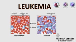
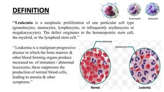
Mr. HIREN GEHLOTH
JG COLLEGE OF NURSING
• The term leukocytosis refers to an increased level of leukocytes in the
circulation. Typically, only one specific cell type is increased. Because the
proportions of several types of leukocytes (eg, eosinophils, basophils,
monocytes) are small, only an increase in neutrophils or lymphocytes can
be great enough to elevate the total leukocyte count.
• Although leukocytosis can be a normal response to increased need (eg, in
acute infection), the elevation in leukocytes should decrease as the
physiologic need decreases.
• A prolonged or progressively increasing elevation in leukocytes is
abnormal and should be evaluated. A significant cause of persistent
leukocytosis is hematologic malignancy.
INTRODUCTION
DEFINITION
‘’Leukemia is a neoplastic proliferation of one particular cell type
(granulocytes, monocytes, lymphocytes, or infrequently erythrocytes or
megakaryocytes). The defect originates in the hematopoietic stem cell,
the myeloid, or the lymphoid stem cell.’’
‘’Leukemia is a malignant progressive
disease in which the bone marrow &
other blood forming organs produce
increased no. of immature / abnormal
leucocytes, these suppresses the
production of normal blood cells,
leading to anemia & other
symptoms.’’
Incidence and Etiology
• Leukemia is the most common malignancy of children less than 15 years of age.
• Incidence in males is greater than in females.
• Its twice more common in white than black children.
• The exact cause of leukemia is unknown. But several etiological factors have
been identified including:
• Virus like Human Papilloma Virus, Epstein Barr virus
• Radiation
• Exposure to chemicals and drugs like benzene and Dilantin
• Familial predisposition
• Chromosomal Abnormalities
CLASSIFICATION
The leukemia's are commonly classified according to the stem cell
line involved, either lymphoid or myeloid.
They are also classified as either acute
or chronic, based on the time
it takes for symptoms to
evolve.
• Acute lymphocytic leukemia (ALL) results from an uncontrolled
proliferation of immature cells (lymphoblasts) from the lymphoid
stem cell. It is most common in young children; boys are affected
more frequently than girls, with a peak incidence at 4 years of age.
• After age 15 years, ALL is uncommon.
• Therapy for this childhood leukemia has improved to the
extent that about 80% of children survive at least 5 years.
1. Acute lymphocytic leukemia
• Chronic lymphocytic leukemia (CLL) is a common cancer of older
adulthood; the average age at diagnosis is 72 years.
• Most of the leukemia cells in CLL are fully mature, so it tends to be a mild
disorder compared with the acute form.
• The disease is usually diagnosed during physical examination or treatment
for another disease.
2. Chronic lymphocytic leukemia
• Acute myeloid leukemia (AML) results from a defect in the hematopoietic
stem cell that differentiates into all myeloid cells: monocytes,
granulocytes (eg, neutrophils, basophils, eosinophils), erythrocytes, and
platelets.
• AML can be further classified into seven different subgroups based on
cytogenetics, histology, and morphology (appearance) of the blasts.
• All age groups are affected; incidence rises with age and peaks at 67 years
of age.
• Death usually occurs secondary to infection or hemorrhage.
3. Acute myeloid leukemia
4. Chronic myeloid leukemia
• Chronic myeloid leukemia (CML) arises from a mutation in the myeloid stem
cells. A wide spectrum of cell types exists within the blood, from blast forms
through mature neutrophils.
• A cytogenetic abnormality termed the Philadelphia chromosome is found in
90% to 95% of patients. CML is uncommon before 20 years of age, but the
incidence increases with age (mean age is 67 years).
• CML has three stages: chronic, transformation, and accelerated or blast crisis.
• Marrow expands into cavities of the long bones, and cells are formed in the
liver and spleen, with resultant painful enlargement problems.
• Infection and bleeding are rare until the disease transforms to the acute phase.
CLINICAL MANIFESTATION
• Tire easily, little energy, weakness.
• Pale skin tone.
• Fever.
• Easy bruising and bleeding. Nosebleeds and bleeding gums. Tiny red
spots in skin (called petechiae). Purplish patches in the skin.
• Bone or joint pain and/or tenderness.
• Swollen lymph nodes in the neck, underarm, groin or stomach; enlarged
spleen or liver.
• Frequent infections.
• Unplanned weight loss.
• Night sweats.
• Shortness of breath.
• Pain or full feeling under the ribs
on the left side .
NURSING MANAGEMENT
ASSESSMENT
• Identify range of signs and symptoms reported by patient in nursing history
and physical examination.
• Assess results of blood studies, and report alterations of WBCs, absolute
neutrophil count (ANC), hematocrit, platelet, creatinine and electrolyte
levels, hepatic function tests, and culture results.
NURSING DIAGNOSIS
• Risk for infection and bleeding.
• Risk for impaired skin integrity related to toxic effects of chemotherapy,
alteration in nutrition, and impaired mobility.
• Impaired gas exchange.
• Impaired mucous membranes from changes in epithelial lining of the
gastrointestinal (GI) tract from chemotherapy or antimicrobial medications.
• Imbalanced nutrition: less than body requirements related to
hypermetabolic state, anorexia, mucositis, pain, and nausea.
• Acute pain and discomfort related to mucositis, leukocytic, infiltration of
systemic tissues, fever, and infection
• Hyperthermia related to tumor lysis and infection
• Fatigue and activity intolerance related to anemia, infection, and
deconditioning
• Impaired physical mobility due to anemia, malaise, discomfort, and
protective isolation
• Risk for excess fluid volume related to renal dysfunction, hypoproteinemia,
need for multiple intravenous (IV) medications and blood products.
• Diarrhea due to altered GI flora, mucosal denudation, prolonged use of
broad-spectrum antibiotics.
• Risk for deficient fluid volume related to potential for diarrhea, bleeding,
infection, and increased metabolic rate.
Leukemia

Leukemia

  • 1.
    Mr. HIREN GEHLOTH JGCOLLEGE OF NURSING
  • 2.
    • The termleukocytosis refers to an increased level of leukocytes in the circulation. Typically, only one specific cell type is increased. Because the proportions of several types of leukocytes (eg, eosinophils, basophils, monocytes) are small, only an increase in neutrophils or lymphocytes can be great enough to elevate the total leukocyte count. • Although leukocytosis can be a normal response to increased need (eg, in acute infection), the elevation in leukocytes should decrease as the physiologic need decreases. • A prolonged or progressively increasing elevation in leukocytes is abnormal and should be evaluated. A significant cause of persistent leukocytosis is hematologic malignancy. INTRODUCTION
  • 3.
    DEFINITION ‘’Leukemia is aneoplastic proliferation of one particular cell type (granulocytes, monocytes, lymphocytes, or infrequently erythrocytes or megakaryocytes). The defect originates in the hematopoietic stem cell, the myeloid, or the lymphoid stem cell.’’ ‘’Leukemia is a malignant progressive disease in which the bone marrow & other blood forming organs produce increased no. of immature / abnormal leucocytes, these suppresses the production of normal blood cells, leading to anemia & other symptoms.’’
  • 4.
    Incidence and Etiology •Leukemia is the most common malignancy of children less than 15 years of age. • Incidence in males is greater than in females. • Its twice more common in white than black children. • The exact cause of leukemia is unknown. But several etiological factors have been identified including: • Virus like Human Papilloma Virus, Epstein Barr virus • Radiation • Exposure to chemicals and drugs like benzene and Dilantin • Familial predisposition • Chromosomal Abnormalities
  • 5.
    CLASSIFICATION The leukemia's arecommonly classified according to the stem cell line involved, either lymphoid or myeloid. They are also classified as either acute or chronic, based on the time it takes for symptoms to evolve.
  • 8.
    • Acute lymphocyticleukemia (ALL) results from an uncontrolled proliferation of immature cells (lymphoblasts) from the lymphoid stem cell. It is most common in young children; boys are affected more frequently than girls, with a peak incidence at 4 years of age. • After age 15 years, ALL is uncommon. • Therapy for this childhood leukemia has improved to the extent that about 80% of children survive at least 5 years. 1. Acute lymphocytic leukemia
  • 9.
    • Chronic lymphocyticleukemia (CLL) is a common cancer of older adulthood; the average age at diagnosis is 72 years. • Most of the leukemia cells in CLL are fully mature, so it tends to be a mild disorder compared with the acute form. • The disease is usually diagnosed during physical examination or treatment for another disease. 2. Chronic lymphocytic leukemia
  • 10.
    • Acute myeloidleukemia (AML) results from a defect in the hematopoietic stem cell that differentiates into all myeloid cells: monocytes, granulocytes (eg, neutrophils, basophils, eosinophils), erythrocytes, and platelets. • AML can be further classified into seven different subgroups based on cytogenetics, histology, and morphology (appearance) of the blasts. • All age groups are affected; incidence rises with age and peaks at 67 years of age. • Death usually occurs secondary to infection or hemorrhage. 3. Acute myeloid leukemia
  • 11.
    4. Chronic myeloidleukemia • Chronic myeloid leukemia (CML) arises from a mutation in the myeloid stem cells. A wide spectrum of cell types exists within the blood, from blast forms through mature neutrophils. • A cytogenetic abnormality termed the Philadelphia chromosome is found in 90% to 95% of patients. CML is uncommon before 20 years of age, but the incidence increases with age (mean age is 67 years). • CML has three stages: chronic, transformation, and accelerated or blast crisis. • Marrow expands into cavities of the long bones, and cells are formed in the liver and spleen, with resultant painful enlargement problems. • Infection and bleeding are rare until the disease transforms to the acute phase.
  • 12.
    CLINICAL MANIFESTATION • Tireeasily, little energy, weakness. • Pale skin tone. • Fever. • Easy bruising and bleeding. Nosebleeds and bleeding gums. Tiny red spots in skin (called petechiae). Purplish patches in the skin. • Bone or joint pain and/or tenderness. • Swollen lymph nodes in the neck, underarm, groin or stomach; enlarged spleen or liver.
  • 13.
    • Frequent infections. •Unplanned weight loss. • Night sweats. • Shortness of breath. • Pain or full feeling under the ribs on the left side .
  • 20.
    NURSING MANAGEMENT ASSESSMENT • Identifyrange of signs and symptoms reported by patient in nursing history and physical examination. • Assess results of blood studies, and report alterations of WBCs, absolute neutrophil count (ANC), hematocrit, platelet, creatinine and electrolyte levels, hepatic function tests, and culture results.
  • 21.
    NURSING DIAGNOSIS • Riskfor infection and bleeding. • Risk for impaired skin integrity related to toxic effects of chemotherapy, alteration in nutrition, and impaired mobility. • Impaired gas exchange. • Impaired mucous membranes from changes in epithelial lining of the gastrointestinal (GI) tract from chemotherapy or antimicrobial medications. • Imbalanced nutrition: less than body requirements related to hypermetabolic state, anorexia, mucositis, pain, and nausea.
  • 22.
    • Acute painand discomfort related to mucositis, leukocytic, infiltration of systemic tissues, fever, and infection • Hyperthermia related to tumor lysis and infection • Fatigue and activity intolerance related to anemia, infection, and deconditioning • Impaired physical mobility due to anemia, malaise, discomfort, and protective isolation • Risk for excess fluid volume related to renal dysfunction, hypoproteinemia, need for multiple intravenous (IV) medications and blood products.
  • 23.
    • Diarrhea dueto altered GI flora, mucosal denudation, prolonged use of broad-spectrum antibiotics. • Risk for deficient fluid volume related to potential for diarrhea, bleeding, infection, and increased metabolic rate.