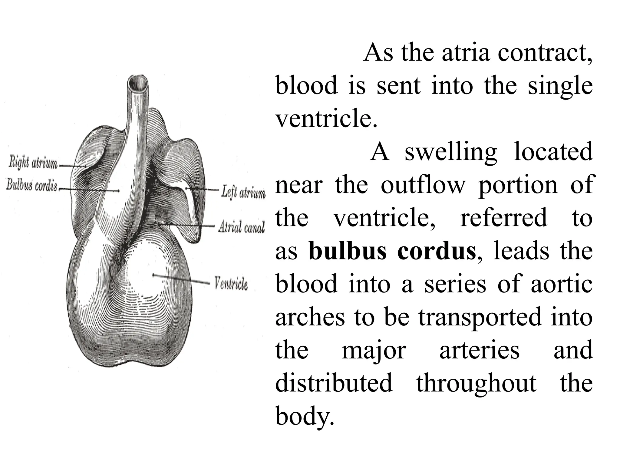
ARTERIAL SYSTEM OF
FROG
Dr. G. D.HANDE
MAJOR ARTERIES
Artery carries blood away from the
ventricle into branches that lead to all parts
of the body.
The atrium receives deoxygenated
blood from the blood vessels (veins) that
drain the various organs of the body.
Deoxygenated blood from the right
atrium is sent, relatively pure, to the
pulmocutaneous arteries taking blood to the
skin and lungs where fresh oxygen can be
picked up.
Frog Artery
The primary function of the heart is to
pump oxygen rich blood to organs such as the
brain, liver, and kidneys as well as all other
tissue.
The heart of the frog is different from
the hearts of warm-blood animals such as
humans.
Although mammals have four
chambers, amphibians, which are cold-blood
animals, have only three consisting of two upper
compartments referred to as the atria and only
one lower vault known as the ventricle.
As the atria contract,
blood is sent into the single
ventricle.
A swelling located
near the outflow portion of
the ventricle, referred to
as bulbus cordus, leads the
blood into a series of aortic
arches to be transported into
the major arteries and
distributed throughout the
body.
They are the carotid (the third),
systemic (the fourth), and pulmonary (the
sixth) arches.
Blood to the lungs (and skin in frogs) is
always carried by the sixth arterial arch,
which loses its connection to the dorsal aorta.
All land vertebrates supply their
lungs with deoxygenated blood from this
source.
Amphibians have a juvenile stage and an
adult stage, and the circulatory systems of the
two are distinct.
In the juvenile (or tadpole) stage, the
circulation is similar to that of a fish; the two-
chambered heart pumps the blood through the
gills where it is oxygenated, and is spread
around the body and back to the heart in a single
loop.
In the adult stage, amphibians (especially
frogs) lose their gills and develop lungs.
They have a heart that consists of a single
ventricle and two atria.
When the ventricle starts contracting,
deoxygenated blood is pumped through
the pulmonary artery to the lungs.
Continued contraction then pumps
oxygenated blood around the rest of the
body.
Mixing of the two bloodstreams is
minimized by the anatomy of the chambers.
Amphibian Circulatory Systems
In amphibians, blood flow is directed in two
circuits: one through the lungs and back to the
heart (pulmonary circulation) and the other
throughout the rest of the body and its organs,
including the brain (systemic circulation).
Amphibians have a three-chambered heart
that has two atria and one ventricle rather than
the two-chambered heart of fish . The two atria
receive blood from the two different circuits
(the lungs and the systems).
There is some mixing of the blood
in the heart’s ventricle, which reduces the
efficiency of oxygen.
The advantage to this arrangement is
that high pressure in the vessels pushes
blood to the lungs and body.
The mixing is mitigated by a ridge
within the ventricle that diverts oxygen-rich
blood through the systemic circulatory
system.
Deoxygenated blood to
pulmocutaneous circuit where gas
exchange occurs in the lungs and
through the skin.
For this reason, amphibians
are often described as having double
circulation.
Carotid Arch:
The carotid arch of each side also divides
into two, an external carotid or lingual going to
the lower jaw and tongue and an internal carotid
to the orbit and brain.
Each internal carotid at its
commencement bears a swollen carotid body or
labyrinth formed from the second pair of gill-
slit. Carotid bodies or labyrinths have a network
of capillaries and chromoffin cells. They detect
the pressure of oxygen and CO2 in the blood.
External Carotid
Internal Carotid
Systemic Arch:
The two systemic arches as pass outward and
curve around the oesophagus to join to form a
median dorsal aorta going backward beneath
the vertebral column.
Each systemic arch gives out three arteries:
(i) Oesophageal artery to the oesophagus,
(ii) Occipitovertebral artery to the head, vertebral
column and spinal cord and
(iii) A large subclavian to the forelimbs.
The oesophageal artery very often arises from the
occipitovertebral artery.
Pulmocutaneous Arch:
Each pulmocutaneous arch divides into two
arteries a pulmonary artery going to a lung
and a cutaneous artery to the skin and buccal
cavity.
Dorsal Aorta:
It is an unpaired artery formed by the
union of two systemic arches.
It runs posteriorly mid-dorsally just
beneath the vertebral column.
(i) Coeliaco-Mesenteric Artery:
A large but unpaired coeliaco-
mesenteric artery arises at the point where
the dorsal aorta is formed by the union of the
right and left systemic arteries.
Coeliaco-mesenteric passing through the
mesentery divides into two main branches- a
small coeliac artery going to the stomach
(gastric).
It gives off a number of arteries:
From the anterior mesenteric artery
arises an impaired posterior mesenteric
artery going to the large intestine.
It sometimes arises from the posterior
end of dorsal aorta.
and liver (hepatic) and a large anterior
mesenteric sending branches to the duodenum
(duodenal), spleen (splenic) and ileum
(intestinal).
(ii) Gonadial:
A pair of small gonadial arteries to the gonads,
called spermatic in male and ovarian in female.
(iii) Renal:
Dorsal aorta runs backward between the
kidneys and gives off 5 to 6 pairs of renal
arteries to both the kidneys.
(iv) Common Iliacs:
Posteriorly dorsal aorta divides into two large
iliac arteries supplying the hindlimbs.
Each iliac as it enters the leg gives
off a branch which divides into an
epigastric supplying the central body
wall, and a recto-visceral artery to the
rectum and bladder.
A femoral artery to the skin and the
muscles of the anterior part of the thigh
and an iliac artery continues further as
the sciatic artery going to the shank.

Major Arteries AS.pptx

  • 1.
  • 2.
  • 3.
    Artery carries bloodaway from the ventricle into branches that lead to all parts of the body. The atrium receives deoxygenated blood from the blood vessels (veins) that drain the various organs of the body. Deoxygenated blood from the right atrium is sent, relatively pure, to the pulmocutaneous arteries taking blood to the skin and lungs where fresh oxygen can be picked up.
  • 4.
    Frog Artery The primaryfunction of the heart is to pump oxygen rich blood to organs such as the brain, liver, and kidneys as well as all other tissue. The heart of the frog is different from the hearts of warm-blood animals such as humans. Although mammals have four chambers, amphibians, which are cold-blood animals, have only three consisting of two upper compartments referred to as the atria and only one lower vault known as the ventricle.
  • 5.
    As the atriacontract, blood is sent into the single ventricle. A swelling located near the outflow portion of the ventricle, referred to as bulbus cordus, leads the blood into a series of aortic arches to be transported into the major arteries and distributed throughout the body.
  • 6.
    They are thecarotid (the third), systemic (the fourth), and pulmonary (the sixth) arches. Blood to the lungs (and skin in frogs) is always carried by the sixth arterial arch, which loses its connection to the dorsal aorta. All land vertebrates supply their lungs with deoxygenated blood from this source.
  • 7.
    Amphibians have ajuvenile stage and an adult stage, and the circulatory systems of the two are distinct. In the juvenile (or tadpole) stage, the circulation is similar to that of a fish; the two- chambered heart pumps the blood through the gills where it is oxygenated, and is spread around the body and back to the heart in a single loop. In the adult stage, amphibians (especially frogs) lose their gills and develop lungs.
  • 9.
    They have aheart that consists of a single ventricle and two atria. When the ventricle starts contracting, deoxygenated blood is pumped through the pulmonary artery to the lungs. Continued contraction then pumps oxygenated blood around the rest of the body. Mixing of the two bloodstreams is minimized by the anatomy of the chambers.
  • 11.
    Amphibian Circulatory Systems Inamphibians, blood flow is directed in two circuits: one through the lungs and back to the heart (pulmonary circulation) and the other throughout the rest of the body and its organs, including the brain (systemic circulation). Amphibians have a three-chambered heart that has two atria and one ventricle rather than the two-chambered heart of fish . The two atria receive blood from the two different circuits (the lungs and the systems).
  • 13.
    There is somemixing of the blood in the heart’s ventricle, which reduces the efficiency of oxygen. The advantage to this arrangement is that high pressure in the vessels pushes blood to the lungs and body. The mixing is mitigated by a ridge within the ventricle that diverts oxygen-rich blood through the systemic circulatory system.
  • 14.
    Deoxygenated blood to pulmocutaneouscircuit where gas exchange occurs in the lungs and through the skin. For this reason, amphibians are often described as having double circulation.
  • 17.
    Carotid Arch: The carotidarch of each side also divides into two, an external carotid or lingual going to the lower jaw and tongue and an internal carotid to the orbit and brain. Each internal carotid at its commencement bears a swollen carotid body or labyrinth formed from the second pair of gill- slit. Carotid bodies or labyrinths have a network of capillaries and chromoffin cells. They detect the pressure of oxygen and CO2 in the blood. External Carotid Internal Carotid
  • 19.
    Systemic Arch: The twosystemic arches as pass outward and curve around the oesophagus to join to form a median dorsal aorta going backward beneath the vertebral column. Each systemic arch gives out three arteries: (i) Oesophageal artery to the oesophagus, (ii) Occipitovertebral artery to the head, vertebral column and spinal cord and (iii) A large subclavian to the forelimbs. The oesophageal artery very often arises from the occipitovertebral artery.
  • 20.
    Pulmocutaneous Arch: Each pulmocutaneousarch divides into two arteries a pulmonary artery going to a lung and a cutaneous artery to the skin and buccal cavity. Dorsal Aorta: It is an unpaired artery formed by the union of two systemic arches. It runs posteriorly mid-dorsally just beneath the vertebral column.
  • 21.
    (i) Coeliaco-Mesenteric Artery: Alarge but unpaired coeliaco- mesenteric artery arises at the point where the dorsal aorta is formed by the union of the right and left systemic arteries. Coeliaco-mesenteric passing through the mesentery divides into two main branches- a small coeliac artery going to the stomach (gastric). It gives off a number of arteries:
  • 22.
    From the anteriormesenteric artery arises an impaired posterior mesenteric artery going to the large intestine. It sometimes arises from the posterior end of dorsal aorta. and liver (hepatic) and a large anterior mesenteric sending branches to the duodenum (duodenal), spleen (splenic) and ileum (intestinal).
  • 23.
    (ii) Gonadial: A pairof small gonadial arteries to the gonads, called spermatic in male and ovarian in female. (iii) Renal: Dorsal aorta runs backward between the kidneys and gives off 5 to 6 pairs of renal arteries to both the kidneys. (iv) Common Iliacs: Posteriorly dorsal aorta divides into two large iliac arteries supplying the hindlimbs.
  • 24.
    Each iliac asit enters the leg gives off a branch which divides into an epigastric supplying the central body wall, and a recto-visceral artery to the rectum and bladder. A femoral artery to the skin and the muscles of the anterior part of the thigh and an iliac artery continues further as the sciatic artery going to the shank.