The heart is a muscular organ that pumps blood through the circulatory system. It has four chambers and is located slightly left of center in the chest. The two upper chambers are called atria, which receive blood, and the two lower chambers are called ventricles, which pump blood out of the heart. The right side receives deoxygenated blood and pumps it to the lungs, while the left side receives oxygenated blood from the lungs and pumps it out to the body. Valves ensure blood flows in only one direction through the heart.
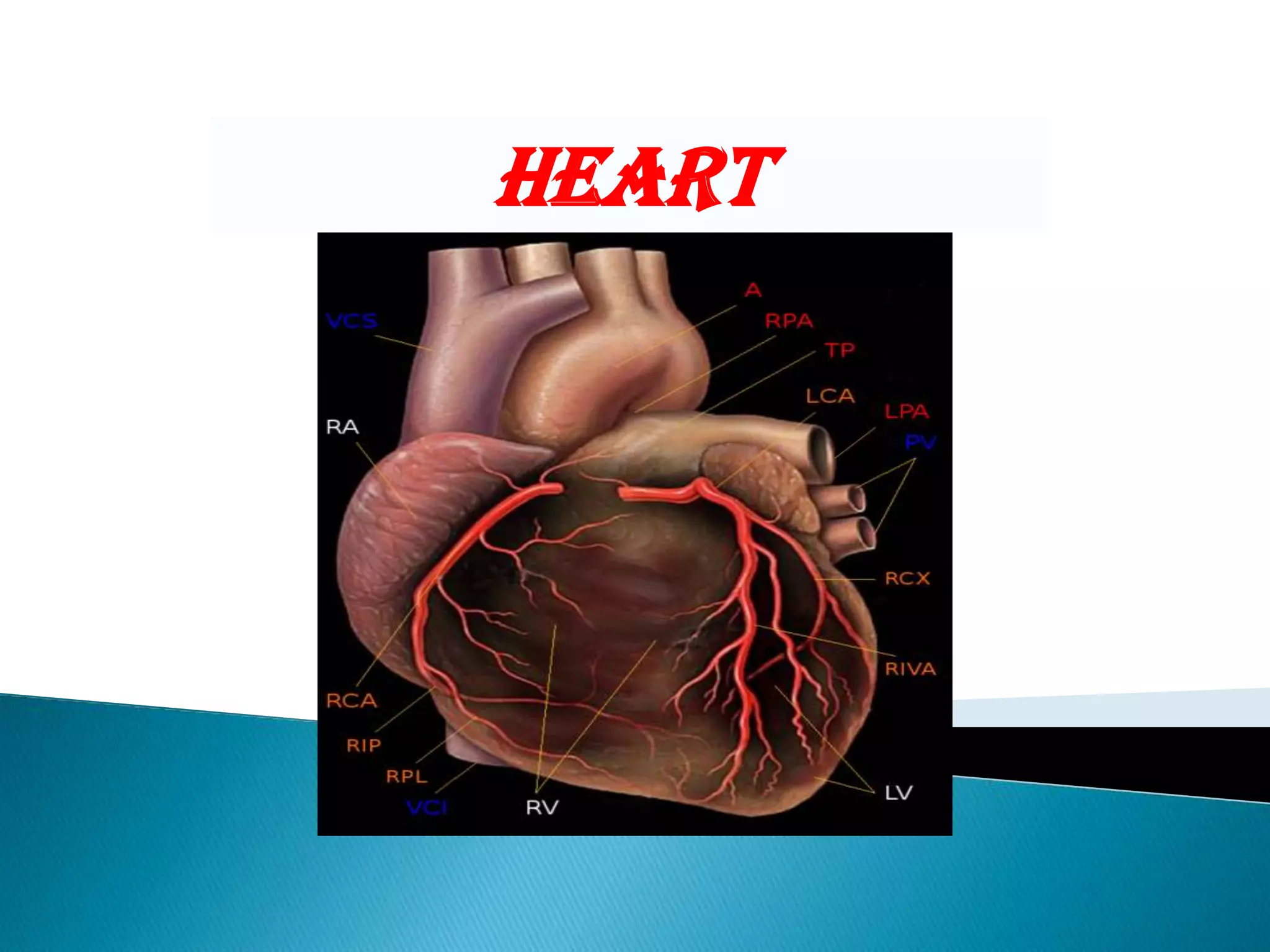
![The structure of the heart varies among the different branches of the animal kingdom. (See Circulatory system.) Cephalopods have two "gill hearts" and one "systemic heart". Fish have a two-chambered heart that pumps the blood to the gills and from there it goes on to the rest of the body. In amphibians and most reptiles, a double circulatory system is used, but the heart is not always completely separated into two pumps. Amphibians have a three-chambered heart.Birds and mammals show complete separation of the heart into two pumps, for a total of four heart chambers; it is thought that the four-chambered heart of birds evolved independently from that of mammals. Human heart removed from a 64-year-old male.In the human body, the heart is usually situated in the middle of the thorax with the largest part of the heart slightly offset to the left (although sometimes it is on the right, see dextrocardia), underneath the breastbone (see diagrams). The heart is usually felt to be on the left side because the left heart (left ventricle) is stronger (it pumps to all body parts). The left lung is smaller than the right lung because the heart occupies more of the left hemithorax. The heart is enclosed by a sac known as the pericardium and is surrounded by the lungs. The pericardium comprises two parts: the fibrous pericardium, made of dense fibrous connective tissue; and a double membrane structure containing a serous fluid to reduce friction during heart contractions (the serious pericardium). The mediastinum, a subdivision of the thoracic cavity, is the name of the heart cavity. [5]The apex is the blunt point situated in an inferior (pointing down and left) direction. A stethoscope can be placed directly over the apex so that the beats can be counted. It is located posterior to the 5th intercostal space in the left mid-clavicular line. In normal adults, the mass of the heart is 250-350 g (9-12 oz), or about three quarters the size of a clenched fist, but extremely diseased hearts can be up to 1000 g (2 lb) in mass due to hypertrophy. It consists of four chambers, the two upper atria (singular: atrium ) and the two lower ventricles. Structure](https://image.slidesharecdn.com/demopresentation-091022232346-phpapp01/75/Demo-Presentation-3-2048.jpg)