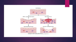
TISSUE REPAIR
AND HEALING
DR. MANAHIL JAMIL
 Tissue repair = restoration of tissue architecture and function
after an injury
 Occurs in two ways:
• Regeneration of injured tissue
• Replacement by connective tissue (scarring)
 Usually, tissue repair involves both processes.
 Involves cell proliferation, and interaction between cells and
extracellular matrix.
REGENERATION
 Some tissues are able to replace the damaged components and
essentially return to a normal state; this process is called regeneration
 Involves;
 Cell proliferation- by growth factors
 Development of mature cells from stem cells
Cell proliferation
 Tissues with high proliferative capacity renew themselves continuously &
can regenerate after injury
 Cell proliferation is driven by signals provided by growth factors and from
extra cellular matrix
 Growth factors are produced by cells near the site of damage
 Sources are; Macrophages, epithelial & stromal cells
Tissue regeneration
 In epithelia of the intestinal tract and skin, injured cells are rapidly
replaced by proliferation & differentiation of cells derived from the tissue
stem cells
 parenchymal organs
 Pancreas, adrenal, thyroid & lung
Liver regeneration
 Occurs by 2 major mechanism;
 Proliferation of remaining hepatocytes
• following partial hepatectomy
• Driven by cytokines such as IL-6 (Kupffer cells), growth factors
(hepatocyte growth factor)
 Liver regeneration from progenitor cells
Repair by
scarring
It occurs by replacement of injured cells with
connective tissue, leading to formation of
scar
or by a combination of regeneration of
some residual cells & scar formation
Steps in scar formation
 Hemostatic plug of platelets is formed – stops bleeding
 INFLAMMATION
• Acute & chronic inflammatory responses
• Mediators are produced that recruit neutrophils & then monocytes
• Macrophages are the central cellular players in the repair process
• M1- clear microbes & necrotic tissue & promote inflammation
• M2 – produce growth factors that stimulate proliferation of many cell
types in the next stage of repair
 CELL PROLIFERATION takes up to 10 days
 Epithelial, endothelial, vascular cells & fibroblasts proliferate & migrate to
close the now-clean wound
 Epithelial cells- respond to locally produced growth factors & migrate
over the wound to cover it
 Endothelial & other vascular cells – proliferate to form new blood vessels
(angiogenesis)
 Fibroblasts- proliferate & migrate to the site of injury & lay down
collagen fibers that form scar
 Combination of proliferating fibroblasts, loose connective tissue, new
blood vessels & scattered chronic inflammatory cells form granulation
tissue
 REMODELLING
• The connective tissue that has been depositd by fibroblasts is reorganized
to produce stable fibrous scar
• Begins 2-3 weeks after injury & may continue for months
HEALING
 Classified into;
1. Healing by first intention(primary union)- epithelial regeneration eith
minimum scarring
2. Healing by second intention(secondary union) – larger wounds that heal
by a combination of regeneration & scarring
ANGIOGENESIS
Development of new blood
vessels from existing blood
vessels
Consists of following steps
1. Vasodilation in response to NO & inc permeability by VEGF
2. Separation of pericytes from the abluminal surface & breakdown of
basement membrane to allow formation of vessel sprout
3. Migration of endothelial cells toward area of tissue injury
4. Proliferation of endothelial cells just behind the leading front of migrating
cells
5. Remodeling into capillary tubes
6. Recruitment of peri endothelial cells to form mature vessel
7. Suppression of endothelial proliferation & migration & deposition of
basement membrane
FACTORS IMPAIRING TISSUE REPAIR
1. Infection
2. Diabetes
3. Nutritional status ( protein and vit. C deficiency)
4. Glucocorticoids (steroids) ( inhbit TGF-beta production and
diminish fibrosis)
5. Mechanical factors
6. Poor perfusion
7. Foreign bodies
8. Type and extent of injury
9. Location of injury
CLINICAL EXAMPELS
 Defects in healing ( chronic wounds):
1. Venous leg ulcers ( deposits of hemosiderin)
2. Arterial ulcers (ischemia)
3. Pressure sores (prolonged compression of tissues against bone)
4. Diabetic ulcers (epithelial ulceration)
 Excessive scarring:
1. Hypertrophic scar (excessive collagen, raised scar, can regress)
2. Keloid (beyond boundaries , does not regress)
Leg ulcer
Diabetic ulcer
Pressure sore
Hypertrophic
scar
keloid
TISSUE REPAIR.pptx

TISSUE REPAIR.pptx

  • 1.
  • 2.
     Tissue repair= restoration of tissue architecture and function after an injury  Occurs in two ways: • Regeneration of injured tissue • Replacement by connective tissue (scarring)  Usually, tissue repair involves both processes.  Involves cell proliferation, and interaction between cells and extracellular matrix.
  • 4.
    REGENERATION  Some tissuesare able to replace the damaged components and essentially return to a normal state; this process is called regeneration  Involves;  Cell proliferation- by growth factors  Development of mature cells from stem cells
  • 5.
    Cell proliferation  Tissueswith high proliferative capacity renew themselves continuously & can regenerate after injury  Cell proliferation is driven by signals provided by growth factors and from extra cellular matrix  Growth factors are produced by cells near the site of damage  Sources are; Macrophages, epithelial & stromal cells
  • 6.
    Tissue regeneration  Inepithelia of the intestinal tract and skin, injured cells are rapidly replaced by proliferation & differentiation of cells derived from the tissue stem cells  parenchymal organs  Pancreas, adrenal, thyroid & lung
  • 7.
    Liver regeneration  Occursby 2 major mechanism;  Proliferation of remaining hepatocytes • following partial hepatectomy • Driven by cytokines such as IL-6 (Kupffer cells), growth factors (hepatocyte growth factor)  Liver regeneration from progenitor cells
  • 8.
    Repair by scarring It occursby replacement of injured cells with connective tissue, leading to formation of scar or by a combination of regeneration of some residual cells & scar formation
  • 9.
    Steps in scarformation  Hemostatic plug of platelets is formed – stops bleeding  INFLAMMATION • Acute & chronic inflammatory responses • Mediators are produced that recruit neutrophils & then monocytes • Macrophages are the central cellular players in the repair process • M1- clear microbes & necrotic tissue & promote inflammation • M2 – produce growth factors that stimulate proliferation of many cell types in the next stage of repair
  • 10.
     CELL PROLIFERATIONtakes up to 10 days  Epithelial, endothelial, vascular cells & fibroblasts proliferate & migrate to close the now-clean wound  Epithelial cells- respond to locally produced growth factors & migrate over the wound to cover it  Endothelial & other vascular cells – proliferate to form new blood vessels (angiogenesis)  Fibroblasts- proliferate & migrate to the site of injury & lay down collagen fibers that form scar  Combination of proliferating fibroblasts, loose connective tissue, new blood vessels & scattered chronic inflammatory cells form granulation tissue
  • 11.
     REMODELLING • Theconnective tissue that has been depositd by fibroblasts is reorganized to produce stable fibrous scar • Begins 2-3 weeks after injury & may continue for months
  • 13.
    HEALING  Classified into; 1.Healing by first intention(primary union)- epithelial regeneration eith minimum scarring 2. Healing by second intention(secondary union) – larger wounds that heal by a combination of regeneration & scarring
  • 15.
    ANGIOGENESIS Development of newblood vessels from existing blood vessels
  • 17.
    Consists of followingsteps 1. Vasodilation in response to NO & inc permeability by VEGF 2. Separation of pericytes from the abluminal surface & breakdown of basement membrane to allow formation of vessel sprout 3. Migration of endothelial cells toward area of tissue injury 4. Proliferation of endothelial cells just behind the leading front of migrating cells 5. Remodeling into capillary tubes 6. Recruitment of peri endothelial cells to form mature vessel 7. Suppression of endothelial proliferation & migration & deposition of basement membrane
  • 18.
    FACTORS IMPAIRING TISSUEREPAIR 1. Infection 2. Diabetes 3. Nutritional status ( protein and vit. C deficiency) 4. Glucocorticoids (steroids) ( inhbit TGF-beta production and diminish fibrosis) 5. Mechanical factors 6. Poor perfusion 7. Foreign bodies 8. Type and extent of injury 9. Location of injury
  • 19.
    CLINICAL EXAMPELS  Defectsin healing ( chronic wounds): 1. Venous leg ulcers ( deposits of hemosiderin) 2. Arterial ulcers (ischemia) 3. Pressure sores (prolonged compression of tissues against bone) 4. Diabetic ulcers (epithelial ulceration)  Excessive scarring: 1. Hypertrophic scar (excessive collagen, raised scar, can regress) 2. Keloid (beyond boundaries , does not regress)
  • 20.
  • 21.
  • 22.
  • 23.
  • 24.