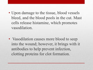
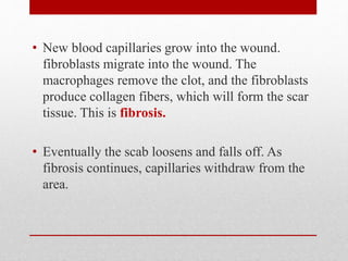
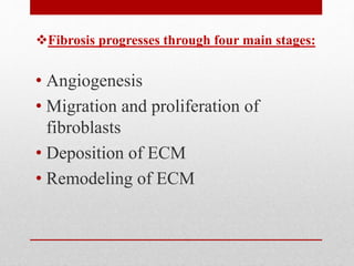
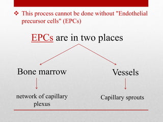
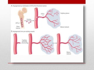
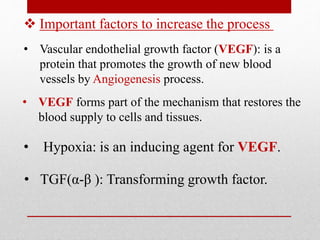
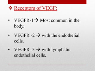
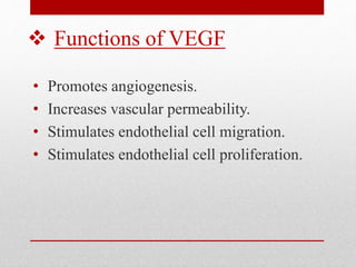
Fibrosis & Scarring
Tissue Repair (Healing)
• Regeneration of injured tissue
(replacement by normal cells of the same kind)
• Replacement by fibrous tissue
(fibrosis, scarring)
• Fibrosis : is the formation of excess fibrous
tissue in an organ or tissue in a reparative
process.
• When one suffers a cut to the skin, the
tissue is repaired by two methods:
regeneration and fibrosis.
• Regeneration replaces dead or damaged
cells with the same type of cell, thus
restoring normal function. Fibrosis
replaces damaged tissue with scar
tissue, which holds the tissue together
but does not restore normal function.
• Upon damage to the tissue, blood vessels
bleed, and the blood pools in the cut. Mast
cells release histamine, which promotes
vasodilation.
• Vasodilation causes more blood to seep
into the wound; however, it brings with it
antibodies to help prevent infection,
clotting proteins for clot formation.
• A clot forms in the tissue. The clot
gently binds together the edges of
the cut to keep and protect the
healthy tissue. The surface of the
clot forms a scab, while
macrophages beneath the scab
remove the damaged tissue.
• New blood capillaries grow into the wound.
fibroblasts migrate into the wound. The
macrophages remove the clot, and the fibroblasts
produce collagen fibers, which will form the scar
tissue. This is fibrosis.
• Eventually the scab loosens and falls off. As
fibrosis continues, capillaries withdraw from the
area.
Fibrosis progresses through four main stages:
• Angiogenesis
• Migration and proliferation of
fibroblasts
• Deposition of ECM
• Remodeling of ECM
Angiogenesis
 This process cannot be done without "Endothelial
precursor cells" (EPCs)
Vessels
EPCs are in two places
Bone marrow
network of capillary
plexus
Capillary sprouts
• Vascular endothelial growth factor (VEGF): is a
protein that promotes the growth of new blood
vessels by Angiogenesis process.
 Important factors to increase the process
• VEGF forms part of the mechanism that restores the
blood supply to cells and tissues.
• Hypoxia: is an inducing agent for VEGF.
• TGF(α-β ): Transforming growth factor.
 Receptors of VEGF:
• VEGFR-1 Most common in the
body.
• VEGFR -2  with the endothelial
cells.
• VEGFR -3  with lymphatic
endothelial cells.
 Functions of VEGF
• Promotes angiogenesis.
• Increases vascular permeability.
• Stimulates endothelial cell migration.
• Stimulates endothelial cell proliferation.
Migration and proliferation of
fibroblasts
• Granulation tissue contains numerous newly formed
blood vessels for transporting of new substances.
• As discussed previously,VEGF promotes angiogenesis
but is also responsible for the increase in vascular
permeability that leads to deposition of plasma proteins,
such as fibrinogen and plasma fibronectin in the ECM
and provides a provisional stroma for fibroblast and
endothelial cell growth.
• Migration of fibroblasts to the site of injury and its
proliferation are caused by multiple growth factors,
including TGF-β, and the cytokines The sources of these
growth factors and cytokines include platelets, is
inflammatory cells and activated endothelium.
• We also have Macrophages are important cellular
constituents , clearing extracellular debris, fibrin,
and other foreign material at the site of repair.
• This factor -> TGF-β, helps in migration of
fibroblasts.
• If the appropriate chemotactic stimuli are
present, mast cells, eosinophils, and lymphocytes
may also accumulate.
• TGF-β appears to be the most important because
of the effects on fibrous tissue deposition in
grannular tissue.
• TGF-β is produced by most of the cells in
granulation tissue and causes fibroblast migration
and proliferation.
• increased synthesis of collagen and fibronectin,
and decreased degradation of ECM by
metalloproteinases (discussed later).
• Increase the permeability of the vessels .
• Increase the deposition of plasma protein .
• Plasma protein will provide provisional
stroma for fibroblasts ingrowth
• Megretion of fibroblasts made under
several factors .
• Macrophages clear the extra cellular
debris, fibrin and some foreign materials
Summary
Deposition of ECM
Major Components of the ECM:
• Collagen
• Elastic fibers
• Proteoglycans
• Fibronectin
• Laminin
• Integrins
• As repair continues, the number of proliferating
endothelial cells and fibroblasts decreases.
• Fibroblasts progressively deposit increased
amounts of ECM. Fibrillar collagens form a
major portion of the connective tissue in repair
sites and are important for the development of
strength in healing wounds.
• collagen synthesis by fibroblasts begins within 3
to 5 days after injury and continues for several
weeks, depending on the size of wound.
• Net collagen accumulation, however, depends not
only on increased collagen synthesis but also on
decreased degradation. Ultimately, the
granulation tissue scaffolding is converted into a
scar composed of spindle-shaped fibroblasts,
dense collagen, fragments of elastic tissue, and
other ECM components. As the scar matures,
vascular regression continues, eventually
transforming the richly vascularized granulation
tissue into a pale, avascular scar.
Remodeling of ECM
• The replacement of granulation tissue with a scar
involves transitions in the composition of the
ECM.
• Some of the growth factors that stimulate
synthesis of collagen and other connective tissue
molecules also modulate the synthesis and
activation of metalloproteinases, enzymes that
degrade these ECM components.
• The balance between ECM synthesis and
degradation results in remodeling of the
connective tissue framework an important feature
of both chronic inflammation and wound repair.
Thank You
Presented by:
Ahmed Kamal Abdallah Hammad
Ahmed Reda Mohamed Edrees
Ahmed Alaa El-Din Mokhtar
Ahmed Emad Mofeed
Ahmed Emad Mounir
Reference : Tissue Repair
and
healing, ROBBINS
Chapter 3.

Fibrosis and scarring

  • 1.
  • 2.
    Tissue Repair (Healing) •Regeneration of injured tissue (replacement by normal cells of the same kind) • Replacement by fibrous tissue (fibrosis, scarring) • Fibrosis : is the formation of excess fibrous tissue in an organ or tissue in a reparative process.
  • 3.
    • When onesuffers a cut to the skin, the tissue is repaired by two methods: regeneration and fibrosis. • Regeneration replaces dead or damaged cells with the same type of cell, thus restoring normal function. Fibrosis replaces damaged tissue with scar tissue, which holds the tissue together but does not restore normal function.
  • 4.
    • Upon damageto the tissue, blood vessels bleed, and the blood pools in the cut. Mast cells release histamine, which promotes vasodilation. • Vasodilation causes more blood to seep into the wound; however, it brings with it antibodies to help prevent infection, clotting proteins for clot formation.
  • 5.
    • A clotforms in the tissue. The clot gently binds together the edges of the cut to keep and protect the healthy tissue. The surface of the clot forms a scab, while macrophages beneath the scab remove the damaged tissue.
  • 6.
    • New bloodcapillaries grow into the wound. fibroblasts migrate into the wound. The macrophages remove the clot, and the fibroblasts produce collagen fibers, which will form the scar tissue. This is fibrosis. • Eventually the scab loosens and falls off. As fibrosis continues, capillaries withdraw from the area.
  • 7.
    Fibrosis progresses throughfour main stages: • Angiogenesis • Migration and proliferation of fibroblasts • Deposition of ECM • Remodeling of ECM
  • 8.
  • 9.
     This processcannot be done without "Endothelial precursor cells" (EPCs) Vessels EPCs are in two places Bone marrow network of capillary plexus Capillary sprouts
  • 11.
    • Vascular endothelialgrowth factor (VEGF): is a protein that promotes the growth of new blood vessels by Angiogenesis process.  Important factors to increase the process • VEGF forms part of the mechanism that restores the blood supply to cells and tissues. • Hypoxia: is an inducing agent for VEGF. • TGF(α-β ): Transforming growth factor.
  • 12.
     Receptors ofVEGF: • VEGFR-1 Most common in the body. • VEGFR -2  with the endothelial cells. • VEGFR -3  with lymphatic endothelial cells.
  • 13.
     Functions ofVEGF • Promotes angiogenesis. • Increases vascular permeability. • Stimulates endothelial cell migration. • Stimulates endothelial cell proliferation.
  • 14.
  • 15.
    • Granulation tissuecontains numerous newly formed blood vessels for transporting of new substances. • As discussed previously,VEGF promotes angiogenesis but is also responsible for the increase in vascular permeability that leads to deposition of plasma proteins, such as fibrinogen and plasma fibronectin in the ECM and provides a provisional stroma for fibroblast and endothelial cell growth. • Migration of fibroblasts to the site of injury and its proliferation are caused by multiple growth factors, including TGF-β, and the cytokines The sources of these growth factors and cytokines include platelets, is inflammatory cells and activated endothelium.
  • 16.
    • We alsohave Macrophages are important cellular constituents , clearing extracellular debris, fibrin, and other foreign material at the site of repair. • This factor -> TGF-β, helps in migration of fibroblasts. • If the appropriate chemotactic stimuli are present, mast cells, eosinophils, and lymphocytes may also accumulate.
  • 17.
    • TGF-β appearsto be the most important because of the effects on fibrous tissue deposition in grannular tissue. • TGF-β is produced by most of the cells in granulation tissue and causes fibroblast migration and proliferation. • increased synthesis of collagen and fibronectin, and decreased degradation of ECM by metalloproteinases (discussed later).
  • 18.
    • Increase thepermeability of the vessels . • Increase the deposition of plasma protein . • Plasma protein will provide provisional stroma for fibroblasts ingrowth • Megretion of fibroblasts made under several factors . • Macrophages clear the extra cellular debris, fibrin and some foreign materials Summary
  • 19.
  • 20.
    Major Components ofthe ECM: • Collagen • Elastic fibers • Proteoglycans • Fibronectin • Laminin • Integrins
  • 21.
    • As repaircontinues, the number of proliferating endothelial cells and fibroblasts decreases. • Fibroblasts progressively deposit increased amounts of ECM. Fibrillar collagens form a major portion of the connective tissue in repair sites and are important for the development of strength in healing wounds. • collagen synthesis by fibroblasts begins within 3 to 5 days after injury and continues for several weeks, depending on the size of wound.
  • 22.
    • Net collagenaccumulation, however, depends not only on increased collagen synthesis but also on decreased degradation. Ultimately, the granulation tissue scaffolding is converted into a scar composed of spindle-shaped fibroblasts, dense collagen, fragments of elastic tissue, and other ECM components. As the scar matures, vascular regression continues, eventually transforming the richly vascularized granulation tissue into a pale, avascular scar.
  • 23.
  • 24.
    • The replacementof granulation tissue with a scar involves transitions in the composition of the ECM. • Some of the growth factors that stimulate synthesis of collagen and other connective tissue molecules also modulate the synthesis and activation of metalloproteinases, enzymes that degrade these ECM components.
  • 25.
    • The balancebetween ECM synthesis and degradation results in remodeling of the connective tissue framework an important feature of both chronic inflammation and wound repair.
  • 26.
  • 27.
    Presented by: Ahmed KamalAbdallah Hammad Ahmed Reda Mohamed Edrees Ahmed Alaa El-Din Mokhtar Ahmed Emad Mofeed Ahmed Emad Mounir
  • 28.
    Reference : TissueRepair and healing, ROBBINS Chapter 3.