Mechanism of Healing and factors influencing wound healing.pptx
The topic covers brief mechanism of healing and the factors influencing healing. The slide is basically designed for PharmD second year students for pathophysiology.
Mechanism of Healing and factors influencing wound healing.pptx
1.
Repairs of woundsin the skin, factors
influencing healing of wounds
DOCTOR OF PHARMACY
2nd
YEAR
PATHOPHYSIOLOGY
PREPARED BY:
DR. ARSHITA KUMARI
ASSISTANT PROFESSOR
2.
Healing, Regeneration, andRepair
Healing is the body’s response to injury aiming to restore normal structure and function.
It occurs through two processes, often acting together:
Regeneration – Replacement by same type of parenchymal cells → complete restoration.
REPAIR: Replacement by connective (fibrous) tissue → scar formation (fibrosis)
REGENERATION: Involves proliferation of surviving parenchymal cells at the margins
of injury. Controlled by growth factors: Epidermal growth factor (EGF), Fibroblast
growth factor (FGF), Platelet-derived growth factor (PDGF), Endothelial growth factor
(EGF), Transforming growth factor-β (TGF-β).
3.
The Cell Cycle
CellCycle: The period between two successive cell divisions.
Phases:
Phases Function
M phase Mitosis (cell division)
G1 phase Post-mitotic growth phase
S phase DNA synthesis
G2 phase Pre - mitotic phase
G0 phase Resting (non-dividing) phase
The interphase G1 + S + G2.
4.
Types of Cellsby Regenerative Capacity
Type Dividing Capacity Examples
Labile Cells
Continuously divide; remain in active
cell cycle
Surface epithelia (skin, GIT,
respiratory, urinary tracts), bone
marrow cells, lymphoid tissue
Stable Cells
Quiescent (in G0 phase) but can re-
enter cycle on stimulation
Liver, pancreas, kidney, adrenal,
thyroid, fibroblasts, endothelium,
bone, cartilage
Permanent Cells
Non-dividing; lost regenerative
ability
Neurons, cardiac muscle, skeletal
muscle
5.
Mechanism of Regeneration
A.Proliferation and migration of surviving cells at the edge of injury.
B. Differentiation and maturation of new cells to restore normal tissue
architecture.
Cell Cycle Regulation
Controlled by cyclins (A, B, E) and cyclin-dependent kinases (CDKs).
Cyclins activate CDKs → promote mitosis.
After division, cyclins are degraded by ubiquitin system to maintain control.
REPAIR (By Connective Tissue / Fibrosis)
Replacement of injured tissue by fibrous connective tissue.
Occurs when regeneration is incomplete or parenchymal cells are permanent.
6.
Two Main Processes
A.Granulation tissue formation
Appearance: Pink, soft, granular tissue at the wound site.
Composition: New capillaries, fibroblasts, and loose collagen.
Purpose: Forms the framework for scar formation.
Phases of granulation tissue formation
Phase Key Events
1. Inflammatory Phase
- Blood clot and acute inflammation within 24 hrs. - Influx of plasma, neutrophils,
and monocytes.
2. Clearance Phase - Necrotic tissue and debris removed by enzymes from neutrophils and macrophages.
3. Ingrowth Phase (Granulation
Tissue Formation)
Two processes:
i)Angiogenesis: New capillaries from existing vessels under influence of VEGF,
PDGF, TGF-β, FGF.
ii) Fibrogenesis: Proliferation of fibroblasts → collagen synthesis. Myofibroblasts
help in contraction.
Note: Newly formed vessels are leaky, causing edema in fresh granulation tissue.
7.
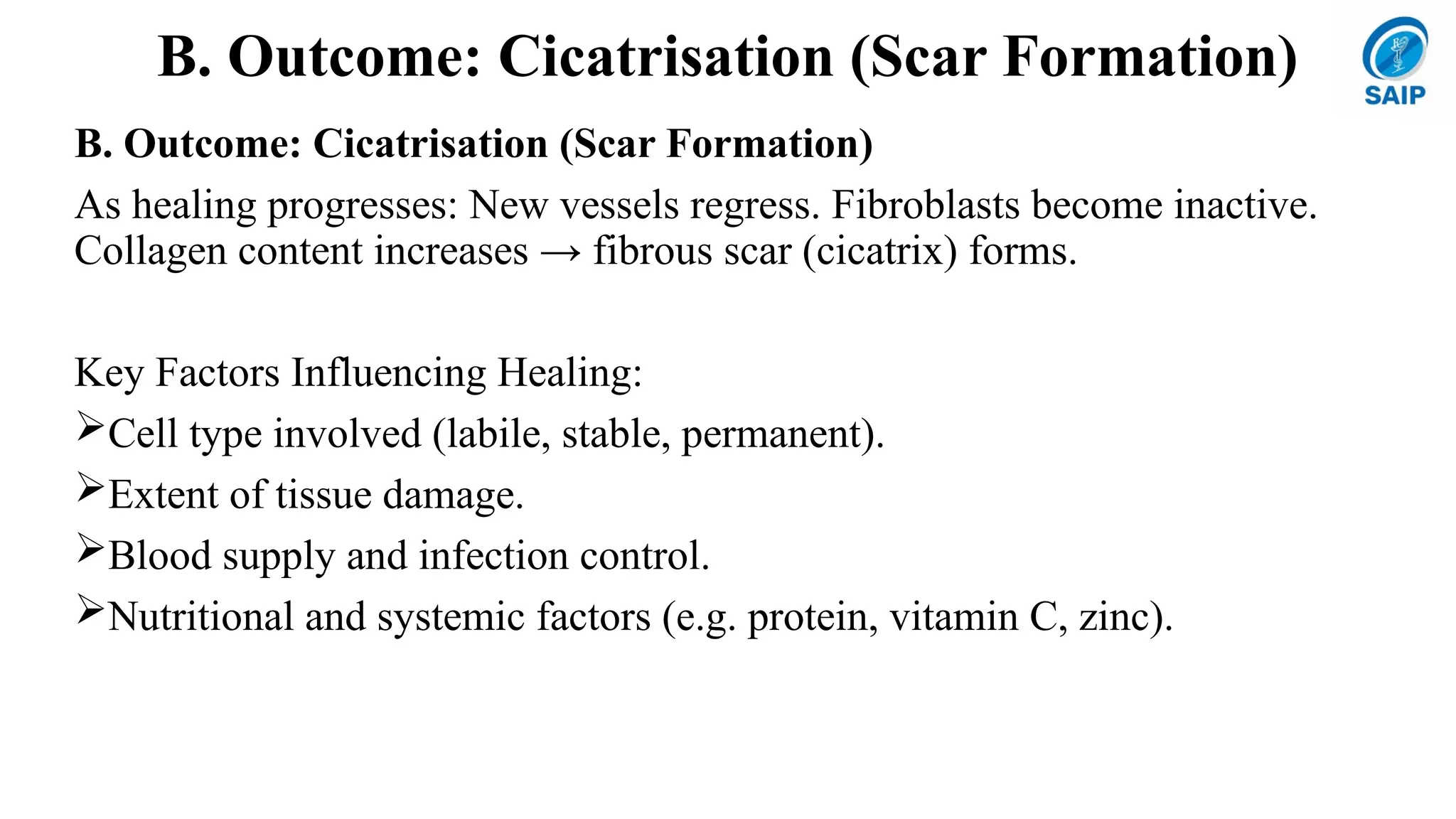
B. Outcome: Cicatrisation(Scar Formation)
B. Outcome: Cicatrisation (Scar Formation)
As healing progresses: New vessels regress. Fibroblasts become inactive.
Collagen content increases → fibrous scar (cicatrix) forms.
Key Factors Influencing Healing:
Cell type involved (labile, stable, permanent).
Extent of tissue damage.
Blood supply and infection control.
Nutritional and systemic factors (e.g. protein, vitamin C, zinc).
8.
Summary Table: Regenerationvs Repair
Feature Regeneration Repair
Definition
Replacement by same cell
type
Replacement by fibrous
tissue
Result Complete restoration Scar formation
Cell types involved Labile and stable cells
Fibroblasts, macrophages,
endothelial cells
Growth factors EGF, FGF, PDGF, TGF-β VEGF, PDGF, TGF-β, FGF
Example
Healing of skin, liver
regeneration
Myocardial infarction scar,
deep ulcer healing
9.
HEALING OF SKINWOUNDS
• Two types:
1. Healing by First Intention (Primary Union)
• Clean, uninfected, surgically incised wound with minimal tissue loss.
• Wound edges are closely approximated by sutures.
Sequence:
• Initial haemorrhage → Clot formation seals the wound.
• Acute inflammation → Polymorphs appear in 24 hrs; replaced by macrophages by 3rd day.
• Epithelial changes → Epidermal cells proliferate; new multilayered epithelium forms by 5th day.
• Organisation → Fibroblast invasion and collagen deposition → scar forms in 4 weeks.
• Suture tracks → Each suture acts as a small wound; may cause infection or cyst if epithelial cells
persist.
• Healing is rapid, scar is neat and small.
10.
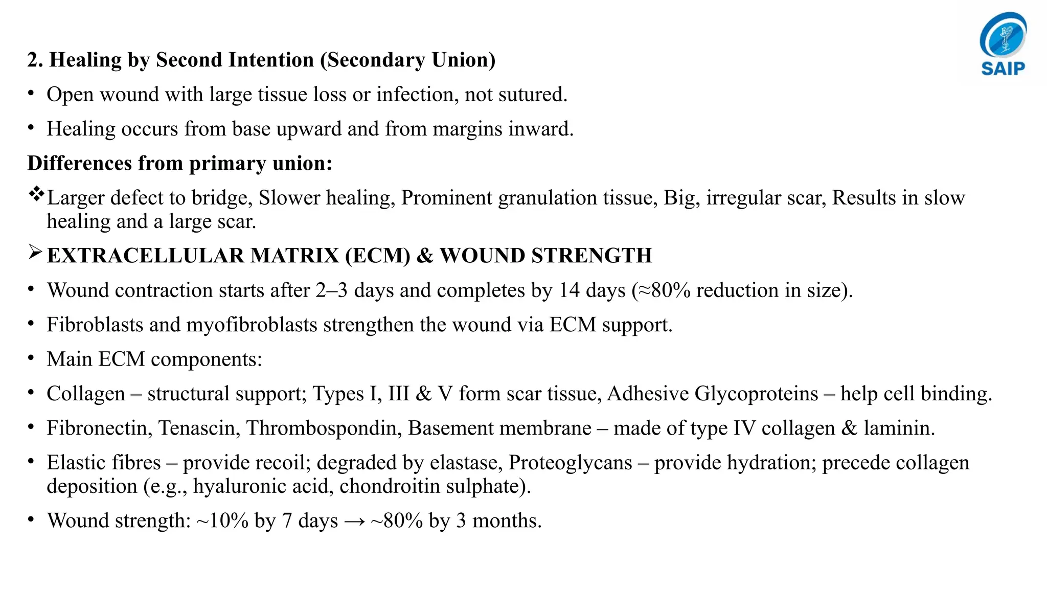
2. Healing bySecond Intention (Secondary Union)
• Open wound with large tissue loss or infection, not sutured.
• Healing occurs from base upward and from margins inward.
Differences from primary union:
Larger defect to bridge, Slower healing, Prominent granulation tissue, Big, irregular scar, Results in slow
healing and a large scar.
EXTRACELLULAR MATRIX (ECM) & WOUND STRENGTH
• Wound contraction starts after 2–3 days and completes by 14 days (≈80% reduction in size).
• Fibroblasts and myofibroblasts strengthen the wound via ECM support.
• Main ECM components:
• Collagen – structural support; Types I, III & V form scar tissue, Adhesive Glycoproteins – help cell binding.
• Fibronectin, Tenascin, Thrombospondin, Basement membrane – made of type IV collagen & laminin.
• Elastic fibres – provide recoil; degraded by elastase, Proteoglycans – provide hydration; precede collagen
deposition (e.g., hyaluronic acid, chondroitin sulphate).
• Wound strength: ~10% by 7 days → ~80% by 3 months.
11.
FACTORS INFLUENCING WOUNDHEALING
A. Local Factors: Infection (most important), Poor blood supply (e.g. leg ulcers
heal slowly), Foreign bodies (sutures, debris), Movement at wound site, Type/size
of injury, UV exposure promotes healing
B. Systemic Factors: Age (faster in young), Nutrition (↓ protein, Vit C, A, Zn →
delayed healing), Infection, Glucocorticoids (anti-inflammatory effect delays
repair), Diabetes mellitus, Blood disorders (neutropenia, poor clotting).
COMPLICATIONS OF WOUND HEALING
Infection, Inclusion cyst formation, Pigmentation, Incisional hernia, Hypertrophied
scar / Keloi, Excessive contraction (Contracture, e.g., Dupuytren’s), Neoplasia
(Marjolin’s ulcer → squamous carcinoma)
12.
HEALING IN SPECIALIZEDTISSUES
1. Fracture Healing
• Types:
A.Primary union – Surgical fixation (plates, clamps); direct bone healing without
periosteal callus.
B.Secondary union – Natural healing with callus formation (most common).
• Steps:
1. Procallus Formation: Haematoma → inflammation → granulation tissue → soft
callus of woven bone/cartilage.
2. Osseous Callus Formation: Replacement by lamellar bone and Haversian
system formation.
3. Remodelling: Normal bone structure restored; marrow cavity reappears.
4. Complications: Fibrous union, non-union, delayed union.
13.
2. Nervous TissueHealing
a) CNS (brain, spinal cord): Neurons = permanent → no regeneration; astrocytic proliferation (gliosis) occurs.
b) PNS (peripheral nerves): Limited regeneration via Schwann cell proliferation and axonal sprouting
(Wallerian degeneration and regrowth).
3. Muscle Healing
a) Skeletal muscle:
If sheath intact → regeneration via sarcolemmal tubes (normal muscle fibre regrowth).
If sheath damaged → disorganized fibrosis (e.g. Volkmann’s contracture).
b) Smooth muscle: limited regeneration, replaced by scar in large lesions.
c) Cardiac muscle: no regeneration → fibrous scar (except mild repair in young if endomysium intact).
14.
4. Mucosal SurfaceHealing
Rapid regeneration from epithelial margins, continuous cell turnover (e.g. GIT,
urinary tract, endometrium).
5. Healing of Solid Epithelial Organs
a. By fibrosis in gross injury (e.g. chronic pyelonephritis, cirrhosis).
b. By regeneration if basement membrane is intact:
c. Renal tubular necrosis → tubular regeneration.
d. Viral hepatitis → hepatocyte proliferation if stromal framework intact.
STEM CELL CONCEPT OF HEALING (Regenerative Medicine)
Properties: Self-renewal
Multilineage differentiation (can form any of ~220 cell types)
Types:
I. Embryonic stem cells: Organ and tissue formation.
II. Adult (somatic) stem cells: Maintain normal cell turnover (e.g., bone
marrow).
15.
Key Points
a. Foundin most adult tissues (bone marrow, skin, liver, etc.)
b. Can be grown and transdifferentiated in labs.
c. Homing – ability to migrate and engraft at the injury site.
d. Major Clinical Applications (experimental except bone marrow therapy):
e. Bone marrow stem cells – for blood cancers & disorders.
f. Neuronal stem cells – potential for Parkinson’s, Alzheimer’s, spinal injury.
g. Islet cell stem cells – for Type 1 diabetes.
h. Cardiac stem cells – post-MI repair.
i. Skeletal muscle stem cells – muscle regeneration.
j. Corneal (limbal) stem cells – for corneal repair.
k. Skin stem cells – from hair follicle/sebaceous glands, may enable scar-free healing.
l. Liver stem cells – canal of Hering, for hepatic regeneration.
m. Intestinal stem cells – in crypts, regenerate villi.
n.Lung tissue stem cells – potential for COPD repair.