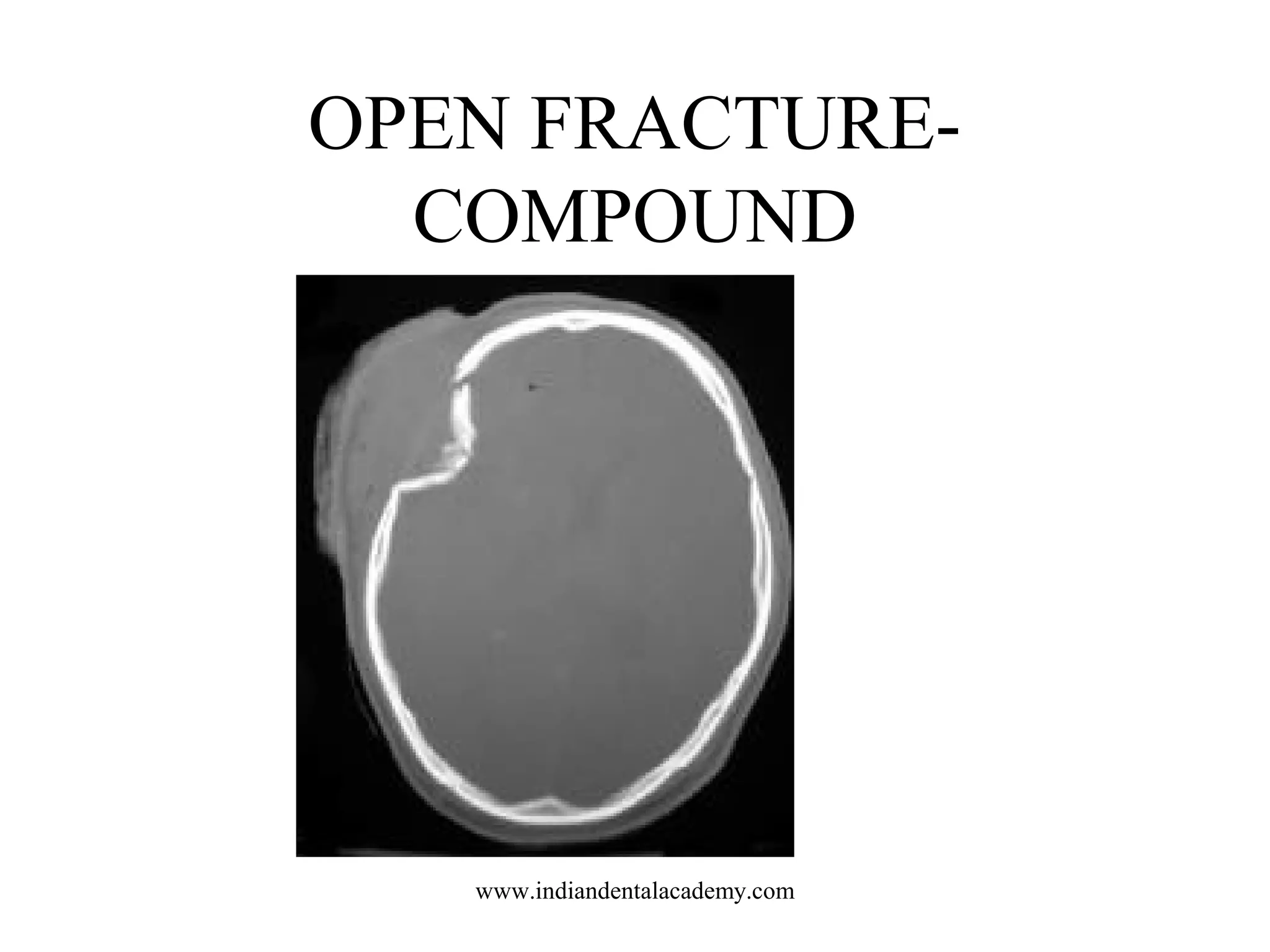
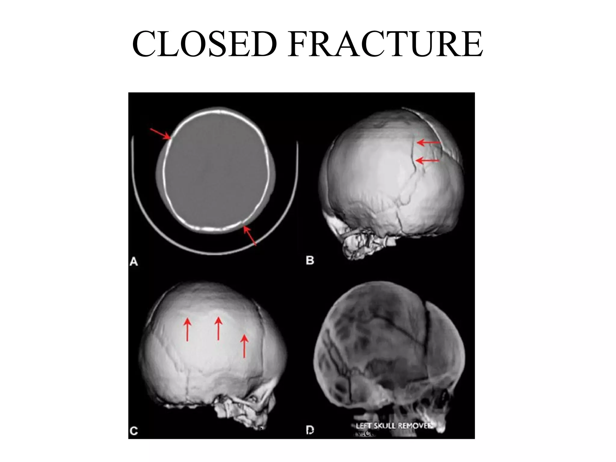
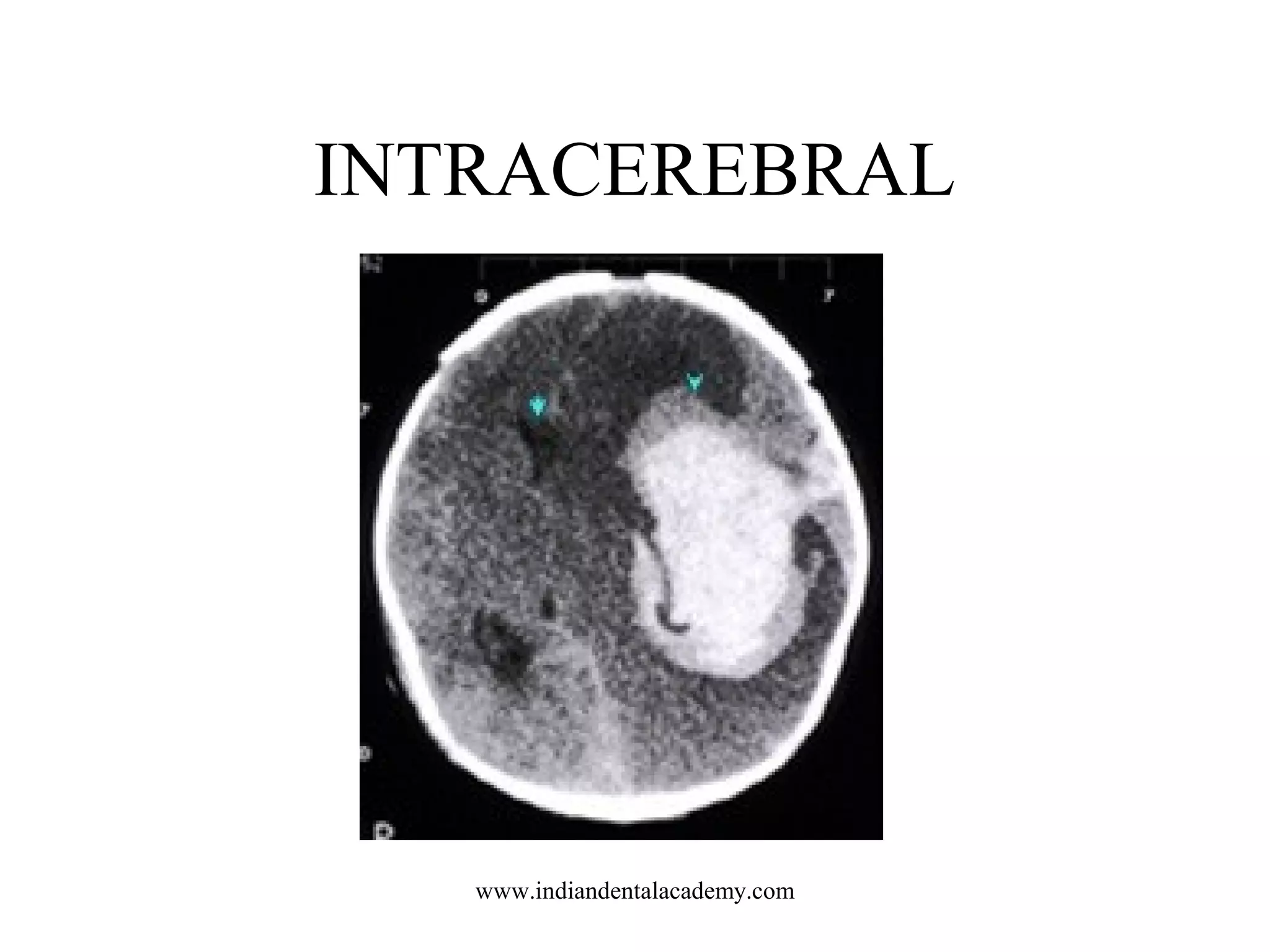
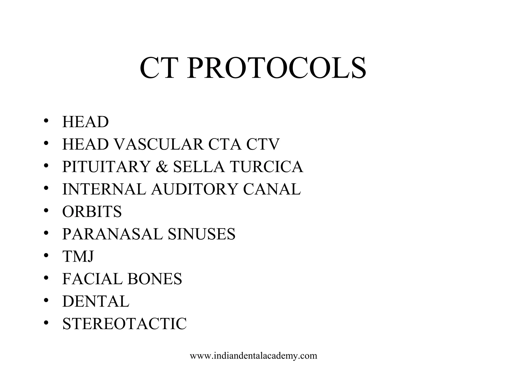
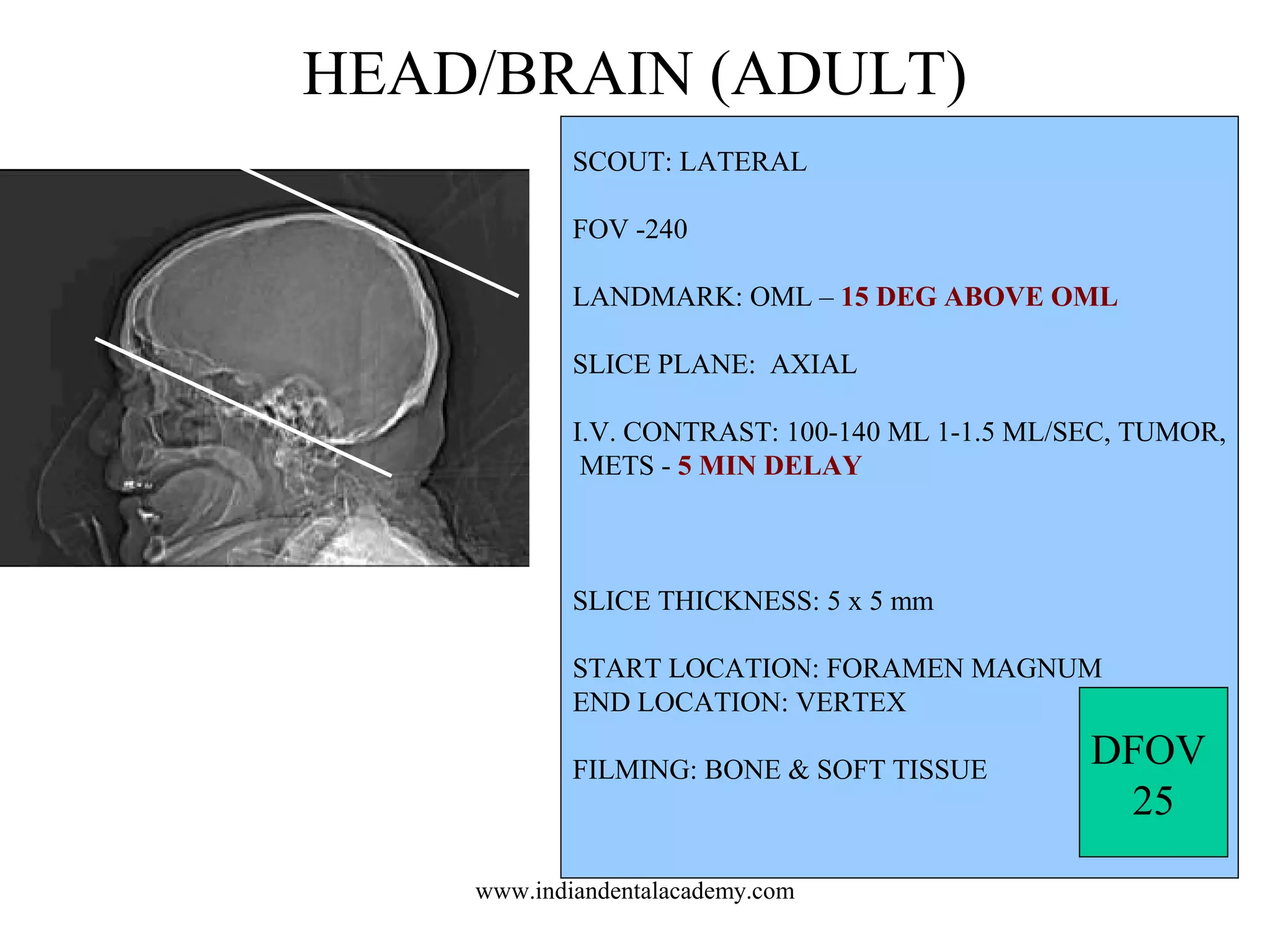
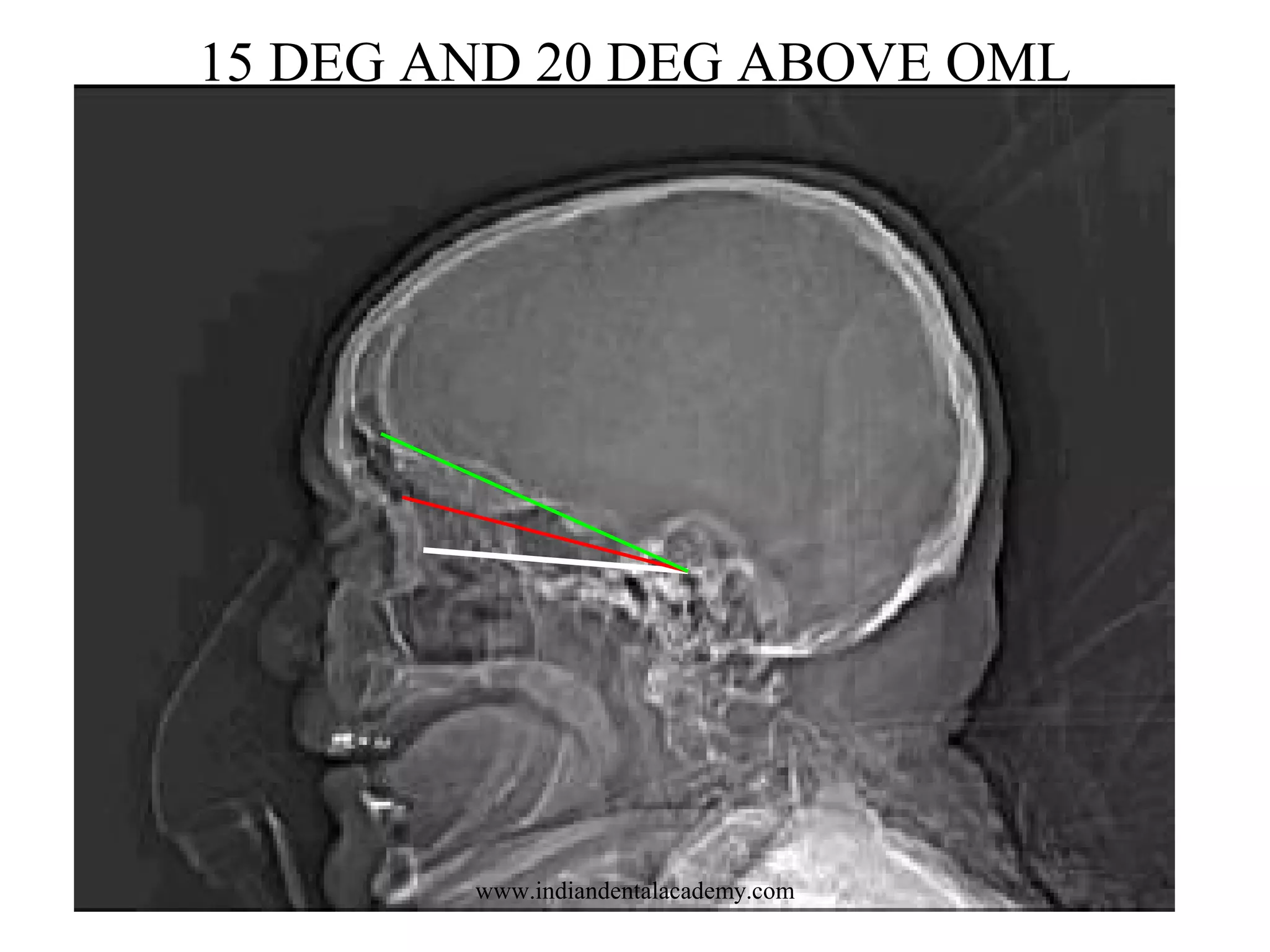
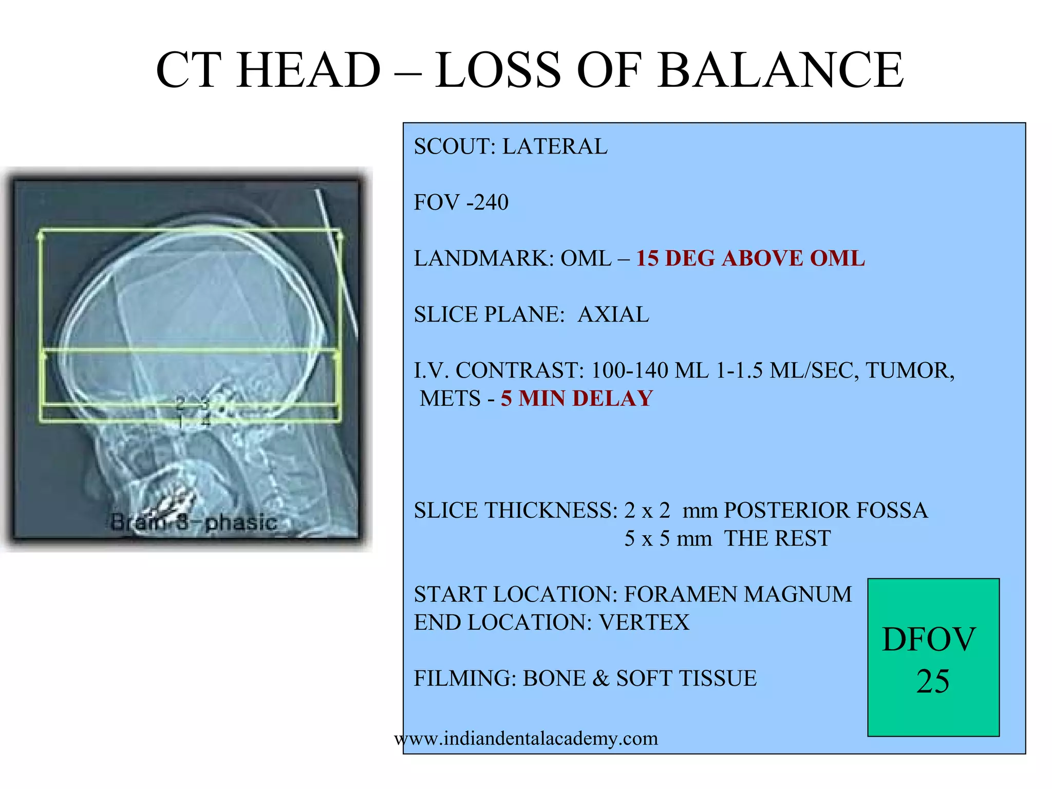
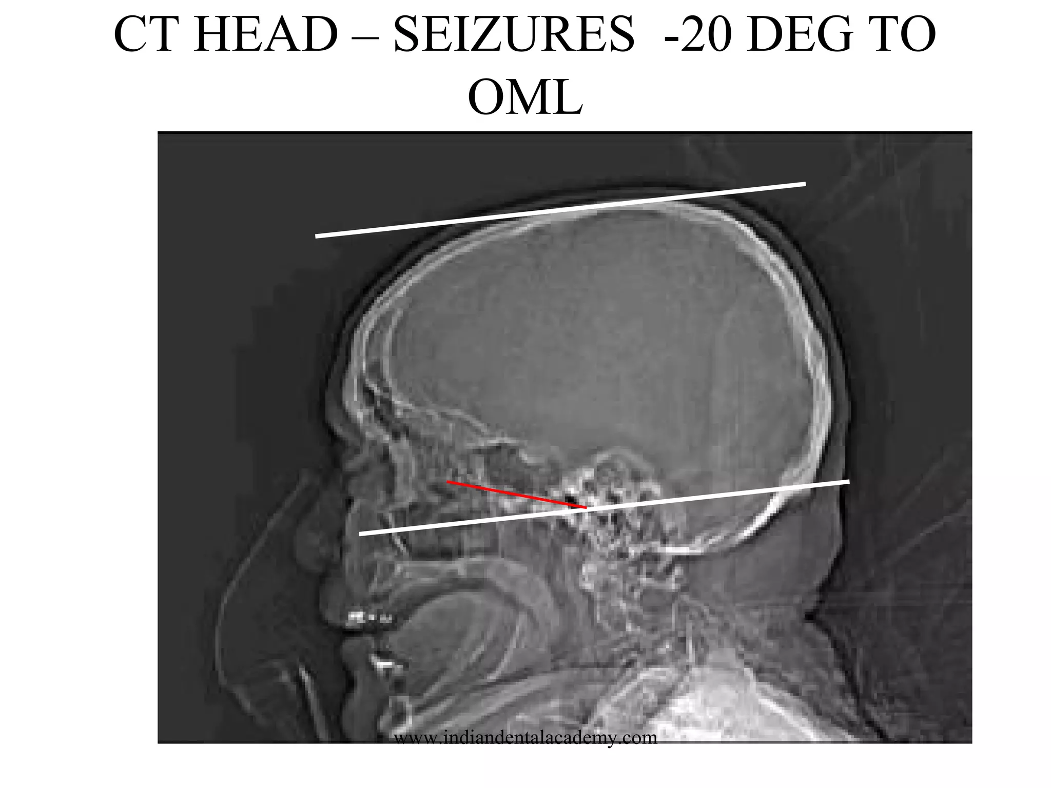
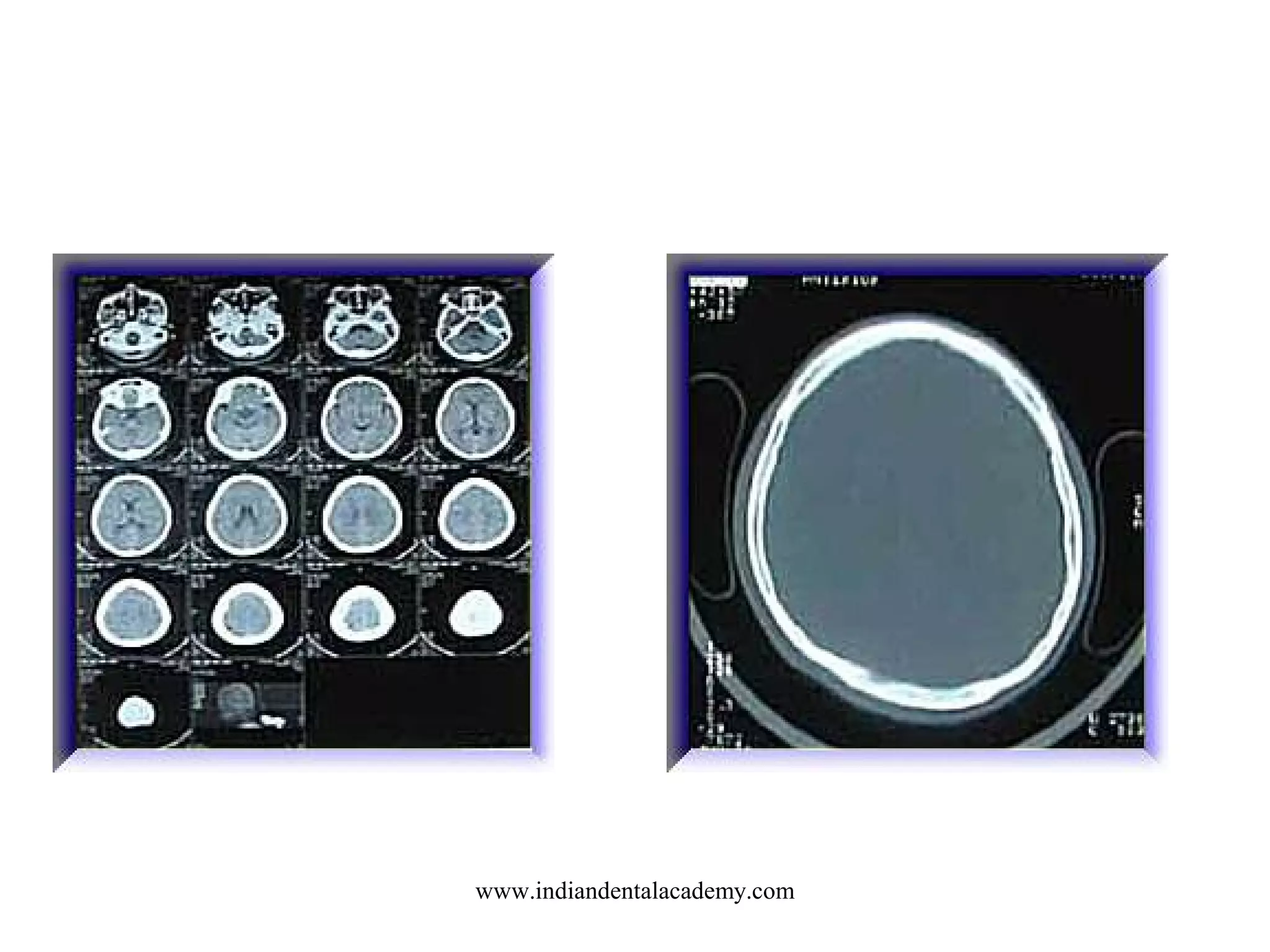
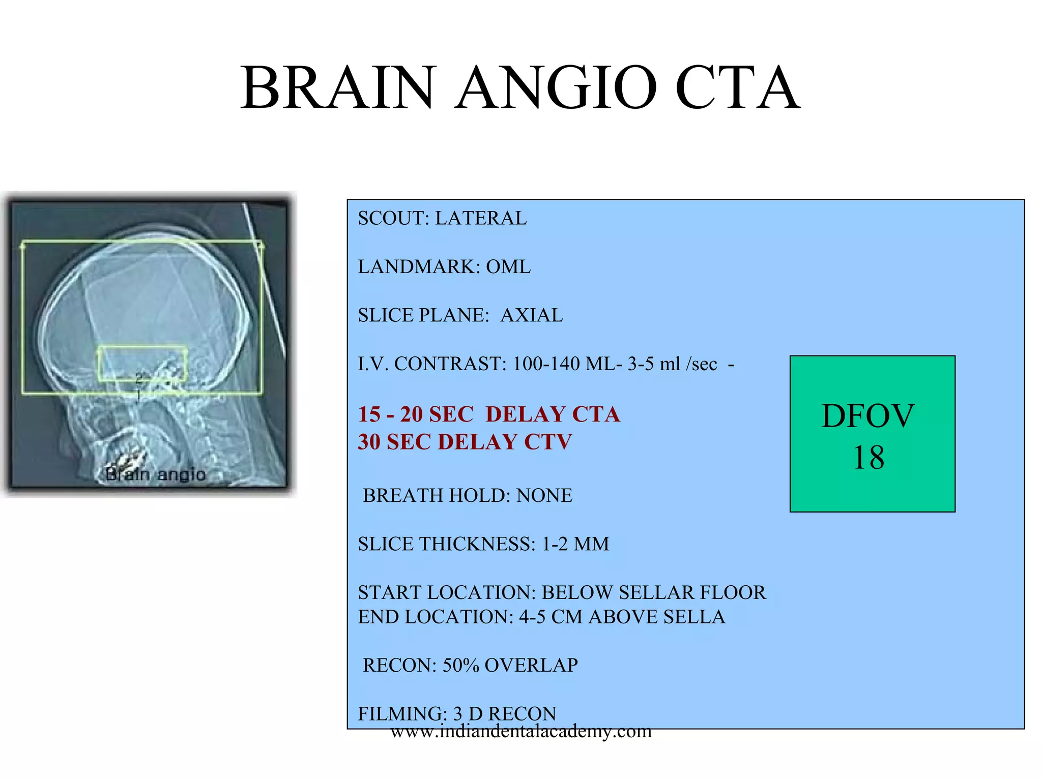
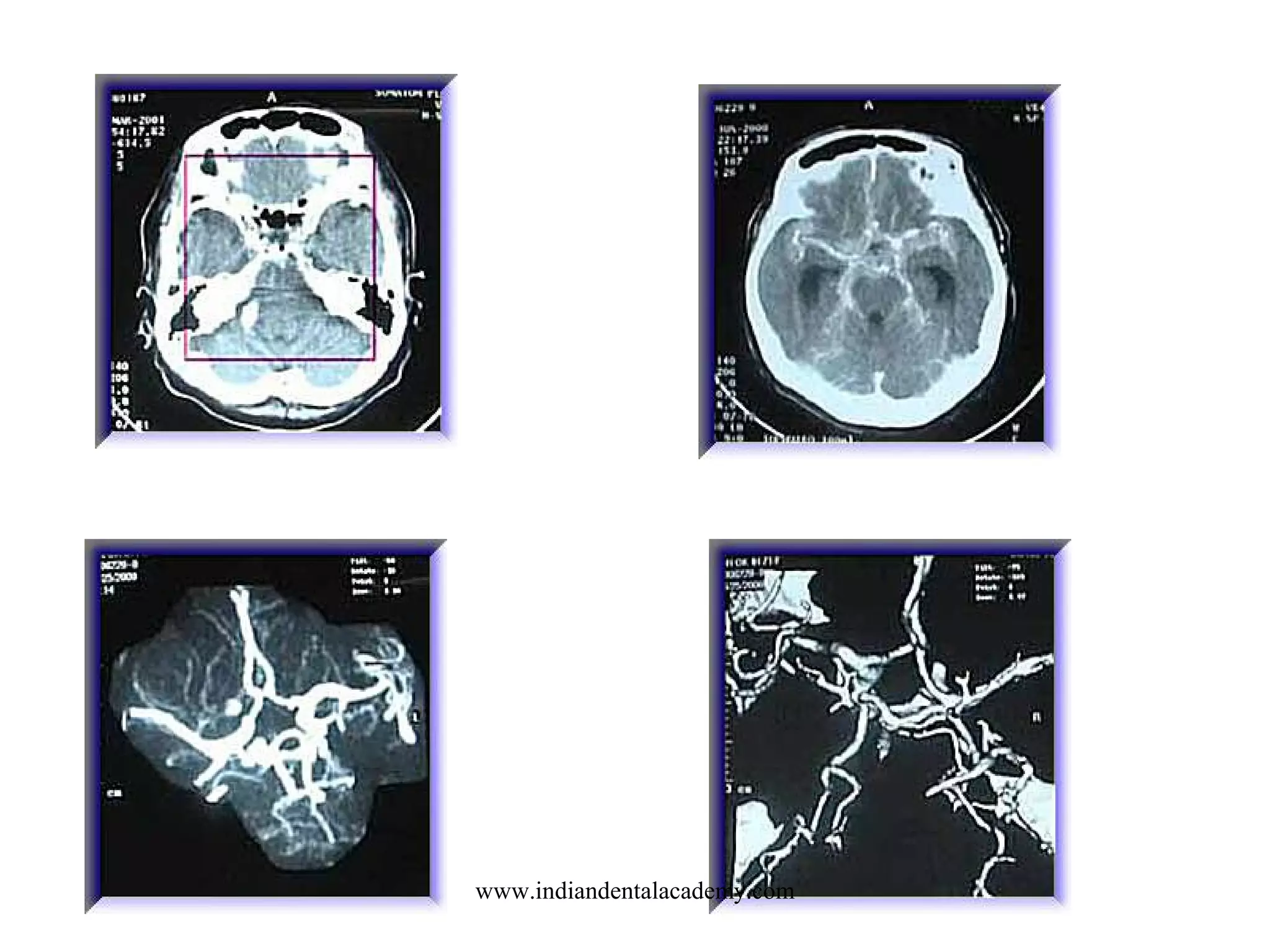
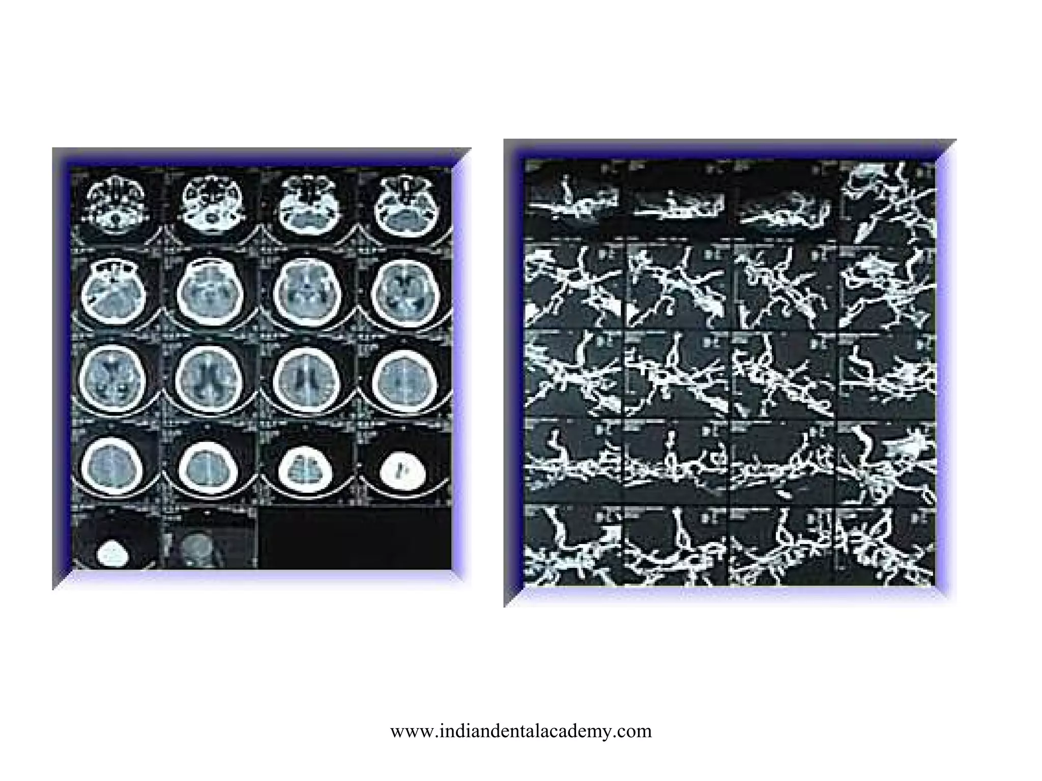
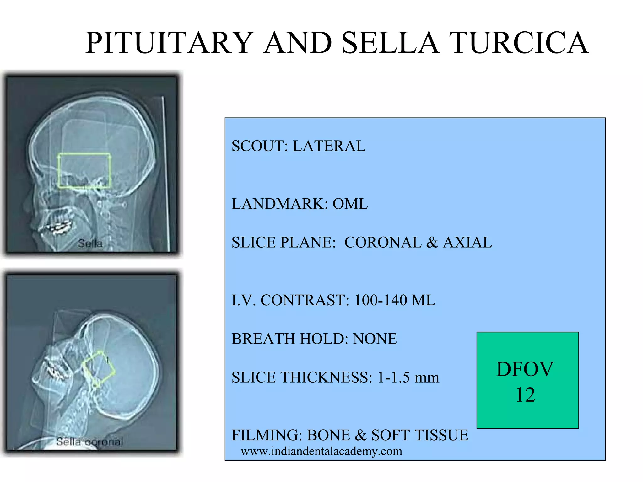
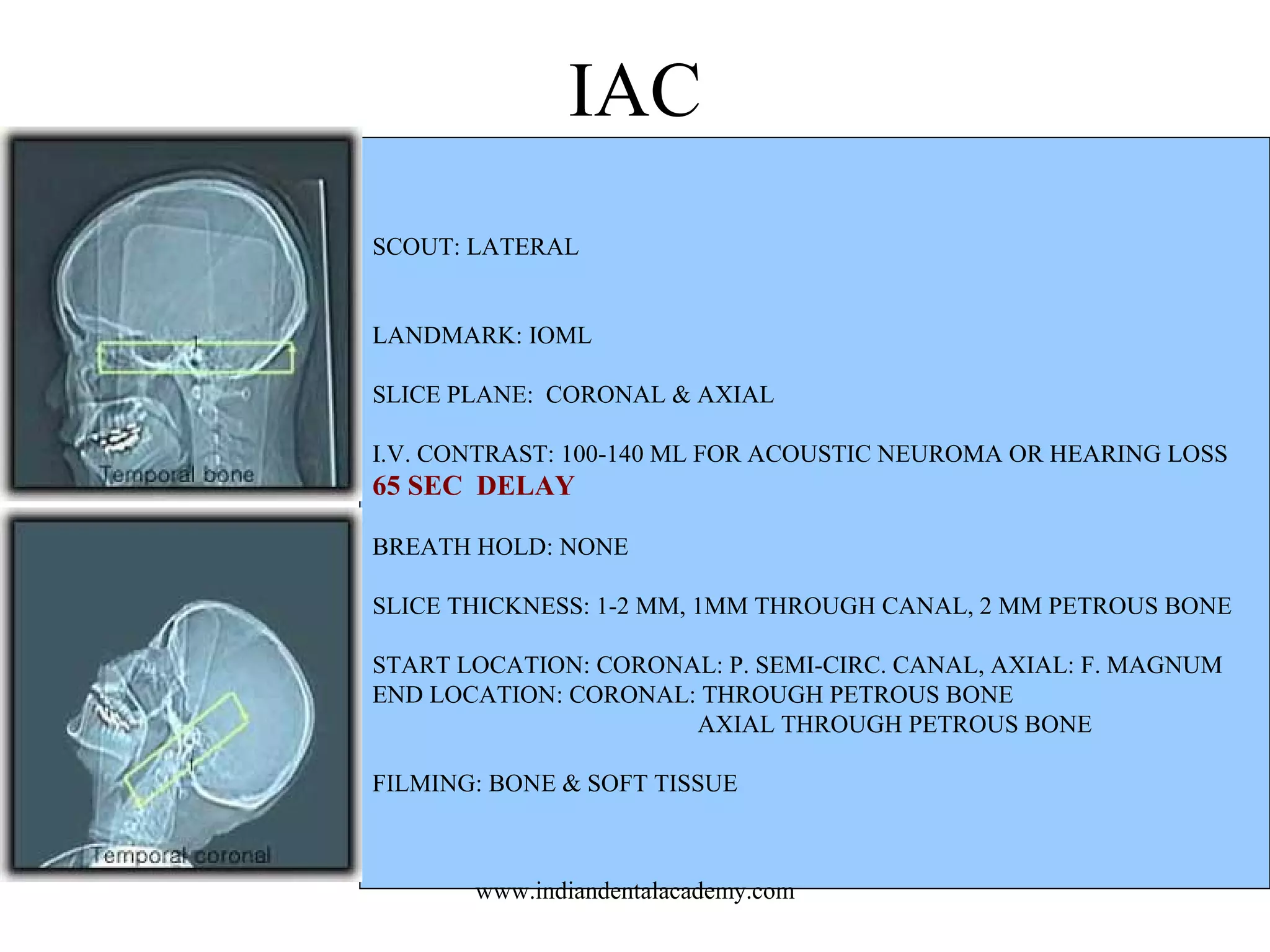
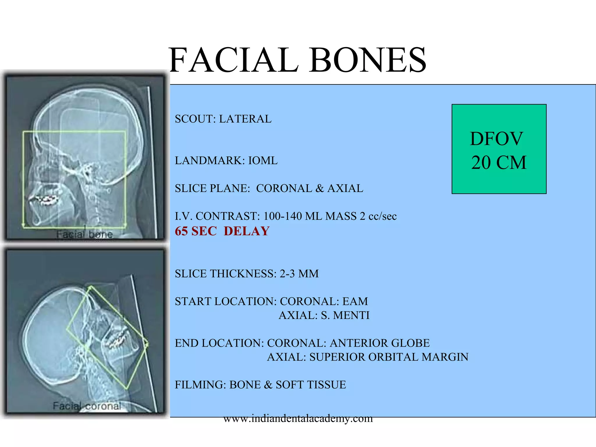
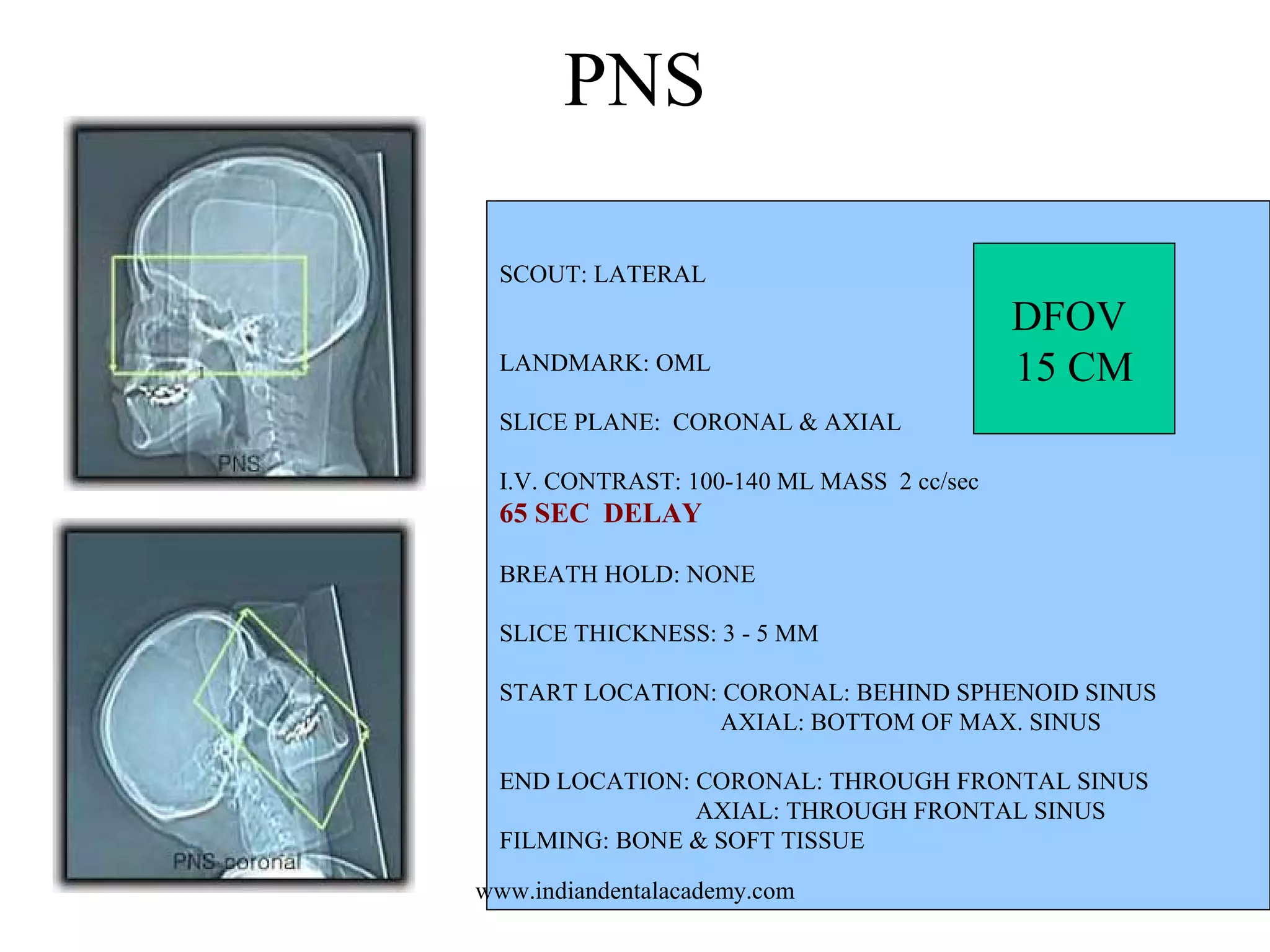
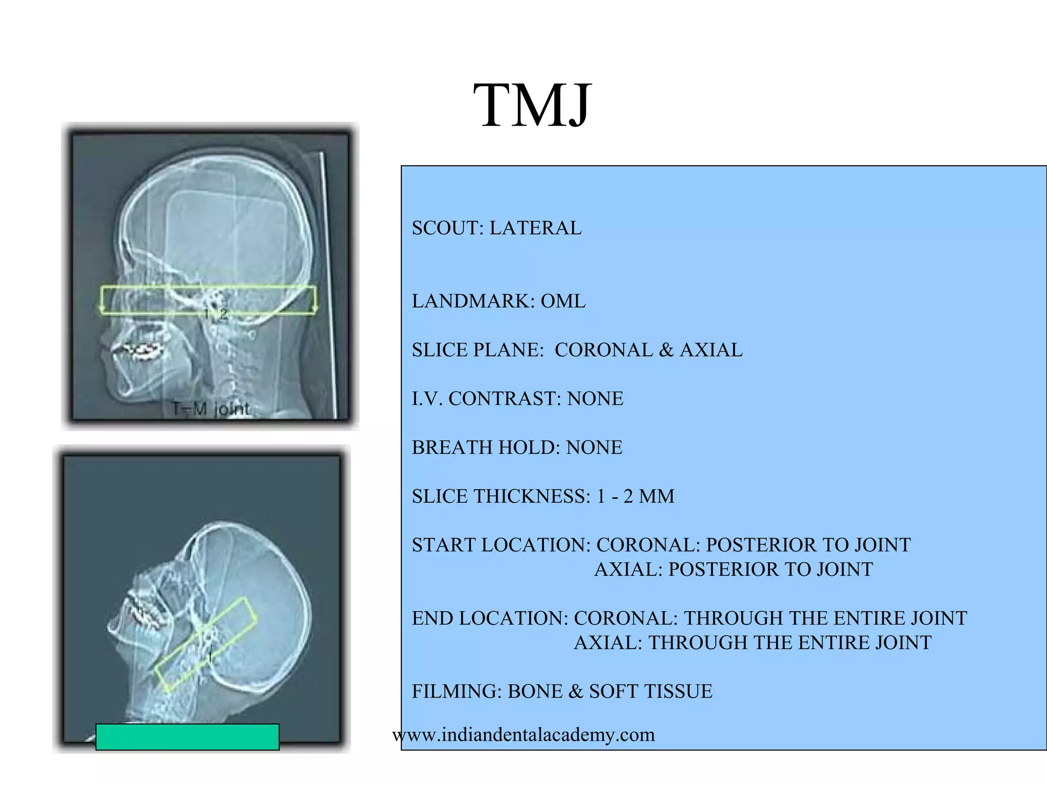
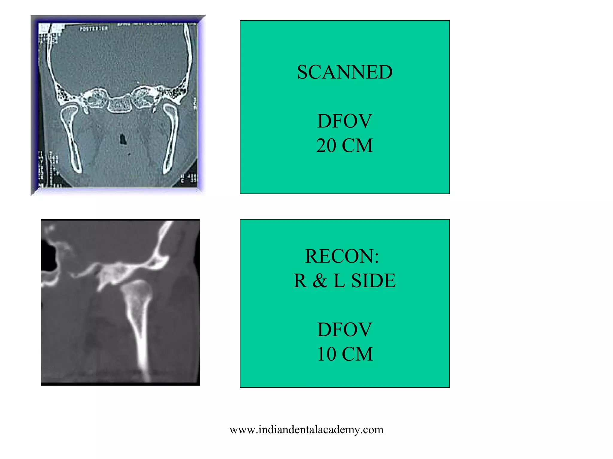
CT OF THE HEAD
PATHOLOGIES AND
PROTOCOLS

www.indiandentalacademy.com
INDIAN DENTAL ACADEMY
Leader in continuing dental education
www.indiandentalacademy.com

www.indiandentalacademy.com
PATHOLOGIES

www.indiandentalacademy.com
SINUSITIS
Sinusitis is the
name given when
the lining of one or
more of these
sinuses is red,
swollen, and tender,
the opening is
blocked, and the
sinus is at least
partially filled with
fluid (mucus and/or
pus).
www.indiandentalacademy.com
SINUS POLYP

www.indiandentalacademy.com
ORBITAL FRACTURE
Demonstrates a
superior orbital
fracture on the
left with
fragments of
bone extending
toward the
frontal lobe.
There was no
evidence of an
optic canal
fracture.
www.indiandentalacademy.com
ORBITAL ROOF FRACTURE

www.indiandentalacademy.com
TRIPOD FRACTURE

www.indiandentalacademy.com
The tripod fracture, also called
the zygomaticomaxillary
complex, is composed of a set of
fractures including the lateral
orbital wall, inferior orbital floor,
and the zygomatic arch.

www.indiandentalacademy.com
Blowout fracture

www.indiandentalacademy.com
MANDIBULAR FRACTURE

www.indiandentalacademy.com
SKULL FRACTURES
• OPEN
• CLOSED

www.indiandentalacademy.com
Although the skull is tough, resilient, and provides excellent protection for the brain, a severe impact or blow can
result in fracture of the skull and may be accompanied by injury to the brain. Some of the different types of skull
fracture include: Simple: a break in the bone without damage to the skinLinear or hairline: a break in a cranial
bone resembling a thin line, without splintering, depression, or distortion of boneDepressed: a break in a cranial
bone (or "crushed" portion of skull) with depression of the bone in toward the brainCompound: a break in or loss
of skin and splintering of the bone. Along with the fracture, brain injury, such as subdural hematoma (bleeding)
may occur.
www.indiandentalacademy.com
OPEN FRACTURECOMPOUND

www.indiandentalacademy.com
CLOSED FRACTURE

www.indiandentalacademy.com
HEMORRHAGE
•
•
•
•

INTRACEREBRAL
SUBDURAL
EPIDURAL
SUBARACHNOID

www.indiandentalacademy.com
INTRACEREBRAL

www.indiandentalacademy.com
SUBDURAL

www.indiandentalacademy.com
SDH
• form of traumatic brain injury in which
blood gathers between the dura (the outer
protective covering of the brain) and the
arachnoid (the middle layer of the
meninges).

www.indiandentalacademy.com
EPIDURAL

www.indiandentalacademy.com
EDH
• buildup of blood occurring between the
dura mater (the brain's tough outer
membrane) and the skull.

www.indiandentalacademy.com
SUBARACHNOID

www.indiandentalacademy.com
SAH
• is bleeding into the subarachnoid space
surrounding the brain, the area between the
arachnoid membrane and the pia mater.

www.indiandentalacademy.com
Rupture of an intracranial aneurysm is
the most common cause of nontraumatic
subarachnoid hemorrhage.
90-95% of all intracranial aneurysms are
located in the carotid system. The
anterior communicating artery is the
most common site (30%), followed by
the posterior communicating artery
(25%) and the middle cerebral artery
(20%).

www.indiandentalacademy.com
CEREBRAL INFARCT

www.indiandentalacademy.com
BRAIN CYST

www.indiandentalacademy.com
HYDROCEPHALUS

www.indiandentalacademy.com
ARTERIO-VENOUS
MALFORMATION

www.indiandentalacademy.com
What is a brain AVM?
Normally, arteries carry
blood containing oxygen
from the heart to the brain,
and veins carry blood with
less oxygen away from the
brain and back to the
heart. When an
arteriovenous
malformation (AVM)
occurs, a tangle of blood
vessels in the brain or on
its surface bypasses
normal brain tissue and
directly diverts blood from
the arteries to the veins
www.indiandentalacademy.com
BRAIN METS

www.indiandentalacademy.com
CVA

www.indiandentalacademy.com
This is a CAT scan of a patient with a bleeding stroke
caused by CAA. The two bright areas represent recent
areas of bleeding into the brain. Both areas are in the
outer part of the brain that is characteristic for CAAwww.indiandentalacademy.com
related strokes.
BRAIN INFECTIONS
• MENINGITIS
• ENCEPHALITIS
• ABSCESS

www.indiandentalacademy.com
MENINGITIS

Subdural empyema and diffuse cerebral edema in a patient with
bacterial meningitis (same patient as in Image 18). Obtained 1
week after Image 18, this contrast-enhanced CT scan shows
www.indiandentalacademy.com
diffuse cerebral edema and lacunar infarcts in the thalamus.
ENCEPHALITIS

Encephalitis
Encephalitis is an inflammation (irritation and swelling with presence of
www.indiandentalacademy.com
extra immune cells) of the brain, usually caused by infections.
BRAIN ABSCESS

www.indiandentalacademy.com
BRAIN TUMORS
•
•
•
•
•
•

ASTROCYTOMAS
GLIOMAS
PINEAL REGION TUMORS
LIPOMA
ACOUSTIC NEUROMA
MENINGIOMA
www.indiandentalacademy.com
•astrocytomas
Astrocytomas are glial cell tumors that are
derived from connective tissue cells called
astrocytes. These cells can be found anywhere in
the brain or spinal cord. Astrocytomas are the
most common type of childhood brain tumor.

www.indiandentalacademy.com
•Brain stem gliomas
are tumors found in the brain stem. Most brain stem tumors
cannot be surgically removed because of the remote location and
delicate and complex function this area controls. Brain stem
gliomas occur almost exclusively in children; the group most
often affected is the school-age child. The child usually does not
have increased intracranial pressure, but may have problems with
double vision, movement of the face or one side of the body, or
difficulty with walking and coordination

www.indiandentalacademy.com
•optic nerve gliomas
Optic nerve gliomas are found in or around the nerves that send
messages from the eyes to the brain. They are frequently found in
persons who have neurofibromatosis, a condition a child is born
with that makes him/her more likely to develop tumors in the
brain. Persons usually experience loss of vision, as well as
hormone problems, since these tumors are usually located at the
base of the brain where hormonal control is located. These are
typically difficult to treat due to the surrounding sensitive brain
structures.
www.indiandentalacademy.com
•medulloblastomas
Medulloblastomas are one type of PNET that are found near the
midline of the cerebellum. This tumor is rapidly growing and
often blocks drainage of the CSF (cerebral spinal fluid, which
bathes the brain and spinal cord), causing symptoms associated
with increased ICP. Medulloblastoma cells can spread
(metastasize) to other areas of the central nervous system,
especially around the spinal cord. A combination of surgery,
radiation, and chemotherapy is usually needed to control these
tumors
www.indiandentalacademy.com
•pineal region tumors
Many different tumors can arise near the pineal gland, a gland which
helps control sleep and wake cycles. Gliomas are common in this
region, as are pineal blastomas (PNET). In addition, germ cell
tumors, another form of malignant tumor, can be found in this area.
Tumors in this region are more common in children than adults, and
make up 3 to 8 percent of pediatric brain tumors. Benign pineal gland
cysts are also seen in this location, which makes the diagnosis
difficult between what is malignant and what is benign. Biopsy or
removal of the tumor is frequently necessary to tell the different
types of tumors apart. Persons with tumors in this region frequently
experience headaches or symptoms of increased intracranial
pressure. Treatment depends on the tumor type and size.
www.indiandentalacademy.com
GLIOMA

www.indiandentalacademy.com
ACOUSTIC NEUROMA

www.indiandentalacademy.com
ACOUSTIC NEUROMA

www.indiandentalacademy.com
PITUITARY GLAND TUMOR

www.indiandentalacademy.com
CT PROTOCOLS
•
•
•
•
•
•
•
•
•
•

HEAD
HEAD VASCULAR CTA CTV
PITUITARY & SELLA TURCICA
INTERNAL AUDITORY CANAL
ORBITS
PARANASAL SINUSES
TMJ
FACIAL BONES
DENTAL
STEREOTACTIC
www.indiandentalacademy.com
OML
CML

IOML

www.indiandentalacademy.com
HEAD/BRAIN (ADULT)
SCOUT: LATERAL
FOV -240
LANDMARK: OML – 15 DEG ABOVE OML
SLICE PLANE: AXIAL
I.V. CONTRAST: 100-140 ML 1-1.5 ML/SEC, TUMOR,
METS - 5 MIN DELAY

SLICE THICKNESS: 5 x 5 mm
START LOCATION: FORAMEN MAGNUM
END LOCATION: VERTEX
FILMING: BONE & SOFT TISSUE

www.indiandentalacademy.com

DFOV
25
15 DEG AND 20 DEG ABOVE OML

www.indiandentalacademy.com
CT HEAD – LOSS OF BALANCE
SCOUT: LATERAL
FOV -240
LANDMARK: OML – 15 DEG ABOVE OML
SLICE PLANE: AXIAL
I.V. CONTRAST: 100-140 ML 1-1.5 ML/SEC, TUMOR,
METS - 5 MIN DELAY

SLICE THICKNESS: 2 x 2 mm POSTERIOR FOSSA
5 x 5 mm THE REST
START LOCATION: FORAMEN MAGNUM
END LOCATION: VERTEX
FILMING: BONE & SOFT TISSUE
www.indiandentalacademy.com

DFOV
25
CT HEAD – SEIZURES -20 DEG TO
OML

www.indiandentalacademy.com
BRAIN ANGIO CTA
SCOUT: LATERAL
LANDMARK: OML
SLICE PLANE: AXIAL
I.V. CONTRAST: 100-140 ML- 3-5 ml /sec -

15 - 20 SEC DELAY CTA
30 SEC DELAY CTV
BREATH HOLD: NONE
SLICE THICKNESS: 1-2 MM
START LOCATION: BELOW SELLAR FLOOR
END LOCATION: 4-5 CM ABOVE SELLA
RECON: 50% OVERLAP
FILMING: 3 D RECON
www.indiandentalacademy.com

DFOV
18
www.indiandentalacademy.com
PITUITARY AND SELLA TURCICA
SCOUT: LATERAL
LANDMARK: OML
SLICE PLANE: CORONAL & AXIAL
I.V. CONTRAST: 100-140 ML
BREATH HOLD: NONE
SLICE THICKNESS: 1-1.5 mm
FILMING: BONE & SOFT TISSUE
www.indiandentalacademy.com

DFOV
12
www.indiandentalacademy.com
IAC
SCOUT: LATERAL
LANDMARK: IOML
SLICE PLANE: CORONAL & AXIAL
I.V. CONTRAST: 100-140 ML FOR ACOUSTIC NEUROMA OR HEARING LOSS

65 SEC DELAY
BREATH HOLD: NONE
SLICE THICKNESS: 1-2 MM, 1MM THROUGH CANAL, 2 MM PETROUS BONE
START LOCATION: CORONAL: P. SEMI-CIRC. CANAL, AXIAL: F. MAGNUM
END LOCATION: CORONAL: THROUGH PETROUS BONE
AXIAL THROUGH PETROUS BONE
FILMING: BONE & SOFT TISSUE

www.indiandentalacademy.com
SCANNED
DFOV
20 CM

RECON:
R & L SIDE
DFOV
10 CM
www.indiandentalacademy.com
ORBITS
SCOUT: LATERAL
LANDMARK: IOML

DFOV
15CM

SLICE PLANE: CORONAL & AXIAL
I.V. CONTRAST: 100-140 ML MASS OR VISUAL DISTURBANCE 2 CC/SEC

65 SEC DELAY
BREATH HOLD: NONE
SLICE THICKNESS: 2-3 MM
START LOCATION: CORONAL: SPH. SINUS, AXIAL: TOP OF MAX. SINUS
END LOCATION: CORONAL: ANTERIOR GLOBE
AXIAL:UPPER ORBITAL RIM
FILMING: BONE & SOFT TISSUE
www.indiandentalacademy.com
OPTIC NERVE PROTOCOL

www.indiandentalacademy.com
PATIENT CAN’T ASSUME
PRONE POSITION

SUPINE-CORONAL

www.indiandentalacademy.com
DENTAL ARTIFACT OMISSIONMULTIANGULATION

www.indiandentalacademy.com
FACIAL BONES
SCOUT: LATERAL
LANDMARK: IOML
SLICE PLANE: CORONAL & AXIAL
I.V. CONTRAST: 100-140 ML MASS 2 cc/sec

65 SEC DELAY
SLICE THICKNESS: 2-3 MM
START LOCATION: CORONAL: EAM
AXIAL: S. MENTI
END LOCATION: CORONAL: ANTERIOR GLOBE
AXIAL: SUPERIOR ORBITAL MARGIN
FILMING: BONE & SOFT TISSUE
www.indiandentalacademy.com

DFOV
20 CM
FACIAL BONES

INCLUDE MANDIBLE!!!!!!

www.indiandentalacademy.com
PNS
SCOUT: LATERAL
LANDMARK: OML

DFOV
15 CM

SLICE PLANE: CORONAL & AXIAL
I.V. CONTRAST: 100-140 ML MASS 2 cc/sec

65 SEC DELAY
BREATH HOLD: NONE
SLICE THICKNESS: 3 - 5 MM
START LOCATION: CORONAL: BEHIND SPHENOID SINUS
AXIAL: BOTTOM OF MAX. SINUS
END LOCATION: CORONAL: THROUGH FRONTAL SINUS
AXIAL: THROUGH FRONTAL SINUS
FILMING: BONE & SOFT TISSUE
www.indiandentalacademy.com
TMJ
SCOUT: LATERAL
LANDMARK: OML
SLICE PLANE: CORONAL & AXIAL
I.V. CONTRAST: NONE
BREATH HOLD: NONE
SLICE THICKNESS: 1 - 2 MM
START LOCATION: CORONAL: POSTERIOR TO JOINT
AXIAL: POSTERIOR TO JOINT
END LOCATION: CORONAL: THROUGH THE ENTIRE JOINT
AXIAL: THROUGH THE ENTIRE JOINT
FILMING: BONE & SOFT TISSUE
www.indiandentalacademy.com
SCANNED
DFOV
20 CM

RECON:
R & L SIDE
DFOV
10 CM

www.indiandentalacademy.com
DENTAL

www.indiandentalacademy.com
STEREOTACTIC

www.indiandentalacademy.com
Stereotactic system use
•
•
•
•

Biopsy of intracranial lesions
Aspiration of cysts
Laser microsurgery
Aspiration of brain abcess

www.indiandentalacademy.com
Thank you
www.indiandentalacademy.com
Leader in continuing dental education

www.indiandentalacademy.com

Head pathologies and protocols /certified fixed orthodontic courses by Indian dental academy