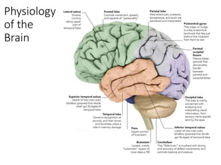
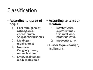
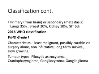
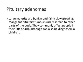
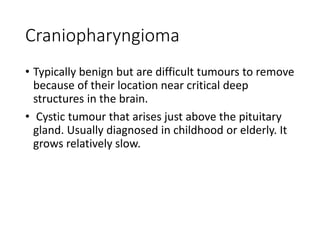
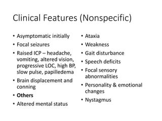
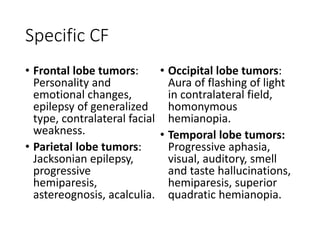
SlideShare a Scribd company logo
Brain Tumors
Presenters: Rolex Maklago & Irene Onyango
Moderator: Dr. Lee Ogutha
Date: 12th July 2021
Outline
• Brain anatomy and physiology
• Risk factors
• Classification
• Specific tumors
• Clinical presentations
• Diagnosis
• Treatment
Brain
Anatomy
Anatomy
• Blood Supply • Lymphatic Drainage
Physiology
of the
Brain
Risk factors
• Exposure to radiation
• Genetics- neurofibromatosis 1 and 2,
retinoblastoma, Li Fraumeni and Turcots syndrome
• Immunosuppression
• Viruses like JC and simian virus
Classification
• According to tissue of
origin
1. Glial cells- gliomas;
astrocytoma,
ependymoma,
5oligodendrogliomas
2. Meninges-
meningiomas
3. Neurons-
Gangliocytomas,
neuroblastoma
4. Embryonal tumors-
medulloblastoma
• According to tumour
location
1. Infratentorial,
supratentorial,
temporal lobe,
posterior fossa,
2. Intraventricular
• Tumor type –Benign,
malignant
Classification cont.
• Primary (from brain) or secondary (metastasis:
Lungs 35% , Breast 20%, Kidney 10%, GIT 5%
2016 WHO classification
WHO Grade I
Characteristics – least malignant, possibly curable via
surgery alone, non infiltrative, long term survival,
slow growing
Tumour types- Pilocytic astrocytoma, ,
Craniopharyngioma, Gangliocytoma, Ganglioglioma
Classification cont
WHO Grade II
Characteristics- relatively slow growing, somewhat
infiltrative, may recur as higher grade
Tumour types- diffuse astrocytoma, Pineocytoma,
Pure oligodendroglioma
WHO Grade III
Characteristics- malignant, infiltrative, tend to recur
as higher grade.
Tumour types- Anaplastic astrocytoma, Anaplastic
ependymoma, Anaplastic oligodendroglioma
WHO Grade IV
Characteristic- most malignant, rapid growth,
aggressive, widely infiltrative, rapid recurrence,
necrosis prone
Tumour types- Glioblastoma multiforme,
Pineoblastoma, Medulloblastoma,
Ependymoblastoma
• Account for 10% of all malignancies. Most common
CNS malignancies are metastatic lesions.
• The most common primary brain tumours are
gliomas.
• In adults two thirds of primary brain tumours arise
from structures above the tentorium
(supratentorial), whereas in children two thirds of
brain tumours arise from structures below the
tentorium (infratentorial)
Primary brain tumours
1.Gliomas
• Astrocytoma
Commonest type, usually malignant. They can occur
anywhere in the cerebral hemispheres, medulla,
brainstem. Peak incidence is 4th decade. Are graded
based on the quantity of adult and primitive cells
Grade I- cystic
Grade II- diffuse
Grade III- anaplastic
Grade IV- glioblastoma multiforme
• Glioblastoma multiforme
Most invasive type of glial tumours. Tend to grow
rapidly and spread to other tissues and have poor
prognosis. May be composed of several types of cells
eg astrocytes, oligodendrocytes. More common in
people ages 50-70 and more prevalent in men than
women.
• Oligodendrogliomas
Slow growing tumours, commonly arising from the
frontal lobes, lasts for years and show calcification
• Medulloblastoma
Highly malignant embryonal tumours grouped as PNET
arising from primitive cell nests. Most common brain
tumour in children. It often spreads within the brain itself
and can extend to the spinal canal
• Ependymoma
Here cells resemble the ependymal cells. Can occur
throughout the hemisphere. Arise from cells lining the
ventricles of the brain and central canal of the spinal cord
Meningiomas
• Usually globular, arising from the arachnoids, the
tumour gets attached to the dura and gets its blood
supply from dural arteries and veins. Along these
veins tumour cells invade bone, causing bone
destruction and reactive hyperostosis. 80% are
supratentorial. Common in females of middle aged.
• Classified as fibroblastic, endothelial and
angioblastic.
• Psammoma bodies seen microscopically
Schwannomas
• Common in auditory nerve, also called acoustic
neuromas. Occur in the internal auditory meatus
which projects into the cerebellopontine angle,
compressing 5,6,7,8th nerves.
• Presents with compressive features like unilateral
deafness, trigeminal neuralgia, squint, cerebellar
compression.
Pituitary adenomas
• Large majority are benign and fairly slow growing.
Malignant pituitary tumours rarely spread to other
parts of the body. They commonly affect people in
their 30s or 40s, although can also be diagnosed in
children.
Craniopharyngioma
• Typically benign but are difficult tumours to remove
because of their location near critical deep
structures in the brain.
• Cystic tumour that arises just above the pituitary
gland. Usually diagnosed in childhood or elderly. It
grows relatively slow.
Clinical Features (Nonspecific)
• Asymptomatic initially
• Focal seizures
• Raised ICP – headache,
vomiting, altered vision,
progressive LOC, high BP,
slow pulse, papilledema
• Brain displacement and
conning
• Others
• Altered mental status
• Ataxia
• Weakness
• Gait disturbance
• Speech deficits
• Focal sensory
abnormalities
• Personality & emotional
changes
• Nystagmus
Specific CF
• Frontal lobe tumors:
Personality and
emotional changes,
epilepsy of generalized
type, contralateral facial
weakness.
• Parietal lobe tumors:
Jacksonian epilepsy,
progressive
hemiparesis,
astereognosis, acalculia.
• Occipital lobe tumors:
Aura of flashing of light
in contralateral field,
homonymous
hemianopia.
• Temporal lobe tumors:
Progressive aphasia,
visual, auditory, smell
and taste hallucinations,
hemiparesis, superior
quadratic hemianopia.
Specific CF
• Midline tumors: Produces
bilateral hydrocephalus.
• Tumors of the third ventricle
(colloid cyst is common):
Causes bilateral
hydrocephalus, progressive
cerebral atrophy, dementia,
sexual precocity, endocrine
disturbances.
• Pineal tumors: Causes
precocious puberty.
• Cerebellar vermis tumors:
Usually medulloblastomas,
occur in young children,
presents with progressive
hydrocephalus and features
of herniation of cerebellar
tonsils
through foramen magnum.
• Cerebellar hemisphere
tumors: Commonly are
astrocytomas, produce
cerebellar syndromes,
nystagmus.
Diagnosis
• X-ray skull
• Calcifications –
meningiomas,
craniopharungiomas
• Separation of sutures
• Beaten silver appearance
• Lateral displacement of
pineal body
• Hyperostosis, skull
destruction, expansion
• CT Scan
• Large meningioma
affecting frontal region
• MRI
• Glioma vs GBM
Diagnosis Cont.
• Isotope Scan
• PET Scan
• Carotid Angiogram
• Ventriculography
• EEG
• Biopsy - CT guided or
other intraoperative
options
• Brain-stem evoked
potentials and
audiometric evaluations
if suspecting acoustic
neuroma
Treatment
• Relief of ↑ICP
• Ventricular tap and
drainage through
posterior parietal burr
hole
• Tapping of cystic
tumor/abscess
• Mannitol
• Emergency
decompression by partial
tumor removal
• Establish pathological Dx
• Burr hole and biopsy
• Craniotomy and biopsy
using brain cannula
• Frozen section biopsy
• CT guided stereotactic
biopsy
• Removal of benign
tumors (*craniotomy
approaches)
• Decompressive surgeries
if malignant
Treatment Cont
• Shunt surgeries to drain
CSF e.g. VP shunt or
ventriculoatrial shunt
• Radiotherapy – ext
radiotherapy can be
used as primary rx or as
an adjuvant therapy
following surgery
• Chemotherapy
occasionally –
Temozolamide
• Corticosteroids e.g
Dexamethasone may be
used for secondary
tumors – reduces
vasogenic edema
• Prognosis – better
prognosis for benign
tumors, and those that
are surgically accessible

More Related Content

What's hot

CNS Tumors
CNS TumorsCNS Tumors
CNS Tumors
Miami Dade
 
MEDULLOBLASTOMA
MEDULLOBLASTOMAMEDULLOBLASTOMA
MEDULLOBLASTOMA
suresh Bishokarma
 
Spinal cord tumor
Spinal cord tumorSpinal cord tumor
Overview of brain tumors
Overview of brain tumorsOverview of brain tumors
Overview of brain tumors
DrAyush Garg
 
Astrocytoma
AstrocytomaAstrocytoma
Astrocytoma
Timothy Zagada
 
2021 WHO Classification of brain tumours.pptx
2021 WHO Classification of brain tumours.pptx2021 WHO Classification of brain tumours.pptx
2021 WHO Classification of brain tumours.pptx
RejoyceAnto
 
Intramedullary tumors
Intramedullary tumorsIntramedullary tumors
Intramedullary tumors
Ritesh Mahajan
 
Brain tumors advanced
Brain tumors advancedBrain tumors advanced
Brain tumors advanced
ranaelsaeedAboelfeto
 
Gliomas - Brain Tumor
Gliomas - Brain TumorGliomas - Brain Tumor
Gliomas - Brain Tumor
suresh Bishokarma
 
Brain tumours
Brain tumoursBrain tumours
Brain tumours
Dr Fakir Mohan Sahu
 
What is brain tumor? symptoms, causes and treatment
What is brain tumor? symptoms, causes and treatmentWhat is brain tumor? symptoms, causes and treatment
What is brain tumor? symptoms, causes and treatment
MediFee
 
Cns tumors and pns
Cns tumors and pnsCns tumors and pns
Cns tumors and pnsMUBOSScz
 
Brain metastasis - Simplified
Brain metastasis - SimplifiedBrain metastasis - Simplified
Brain metastasis - Simplified
suresh Bishokarma
 
Brain stem glioma
Brain stem gliomaBrain stem glioma
Brain stem glioma
suresh Bishokarma
 
Pathology of intracranial tumors lecture
Pathology of intracranial tumors   lecturePathology of intracranial tumors   lecture
Pathology of intracranial tumors lecture
Effiong Akang
 
Presentation1.pptx, imaging of multiple sclerosis.
Presentation1.pptx, imaging of multiple sclerosis.Presentation1.pptx, imaging of multiple sclerosis.
Presentation1.pptx, imaging of multiple sclerosis.Abdellah Nazeer
 
Brain tumor ppt.pptx
Brain tumor ppt.pptxBrain tumor ppt.pptx
Brain tumor ppt.pptx
Sarmila Asif
 
Neoplasia introduction
Neoplasia introductionNeoplasia introduction
Neoplasia introduction
Prasad CSBR
 

What's hot (20)

CNS Tumors
CNS TumorsCNS Tumors
CNS Tumors
 
Brain Tumors
Brain TumorsBrain Tumors
Brain Tumors
 
MEDULLOBLASTOMA
MEDULLOBLASTOMAMEDULLOBLASTOMA
MEDULLOBLASTOMA
 
Spinal cord tumor
Spinal cord tumorSpinal cord tumor
Spinal cord tumor
 
Overview of brain tumors
Overview of brain tumorsOverview of brain tumors
Overview of brain tumors
 
Astrocytoma
AstrocytomaAstrocytoma
Astrocytoma
 
2021 WHO Classification of brain tumours.pptx
2021 WHO Classification of brain tumours.pptx2021 WHO Classification of brain tumours.pptx
2021 WHO Classification of brain tumours.pptx
 
Intramedullary tumors
Intramedullary tumorsIntramedullary tumors
Intramedullary tumors
 
Brain tumors advanced
Brain tumors advancedBrain tumors advanced
Brain tumors advanced
 
Gliomas - Brain Tumor
Gliomas - Brain TumorGliomas - Brain Tumor
Gliomas - Brain Tumor
 
Brain tumours
Brain tumoursBrain tumours
Brain tumours
 
What is brain tumor? symptoms, causes and treatment
What is brain tumor? symptoms, causes and treatmentWhat is brain tumor? symptoms, causes and treatment
What is brain tumor? symptoms, causes and treatment
 
Cns tumors and pns
Cns tumors and pnsCns tumors and pns
Cns tumors and pns
 
Brain metastasis - Simplified
Brain metastasis - SimplifiedBrain metastasis - Simplified
Brain metastasis - Simplified
 
Brain stem glioma
Brain stem gliomaBrain stem glioma
Brain stem glioma
 
Pathology of intracranial tumors lecture
Pathology of intracranial tumors   lecturePathology of intracranial tumors   lecture
Pathology of intracranial tumors lecture
 
Presentation1.pptx, imaging of multiple sclerosis.
Presentation1.pptx, imaging of multiple sclerosis.Presentation1.pptx, imaging of multiple sclerosis.
Presentation1.pptx, imaging of multiple sclerosis.
 
Brain tumor ppt.pptx
Brain tumor ppt.pptxBrain tumor ppt.pptx
Brain tumor ppt.pptx
 
Neoplasia introduction
Neoplasia introductionNeoplasia introduction
Neoplasia introduction
 
Medulloblastomas
MedulloblastomasMedulloblastomas
Medulloblastomas
 

Similar to Brain tumors

Lecture 4 CNS TUMOR 1 2.ppt
Lecture 4 CNS TUMOR 1 2.pptLecture 4 CNS TUMOR 1 2.ppt
Lecture 4 CNS TUMOR 1 2.ppt
ugonnanwoke
 
Brain tumor in children
Brain tumor in childrenBrain tumor in children
Brain tumor in children
SharminNaharNabila
 
Brain tumors in children with Updates- Pranav
Brain tumors in children with Updates- PranavBrain tumors in children with Updates- Pranav
Brain tumors in children with Updates- Pranav
Pranav S
 
CNS malignancies in childhood.pptx
CNS malignancies  in childhood.pptxCNS malignancies  in childhood.pptx
CNS malignancies in childhood.pptx
Ahmed Moharram
 
6.Tumors Presentation.ppt
6.Tumors Presentation.ppt6.Tumors Presentation.ppt
6.Tumors Presentation.ppt
AsmaNoreen13
 
medicine.Intracranial neoplasms.(dr.hawar)
medicine.Intracranial neoplasms.(dr.hawar)medicine.Intracranial neoplasms.(dr.hawar)
medicine.Intracranial neoplasms.(dr.hawar)student
 
brain tumor.pptx
brain tumor.pptxbrain tumor.pptx
brain tumor.pptx
mousaderhem1
 
BRAIN-TUMORS.pptx
BRAIN-TUMORS.pptxBRAIN-TUMORS.pptx
BRAIN-TUMORS.pptx
WengelRedkiss
 
BRAIN-TUMORS.pptx
BRAIN-TUMORS.pptxBRAIN-TUMORS.pptx
BRAIN-TUMORS.pptx
WengelRedkiss
 
Brain tumor
Brain tumorBrain tumor
Brain tumor
Dr. Vyom Jain
 
Brain tumors
Brain tumorsBrain tumors
Brain tumors
Mr. Dipti sorte
 
L75- Pathology of CNS Tumors.ppt
L75- Pathology of CNS Tumors.pptL75- Pathology of CNS Tumors.ppt
L75- Pathology of CNS Tumors.ppt
Gowthun
 
CNS TUMORS.ppt
CNS TUMORS.pptCNS TUMORS.ppt
CNS TUMORS.ppt
LarissaSams1
 
Brain-tumors.pdf
Brain-tumors.pdfBrain-tumors.pdf
Brain-tumors.pdf
KushalathaShetty
 
Clival Pathology.ppt
Clival Pathology.pptClival Pathology.ppt
Clival Pathology.ppt
mhmodsaad2
 
2brain tumors.pptx
2brain tumors.pptx2brain tumors.pptx
2brain tumors.pptx
Adesh Shrivastava
 
Brain tumors - of adults -
Brain tumors - of adults -Brain tumors - of adults -
Brain tumors - of adults -
LeenaMubiden
 
Brain tumors clinical features and causes.ppt
Brain tumors clinical features and causes.pptBrain tumors clinical features and causes.ppt
Brain tumors clinical features and causes.ppt
rajalaxmiphysio1
 

Similar to Brain tumors (20)

Lecture 4 CNS TUMOR 1 2.ppt
Lecture 4 CNS TUMOR 1 2.pptLecture 4 CNS TUMOR 1 2.ppt
Lecture 4 CNS TUMOR 1 2.ppt
 
Brain tumor in children
Brain tumor in childrenBrain tumor in children
Brain tumor in children
 
Brain tumors in children with Updates- Pranav
Brain tumors in children with Updates- PranavBrain tumors in children with Updates- Pranav
Brain tumors in children with Updates- Pranav
 
Brain cancer (tumors)
Brain cancer (tumors)Brain cancer (tumors)
Brain cancer (tumors)
 
CNS malignancies in childhood.pptx
CNS malignancies  in childhood.pptxCNS malignancies  in childhood.pptx
CNS malignancies in childhood.pptx
 
6.Tumors Presentation.ppt
6.Tumors Presentation.ppt6.Tumors Presentation.ppt
6.Tumors Presentation.ppt
 
medicine.Intracranial neoplasms.(dr.hawar)
medicine.Intracranial neoplasms.(dr.hawar)medicine.Intracranial neoplasms.(dr.hawar)
medicine.Intracranial neoplasms.(dr.hawar)
 
brain tumor.pptx
brain tumor.pptxbrain tumor.pptx
brain tumor.pptx
 
BRAIN-TUMORS.pptx
BRAIN-TUMORS.pptxBRAIN-TUMORS.pptx
BRAIN-TUMORS.pptx
 
BRAIN-TUMORS.pptx
BRAIN-TUMORS.pptxBRAIN-TUMORS.pptx
BRAIN-TUMORS.pptx
 
Brain tumor
Brain tumorBrain tumor
Brain tumor
 
Brain tumors
Brain tumorsBrain tumors
Brain tumors
 
L75- Pathology of CNS Tumors.ppt
L75- Pathology of CNS Tumors.pptL75- Pathology of CNS Tumors.ppt
L75- Pathology of CNS Tumors.ppt
 
CNS TUMORS.ppt
CNS TUMORS.pptCNS TUMORS.ppt
CNS TUMORS.ppt
 
Brain-tumors.pdf
Brain-tumors.pdfBrain-tumors.pdf
Brain-tumors.pdf
 
Clival Pathology.ppt
Clival Pathology.pptClival Pathology.ppt
Clival Pathology.ppt
 
2brain tumors.pptx
2brain tumors.pptx2brain tumors.pptx
2brain tumors.pptx
 
Brain tumors
Brain tumorsBrain tumors
Brain tumors
 
Brain tumors - of adults -
Brain tumors - of adults -Brain tumors - of adults -
Brain tumors - of adults -
 
Brain tumors clinical features and causes.ppt
Brain tumors clinical features and causes.pptBrain tumors clinical features and causes.ppt
Brain tumors clinical features and causes.ppt
 

Recently uploaded

Pulmonary Thromboembolism - etilogy, types, medical- Surgical and nursing man...
Pulmonary Thromboembolism - etilogy, types, medical- Surgical and nursing man...Pulmonary Thromboembolism - etilogy, types, medical- Surgical and nursing man...
Pulmonary Thromboembolism - etilogy, types, medical- Surgical and nursing man...
VarunMahajani
 
Lung Cancer: Artificial Intelligence, Synergetics, Complex System Analysis, S...
Lung Cancer: Artificial Intelligence, Synergetics, Complex System Analysis, S...Lung Cancer: Artificial Intelligence, Synergetics, Complex System Analysis, S...
Lung Cancer: Artificial Intelligence, Synergetics, Complex System Analysis, S...
Oleg Kshivets
 
The Normal Electrocardiogram - Part I of II
The Normal Electrocardiogram - Part I of IIThe Normal Electrocardiogram - Part I of II
The Normal Electrocardiogram - Part I of II
MedicoseAcademics
 
KDIGO 2024 guidelines for diabetologists
KDIGO 2024 guidelines for diabetologistsKDIGO 2024 guidelines for diabetologists
KDIGO 2024 guidelines for diabetologists
د.محمود نجيب
 
Ocular injury ppt Upendra pal optometrist upums saifai etawah
Ocular injury  ppt  Upendra pal  optometrist upums saifai etawahOcular injury  ppt  Upendra pal  optometrist upums saifai etawah
Ocular injury ppt Upendra pal optometrist upums saifai etawah
pal078100
 
heat stroke and heat exhaustion in children
heat stroke and heat exhaustion in childrenheat stroke and heat exhaustion in children
heat stroke and heat exhaustion in children
SumeraAhmad5
 
263778731218 Abortion Clinic /Pills In Harare ,
263778731218 Abortion Clinic /Pills In Harare ,263778731218 Abortion Clinic /Pills In Harare ,
263778731218 Abortion Clinic /Pills In Harare ,
sisternakatoto
 
ARTHROLOGY PPT NCISM SYLLABUS AYURVEDA STUDENTS
ARTHROLOGY PPT NCISM SYLLABUS AYURVEDA STUDENTSARTHROLOGY PPT NCISM SYLLABUS AYURVEDA STUDENTS
ARTHROLOGY PPT NCISM SYLLABUS AYURVEDA STUDENTS
Dr. Vinay Pareek
 
Alcohol_Dr. Jeenal Mistry MD Pharmacology.pdf
Alcohol_Dr. Jeenal Mistry MD Pharmacology.pdfAlcohol_Dr. Jeenal Mistry MD Pharmacology.pdf
Alcohol_Dr. Jeenal Mistry MD Pharmacology.pdf
Dr Jeenal Mistry
 
Evaluation of antidepressant activity of clitoris ternatea in animals
Evaluation of antidepressant activity of clitoris ternatea in animalsEvaluation of antidepressant activity of clitoris ternatea in animals
Evaluation of antidepressant activity of clitoris ternatea in animals
Shweta
 
TEST BANK for Operations Management, 14th Edition by William J. Stevenson, Ve...
TEST BANK for Operations Management, 14th Edition by William J. Stevenson, Ve...TEST BANK for Operations Management, 14th Edition by William J. Stevenson, Ve...
TEST BANK for Operations Management, 14th Edition by William J. Stevenson, Ve...
kevinkariuki227
 
NVBDCP.pptx Nation vector borne disease control program
NVBDCP.pptx Nation vector borne disease control programNVBDCP.pptx Nation vector borne disease control program
NVBDCP.pptx Nation vector borne disease control program
Sapna Thakur
 
New Directions in Targeted Therapeutic Approaches for Older Adults With Mantl...
New Directions in Targeted Therapeutic Approaches for Older Adults With Mantl...New Directions in Targeted Therapeutic Approaches for Older Adults With Mantl...
New Directions in Targeted Therapeutic Approaches for Older Adults With Mantl...
i3 Health
 
Superficial & Deep Fascia of the NECK.pptx
Superficial & Deep Fascia of the NECK.pptxSuperficial & Deep Fascia of the NECK.pptx
Superficial & Deep Fascia of the NECK.pptx
Dr. Rabia Inam Gandapore
 
ACUTE SCROTUM.....pdf. ACUTE SCROTAL CONDITIOND
ACUTE SCROTUM.....pdf. ACUTE SCROTAL CONDITIONDACUTE SCROTUM.....pdf. ACUTE SCROTAL CONDITIOND
ACUTE SCROTUM.....pdf. ACUTE SCROTAL CONDITIOND
DR SETH JOTHAM
 
Novas diretrizes da OMS para os cuidados perinatais de mais qualidade
Novas diretrizes da OMS para os cuidados perinatais de mais qualidadeNovas diretrizes da OMS para os cuidados perinatais de mais qualidade
Novas diretrizes da OMS para os cuidados perinatais de mais qualidade
Prof. Marcus Renato de Carvalho
 
Ophthalmology Clinical Tests for OSCE exam
Ophthalmology Clinical Tests for OSCE examOphthalmology Clinical Tests for OSCE exam
Ophthalmology Clinical Tests for OSCE exam
KafrELShiekh University
 
Tom Selleck Health: A Comprehensive Look at the Iconic Actor’s Wellness Journey
Tom Selleck Health: A Comprehensive Look at the Iconic Actor’s Wellness JourneyTom Selleck Health: A Comprehensive Look at the Iconic Actor’s Wellness Journey
Tom Selleck Health: A Comprehensive Look at the Iconic Actor’s Wellness Journey
greendigital
 
Phone Us ❤85270-49040❤ #ℂall #gIRLS In Surat By Surat @ℂall @Girls Hotel With...
Phone Us ❤85270-49040❤ #ℂall #gIRLS In Surat By Surat @ℂall @Girls Hotel With...Phone Us ❤85270-49040❤ #ℂall #gIRLS In Surat By Surat @ℂall @Girls Hotel With...
Phone Us ❤85270-49040❤ #ℂall #gIRLS In Surat By Surat @ℂall @Girls Hotel With...
Savita Shen $i11
 
Maxilla, Mandible & Hyoid Bone & Clinical Correlations by Dr. RIG.pptx
Maxilla, Mandible & Hyoid Bone & Clinical Correlations by Dr. RIG.pptxMaxilla, Mandible & Hyoid Bone & Clinical Correlations by Dr. RIG.pptx
Maxilla, Mandible & Hyoid Bone & Clinical Correlations by Dr. RIG.pptx
Dr. Rabia Inam Gandapore
 

Recently uploaded (20)

Pulmonary Thromboembolism - etilogy, types, medical- Surgical and nursing man...
Pulmonary Thromboembolism - etilogy, types, medical- Surgical and nursing man...Pulmonary Thromboembolism - etilogy, types, medical- Surgical and nursing man...
Pulmonary Thromboembolism - etilogy, types, medical- Surgical and nursing man...
 
Lung Cancer: Artificial Intelligence, Synergetics, Complex System Analysis, S...
Lung Cancer: Artificial Intelligence, Synergetics, Complex System Analysis, S...Lung Cancer: Artificial Intelligence, Synergetics, Complex System Analysis, S...
Lung Cancer: Artificial Intelligence, Synergetics, Complex System Analysis, S...
 
The Normal Electrocardiogram - Part I of II
The Normal Electrocardiogram - Part I of IIThe Normal Electrocardiogram - Part I of II
The Normal Electrocardiogram - Part I of II
 
KDIGO 2024 guidelines for diabetologists
KDIGO 2024 guidelines for diabetologistsKDIGO 2024 guidelines for diabetologists
KDIGO 2024 guidelines for diabetologists
 
Ocular injury ppt Upendra pal optometrist upums saifai etawah
Ocular injury  ppt  Upendra pal  optometrist upums saifai etawahOcular injury  ppt  Upendra pal  optometrist upums saifai etawah
Ocular injury ppt Upendra pal optometrist upums saifai etawah
 
heat stroke and heat exhaustion in children
heat stroke and heat exhaustion in childrenheat stroke and heat exhaustion in children
heat stroke and heat exhaustion in children
 
263778731218 Abortion Clinic /Pills In Harare ,
263778731218 Abortion Clinic /Pills In Harare ,263778731218 Abortion Clinic /Pills In Harare ,
263778731218 Abortion Clinic /Pills In Harare ,
 
ARTHROLOGY PPT NCISM SYLLABUS AYURVEDA STUDENTS
ARTHROLOGY PPT NCISM SYLLABUS AYURVEDA STUDENTSARTHROLOGY PPT NCISM SYLLABUS AYURVEDA STUDENTS
ARTHROLOGY PPT NCISM SYLLABUS AYURVEDA STUDENTS
 
Alcohol_Dr. Jeenal Mistry MD Pharmacology.pdf
Alcohol_Dr. Jeenal Mistry MD Pharmacology.pdfAlcohol_Dr. Jeenal Mistry MD Pharmacology.pdf
Alcohol_Dr. Jeenal Mistry MD Pharmacology.pdf
 
Evaluation of antidepressant activity of clitoris ternatea in animals
Evaluation of antidepressant activity of clitoris ternatea in animalsEvaluation of antidepressant activity of clitoris ternatea in animals
Evaluation of antidepressant activity of clitoris ternatea in animals
 
TEST BANK for Operations Management, 14th Edition by William J. Stevenson, Ve...
TEST BANK for Operations Management, 14th Edition by William J. Stevenson, Ve...TEST BANK for Operations Management, 14th Edition by William J. Stevenson, Ve...
TEST BANK for Operations Management, 14th Edition by William J. Stevenson, Ve...
 
NVBDCP.pptx Nation vector borne disease control program
NVBDCP.pptx Nation vector borne disease control programNVBDCP.pptx Nation vector borne disease control program
NVBDCP.pptx Nation vector borne disease control program
 
New Directions in Targeted Therapeutic Approaches for Older Adults With Mantl...
New Directions in Targeted Therapeutic Approaches for Older Adults With Mantl...New Directions in Targeted Therapeutic Approaches for Older Adults With Mantl...
New Directions in Targeted Therapeutic Approaches for Older Adults With Mantl...
 
Superficial & Deep Fascia of the NECK.pptx
Superficial & Deep Fascia of the NECK.pptxSuperficial & Deep Fascia of the NECK.pptx
Superficial & Deep Fascia of the NECK.pptx
 
ACUTE SCROTUM.....pdf. ACUTE SCROTAL CONDITIOND
ACUTE SCROTUM.....pdf. ACUTE SCROTAL CONDITIONDACUTE SCROTUM.....pdf. ACUTE SCROTAL CONDITIOND
ACUTE SCROTUM.....pdf. ACUTE SCROTAL CONDITIOND
 
Novas diretrizes da OMS para os cuidados perinatais de mais qualidade
Novas diretrizes da OMS para os cuidados perinatais de mais qualidadeNovas diretrizes da OMS para os cuidados perinatais de mais qualidade
Novas diretrizes da OMS para os cuidados perinatais de mais qualidade
 
Ophthalmology Clinical Tests for OSCE exam
Ophthalmology Clinical Tests for OSCE examOphthalmology Clinical Tests for OSCE exam
Ophthalmology Clinical Tests for OSCE exam
 
Tom Selleck Health: A Comprehensive Look at the Iconic Actor’s Wellness Journey
Tom Selleck Health: A Comprehensive Look at the Iconic Actor’s Wellness JourneyTom Selleck Health: A Comprehensive Look at the Iconic Actor’s Wellness Journey
Tom Selleck Health: A Comprehensive Look at the Iconic Actor’s Wellness Journey
 
Phone Us ❤85270-49040❤ #ℂall #gIRLS In Surat By Surat @ℂall @Girls Hotel With...
Phone Us ❤85270-49040❤ #ℂall #gIRLS In Surat By Surat @ℂall @Girls Hotel With...Phone Us ❤85270-49040❤ #ℂall #gIRLS In Surat By Surat @ℂall @Girls Hotel With...
Phone Us ❤85270-49040❤ #ℂall #gIRLS In Surat By Surat @ℂall @Girls Hotel With...
 
Maxilla, Mandible & Hyoid Bone & Clinical Correlations by Dr. RIG.pptx
Maxilla, Mandible & Hyoid Bone & Clinical Correlations by Dr. RIG.pptxMaxilla, Mandible & Hyoid Bone & Clinical Correlations by Dr. RIG.pptx
Maxilla, Mandible & Hyoid Bone & Clinical Correlations by Dr. RIG.pptx
 

Brain tumors

  • 1. Brain Tumors Presenters: Rolex Maklago & Irene Onyango Moderator: Dr. Lee Ogutha Date: 12th July 2021
  • 2. Outline • Brain anatomy and physiology • Risk factors • Classification • Specific tumors • Clinical presentations • Diagnosis • Treatment
  • 4. Anatomy • Blood Supply • Lymphatic Drainage
  • 6. Risk factors • Exposure to radiation • Genetics- neurofibromatosis 1 and 2, retinoblastoma, Li Fraumeni and Turcots syndrome • Immunosuppression • Viruses like JC and simian virus
  • 7. Classification • According to tissue of origin 1. Glial cells- gliomas; astrocytoma, ependymoma, 5oligodendrogliomas 2. Meninges- meningiomas 3. Neurons- Gangliocytomas, neuroblastoma 4. Embryonal tumors- medulloblastoma • According to tumour location 1. Infratentorial, supratentorial, temporal lobe, posterior fossa, 2. Intraventricular • Tumor type –Benign, malignant
  • 8. Classification cont. • Primary (from brain) or secondary (metastasis: Lungs 35% , Breast 20%, Kidney 10%, GIT 5% 2016 WHO classification WHO Grade I Characteristics – least malignant, possibly curable via surgery alone, non infiltrative, long term survival, slow growing Tumour types- Pilocytic astrocytoma, , Craniopharyngioma, Gangliocytoma, Ganglioglioma
  • 9. Classification cont WHO Grade II Characteristics- relatively slow growing, somewhat infiltrative, may recur as higher grade Tumour types- diffuse astrocytoma, Pineocytoma, Pure oligodendroglioma WHO Grade III Characteristics- malignant, infiltrative, tend to recur as higher grade. Tumour types- Anaplastic astrocytoma, Anaplastic ependymoma, Anaplastic oligodendroglioma
  • 10. WHO Grade IV Characteristic- most malignant, rapid growth, aggressive, widely infiltrative, rapid recurrence, necrosis prone Tumour types- Glioblastoma multiforme, Pineoblastoma, Medulloblastoma, Ependymoblastoma
  • 11. • Account for 10% of all malignancies. Most common CNS malignancies are metastatic lesions. • The most common primary brain tumours are gliomas. • In adults two thirds of primary brain tumours arise from structures above the tentorium (supratentorial), whereas in children two thirds of brain tumours arise from structures below the tentorium (infratentorial)
  • 12. Primary brain tumours 1.Gliomas • Astrocytoma Commonest type, usually malignant. They can occur anywhere in the cerebral hemispheres, medulla, brainstem. Peak incidence is 4th decade. Are graded based on the quantity of adult and primitive cells Grade I- cystic Grade II- diffuse Grade III- anaplastic Grade IV- glioblastoma multiforme
  • 13. • Glioblastoma multiforme Most invasive type of glial tumours. Tend to grow rapidly and spread to other tissues and have poor prognosis. May be composed of several types of cells eg astrocytes, oligodendrocytes. More common in people ages 50-70 and more prevalent in men than women.
  • 14. • Oligodendrogliomas Slow growing tumours, commonly arising from the frontal lobes, lasts for years and show calcification • Medulloblastoma Highly malignant embryonal tumours grouped as PNET arising from primitive cell nests. Most common brain tumour in children. It often spreads within the brain itself and can extend to the spinal canal • Ependymoma Here cells resemble the ependymal cells. Can occur throughout the hemisphere. Arise from cells lining the ventricles of the brain and central canal of the spinal cord
  • 15. Meningiomas • Usually globular, arising from the arachnoids, the tumour gets attached to the dura and gets its blood supply from dural arteries and veins. Along these veins tumour cells invade bone, causing bone destruction and reactive hyperostosis. 80% are supratentorial. Common in females of middle aged. • Classified as fibroblastic, endothelial and angioblastic. • Psammoma bodies seen microscopically
  • 16. Schwannomas • Common in auditory nerve, also called acoustic neuromas. Occur in the internal auditory meatus which projects into the cerebellopontine angle, compressing 5,6,7,8th nerves. • Presents with compressive features like unilateral deafness, trigeminal neuralgia, squint, cerebellar compression.
  • 17. Pituitary adenomas • Large majority are benign and fairly slow growing. Malignant pituitary tumours rarely spread to other parts of the body. They commonly affect people in their 30s or 40s, although can also be diagnosed in children.
  • 18. Craniopharyngioma • Typically benign but are difficult tumours to remove because of their location near critical deep structures in the brain. • Cystic tumour that arises just above the pituitary gland. Usually diagnosed in childhood or elderly. It grows relatively slow.
  • 19. Clinical Features (Nonspecific) • Asymptomatic initially • Focal seizures • Raised ICP – headache, vomiting, altered vision, progressive LOC, high BP, slow pulse, papilledema • Brain displacement and conning • Others • Altered mental status • Ataxia • Weakness • Gait disturbance • Speech deficits • Focal sensory abnormalities • Personality & emotional changes • Nystagmus
  • 20. Specific CF • Frontal lobe tumors: Personality and emotional changes, epilepsy of generalized type, contralateral facial weakness. • Parietal lobe tumors: Jacksonian epilepsy, progressive hemiparesis, astereognosis, acalculia. • Occipital lobe tumors: Aura of flashing of light in contralateral field, homonymous hemianopia. • Temporal lobe tumors: Progressive aphasia, visual, auditory, smell and taste hallucinations, hemiparesis, superior quadratic hemianopia.
  • 21. Specific CF • Midline tumors: Produces bilateral hydrocephalus. • Tumors of the third ventricle (colloid cyst is common): Causes bilateral hydrocephalus, progressive cerebral atrophy, dementia, sexual precocity, endocrine disturbances. • Pineal tumors: Causes precocious puberty. • Cerebellar vermis tumors: Usually medulloblastomas, occur in young children, presents with progressive hydrocephalus and features of herniation of cerebellar tonsils through foramen magnum. • Cerebellar hemisphere tumors: Commonly are astrocytomas, produce cerebellar syndromes, nystagmus.
  • 22. Diagnosis • X-ray skull • Calcifications – meningiomas, craniopharungiomas • Separation of sutures • Beaten silver appearance • Lateral displacement of pineal body • Hyperostosis, skull destruction, expansion • CT Scan • Large meningioma affecting frontal region • MRI • Glioma vs GBM
  • 23. Diagnosis Cont. • Isotope Scan • PET Scan • Carotid Angiogram • Ventriculography • EEG • Biopsy - CT guided or other intraoperative options • Brain-stem evoked potentials and audiometric evaluations if suspecting acoustic neuroma
  • 24. Treatment • Relief of ↑ICP • Ventricular tap and drainage through posterior parietal burr hole • Tapping of cystic tumor/abscess • Mannitol • Emergency decompression by partial tumor removal • Establish pathological Dx • Burr hole and biopsy • Craniotomy and biopsy using brain cannula • Frozen section biopsy • CT guided stereotactic biopsy • Removal of benign tumors (*craniotomy approaches) • Decompressive surgeries if malignant
  • 25. Treatment Cont • Shunt surgeries to drain CSF e.g. VP shunt or ventriculoatrial shunt • Radiotherapy – ext radiotherapy can be used as primary rx or as an adjuvant therapy following surgery • Chemotherapy occasionally – Temozolamide • Corticosteroids e.g Dexamethasone may be used for secondary tumors – reduces vasogenic edema • Prognosis – better prognosis for benign tumors, and those that are surgically accessible