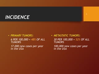
This document discusses intracranial tumors, including:
- Their incidence rates, with primary brain tumors occurring in 6 per 100,000 people and metastatic tumors in 30 per 100,000 people.
- Common tumor types like astrocytomas, oligodendrogliomas, ependymomas, medulloblastomas, meningiomas, and pituitary tumors.
- Risk factors, clinical presentation, investigations including CT, MRI, PET and angiography, pathology classification, and management approaches like surgery, radiation and chemotherapy.
I LOVE NEUROSURGERY INITIATIVE: Spinal Tumorswalid maani
A comprehensive presentation about spinal tumors. Some concentration on anatomy. Discussion of presentation, diagnosis and management. Plenty of images.
I LOVE NEUROSURGERY INITIATIVE: Cranio-cerebral Injuries part 1walid maani
A comprehensive presentation about the primary injuries of the scalp, skull and brain occurring in head injuries. Directed to medical students and junior hospital doctors.
A short talk about two of the traumatic intracranial bleeds, namely extra and subdural hematomas. Directed to med students moving from basic into clinical teaching.
Brain arteriovenous malformations (bAVM) are abnormal connections of arteries and veins in the brain, forming a tangled web of vessels instead of a normal capillary network treated with multimodalities including, SRS, embolisation and Microneurosurgery.
This slides updates the management of AVM highlighting the importance of SM grading, Pollock radiation grading etc.
PINEAL REGION TUMORS DIAGNOSIS & SURGICAL APPROACHES.pptxMedhatMoustafa3
Anatomy and related vascular structures of pineal region.pathological classification and incidence. Clinical Presentations and different diagnostics modalities. Different surgical approaches for pineal region
O artigo fala sobre a reabilitação em pacientes com tumores cerebrais sob uma visão multidisciplinar, visando a funcionalidade e tratamento das sequelas.
CRANIO CEREBRAL INJURIES FOR MEDICAL STUDENTSwalid maani
This is a simple outline of traumatic injuries which occures to the scalp, skull and brain with some simplified classifications and outlined management
Established In 1984 by Prof. Walid Maani, Started with one Neurosurgical Resident. Today it had graduated 20 of the 50 neurosurgeons practicing in Jordan
NVBDCP.pptx Nation vector borne disease control programSapna Thakur
NVBDCP was launched in 2003-2004 . Vector-Borne Disease: Disease that results from an infection transmitted to humans and other animals by blood-feeding arthropods, such as mosquitoes, ticks, and fleas. Examples of vector-borne diseases include Dengue fever, West Nile Virus, Lyme disease, and malaria.
The Gram stain is a fundamental technique in microbiology used to classify bacteria based on their cell wall structure. It provides a quick and simple method to distinguish between Gram-positive and Gram-negative bacteria, which have different susceptibilities to antibiotics
- Video recording of this lecture in English language: https://youtu.be/lK81BzxMqdo
- Video recording of this lecture in Arabic language: https://youtu.be/Ve4P0COk9OI
- Link to download the book free: https://nephrotube.blogspot.com/p/nephrotube-nephrology-books.html
- Link to NephroTube website: www.NephroTube.com
- Link to NephroTube social media accounts: https://nephrotube.blogspot.com/p/join-nephrotube-on-social-media.html
Adv. biopharm. APPLICATION OF PHARMACOKINETICS : TARGETED DRUG DELIVERY SYSTEMSAkankshaAshtankar
MIP 201T & MPH 202T
ADVANCED BIOPHARMACEUTICS & PHARMACOKINETICS : UNIT 5
APPLICATION OF PHARMACOKINETICS : TARGETED DRUG DELIVERY SYSTEMS By - AKANKSHA ASHTANKAR
Flu Vaccine Alert in Bangalore Karnatakaaddon Scans
As flu season approaches, health officials in Bangalore, Karnataka, are urging residents to get their flu vaccinations. The seasonal flu, while common, can lead to severe health complications, particularly for vulnerable populations such as young children, the elderly, and those with underlying health conditions.
Dr. Vidisha Kumari, a leading epidemiologist in Bangalore, emphasizes the importance of getting vaccinated. "The flu vaccine is our best defense against the influenza virus. It not only protects individuals but also helps prevent the spread of the virus in our communities," he says.
This year, the flu season is expected to coincide with a potential increase in other respiratory illnesses. The Karnataka Health Department has launched an awareness campaign highlighting the significance of flu vaccinations. They have set up multiple vaccination centers across Bangalore, making it convenient for residents to receive their shots.
To encourage widespread vaccination, the government is also collaborating with local schools, workplaces, and community centers to facilitate vaccination drives. Special attention is being given to ensuring that the vaccine is accessible to all, including marginalized communities who may have limited access to healthcare.
Residents are reminded that the flu vaccine is safe and effective. Common side effects are mild and may include soreness at the injection site, mild fever, or muscle aches. These side effects are generally short-lived and far less severe than the flu itself.
Healthcare providers are also stressing the importance of continuing COVID-19 precautions. Wearing masks, practicing good hand hygiene, and maintaining social distancing are still crucial, especially in crowded places.
Protect yourself and your loved ones by getting vaccinated. Together, we can help keep Bangalore healthy and safe this flu season. For more information on vaccination centers and schedules, residents can visit the Karnataka Health Department’s official website or follow their social media pages.
Stay informed, stay safe, and get your flu shot today!
Title: Sense of Smell
Presenter: Dr. Faiza, Assistant Professor of Physiology
Qualifications:
MBBS (Best Graduate, AIMC Lahore)
FCPS Physiology
ICMT, CHPE, DHPE (STMU)
MPH (GC University, Faisalabad)
MBA (Virtual University of Pakistan)
Learning Objectives:
Describe the primary categories of smells and the concept of odor blindness.
Explain the structure and location of the olfactory membrane and mucosa, including the types and roles of cells involved in olfaction.
Describe the pathway and mechanisms of olfactory signal transmission from the olfactory receptors to the brain.
Illustrate the biochemical cascade triggered by odorant binding to olfactory receptors, including the role of G-proteins and second messengers in generating an action potential.
Identify different types of olfactory disorders such as anosmia, hyposmia, hyperosmia, and dysosmia, including their potential causes.
Key Topics:
Olfactory Genes:
3% of the human genome accounts for olfactory genes.
400 genes for odorant receptors.
Olfactory Membrane:
Located in the superior part of the nasal cavity.
Medially: Folds downward along the superior septum.
Laterally: Folds over the superior turbinate and upper surface of the middle turbinate.
Total surface area: 5-10 square centimeters.
Olfactory Mucosa:
Olfactory Cells: Bipolar nerve cells derived from the CNS (100 million), with 4-25 olfactory cilia per cell.
Sustentacular Cells: Produce mucus and maintain ionic and molecular environment.
Basal Cells: Replace worn-out olfactory cells with an average lifespan of 1-2 months.
Bowman’s Gland: Secretes mucus.
Stimulation of Olfactory Cells:
Odorant dissolves in mucus and attaches to receptors on olfactory cilia.
Involves a cascade effect through G-proteins and second messengers, leading to depolarization and action potential generation in the olfactory nerve.
Quality of a Good Odorant:
Small (3-20 Carbon atoms), volatile, water-soluble, and lipid-soluble.
Facilitated by odorant-binding proteins in mucus.
Membrane Potential and Action Potential:
Resting membrane potential: -55mV.
Action potential frequency in the olfactory nerve increases with odorant strength.
Adaptation Towards the Sense of Smell:
Rapid adaptation within the first second, with further slow adaptation.
Psychological adaptation greater than receptor adaptation, involving feedback inhibition from the central nervous system.
Primary Sensations of Smell:
Camphoraceous, Musky, Floral, Pepperminty, Ethereal, Pungent, Putrid.
Odor Detection Threshold:
Examples: Hydrogen sulfide (0.0005 ppm), Methyl-mercaptan (0.002 ppm).
Some toxic substances are odorless at lethal concentrations.
Characteristics of Smell:
Odor blindness for single substances due to lack of appropriate receptor protein.
Behavioral and emotional influences of smell.
Transmission of Olfactory Signals:
From olfactory cells to glomeruli in the olfactory bulb, involving lateral inhibition.
Primitive, less old, and new olfactory systems with different path
Tom Selleck Health: A Comprehensive Look at the Iconic Actor’s Wellness Journeygreendigital
Tom Selleck, an enduring figure in Hollywood. has captivated audiences for decades with his rugged charm, iconic moustache. and memorable roles in television and film. From his breakout role as Thomas Magnum in Magnum P.I. to his current portrayal of Frank Reagan in Blue Bloods. Selleck's career has spanned over 50 years. But beyond his professional achievements. fans have often been curious about Tom Selleck Health. especially as he has aged in the public eye.
Follow us on: Pinterest
Introduction
Many have been interested in Tom Selleck health. not only because of his enduring presence on screen but also because of the challenges. and lifestyle choices he has faced and made over the years. This article delves into the various aspects of Tom Selleck health. exploring his fitness regimen, diet, mental health. and the challenges he has encountered as he ages. We'll look at how he maintains his well-being. the health issues he has faced, and his approach to ageing .
Early Life and Career
Childhood and Athletic Beginnings
Tom Selleck was born on January 29, 1945, in Detroit, Michigan, and grew up in Sherman Oaks, California. From an early age, he was involved in sports, particularly basketball. which played a significant role in his physical development. His athletic pursuits continued into college. where he attended the University of Southern California (USC) on a basketball scholarship. This early involvement in sports laid a strong foundation for his physical health and disciplined lifestyle.
Transition to Acting
Selleck's transition from an athlete to an actor came with its physical demands. His first significant role in "Magnum P.I." required him to perform various stunts and maintain a fit appearance. This role, which he played from 1980 to 1988. necessitated a rigorous fitness routine to meet the show's demands. setting the stage for his long-term commitment to health and wellness.
Fitness Regimen
Workout Routine
Tom Selleck health and fitness regimen has evolved. adapting to his changing roles and age. During his "Magnum, P.I." days. Selleck's workouts were intense and focused on building and maintaining muscle mass. His routine included weightlifting, cardiovascular exercises. and specific training for the stunts he performed on the show.
Selleck adjusted his fitness routine as he aged to suit his body's needs. Today, his workouts focus on maintaining flexibility, strength, and cardiovascular health. He incorporates low-impact exercises such as swimming, walking, and light weightlifting. This balanced approach helps him stay fit without putting undue strain on his joints and muscles.
Importance of Flexibility and Mobility
In recent years, Selleck has emphasized the importance of flexibility and mobility in his fitness regimen. Understanding the natural decline in muscle mass and joint flexibility with age. he includes stretching and yoga in his routine. These practices help prevent injuries, improve posture, and maintain mobilit
Basavarajeeyam is a Sreshta Sangraha grantha (Compiled book ), written by Neelkanta kotturu Basavaraja Virachita. It contains 25 Prakaranas, First 24 Chapters related to Rogas& 25th to Rasadravyas.
I LOVE NEUROSURGERY INITIATIVE: INTRACRANIAL TUMORS.ppt
1. INTRACRANIAL
TUMORS
WALID S. MAANI MD., FRCSEd.
EMERITUS PROFESSOR OF NEUROSURGERY
UNIVERSITY OF JORDAN MEDICAL SCHOOL
I_ _NEUROSURGERY INITIATIVE
2. PRELUDE
Intracranial neoplasms is a more suitable name for
these tumors, because it represents tumors
originating in the brain substance and other tumors
of none neural origin, like meningiomas
2
3. INCIDENCE
• PRIMARY TUMORS:
6 PER 100,000 = 48% OF ALL
TUMORS
17,000 new cases per year
in the USA
• METASTATIC TUMORS:
30 PER 100,000 = 52% OF ALL
TUMORS
100,000 new cases per year
in the USA
3
6. RISK FACTORS
• Genetic factors. Around 5% of cases have a positive
family history. Brain tumors were found to occur in
families with a history of von Hippel-Lindau disease
(hemangioblastoma), or Li-Fraumeni syndrome (gliomas)
and tuberous sclerosis (subependymal astrocytoma).
Optic nerve gliomas could be found in NF1
(neurofibromatosis type one) and multiple meningioma
in NF2 (neurofibromatosis type two)
6
7. RISK FACTORS
• Exposure to radiation especially at young age was
noticed to be associated with increased incidence of
brain tumors.
• Certain types of viruses have been shown to cause
cancer in laboratory animals, the only viruses to have
been associated with increased incidence of lymphoma is
the Epstein-Barr virus (EBV) and (CMV) with
medulloblastomas.
7
8. RISK FACTORS
• Trauma was linked to increased incidence of
meningiomas.
• Suppression of the immune system was found to be
associated with increased risk of CNS lymphomas.
• Racial and ethnic differences seem to affect the
incidence of brain tumors. Tumors are twice as common
in north Europe compared to Japan
8
11. PATHOLOGY (contd.)
• The WHO graded all tumors and placed each tumor
in a category of I to IV, the lesser the grade the
more benign the tumor. The grade of a tumor is
used among many other factors to determine
prognosis.
11
12. CLASSIFICATION ACCORDING TO SITE
• CEREBRAL HEMISPHERES:
• EXTRENSIC
• INTRENSIC
• VENTRICULAR SYSTEM
• HYPOTHALMIC
• SELLAR/SUPRACELLAR REGION
• SKULL BASE AND SINUSES
• PINEAL REGION
• POSTERIOR FOSSA:
• EXTRENSIC
• INTRENSIC
12
13. CLINICAL PRESENTATION
• MASS EFFECT:
• RAISED INTRACRANIAL PRESSURE
• BRAIN SHIFTS
• FOCAL DAMAGE:
• EPILEPSY
• FEATURES OF DISTURBED FUNCTION
• HORMONAL
• CSF PATHWAY OBSTRUCTION
13
14. CLINICAL PRESENTATION
• Most of the time, brain tumors follow a course
dependent on their nature wither benign or
malignant, and the malignant if low grade or high
grade. However, some of these tumors may present
acutely when they are complicated by hemorrhage
or produce hydrocephalus.
14
16. INVESTIGATIONS
• SKULL X-RAYS
• COMPUTERIZED TOMOGRAPHY (CT) WITH AND WITHOUT CONTRAST
• HIGH DEFINITION SCANS AND RECONSTRUCTION
• MAGNETIC RESONANCE IMAGING (MRI)
• ANGIOGRAPHY / CTA / MRA
• CSF EXAMINATION
• CHEST X-RAYS, ESR, CRP
• HORMONAL STUDIES
16
17. PLAIN X-RAYS
Enlarged sella turcica due to pituitary
tumor.
• Are of limited help, however,
they may show widened sutures
in children, signs of increased
ICP, or calcifications. They may
show osteolytic lesions of the
skull like metastases, multiple
myeloma or enlarged sella
turcica.
17
18. COMPUTERIZED TOMOGRAPHY
(CT)
Skull destruction due to bone
tumor
Usually used to determine
the structure of the bone
in case of destruction or
detect calcifications. It is
inferior to MRI for soft
tissue recognition.
18
21. POSITRON EMISSION TOMOGRAPHY
(PET)
• Usually combined with CT. So, a PET-CT is used for
following the results of treatment, mainly
chemotherapy, or in cases when a recurrence is
suspected. It works by detecting the metabolic activity
of the brain through the use of tracer substances.
Therefore, it is used to differentiate between a
recurrent tumor and radionecrosis after radiotherapy.
21
22. CEREBRAL ANGIOGRAPHY
• This is the visualization of the vessels of the brain
especially those in relation to the tumor. Sometimes it
is important to know which vessel supplies the tumor so
that measures could be taken to deal with it first
(cavernoma, meningioma and hemangioblastoma), or to
show the anatomy of a vessel to avoid it during surgery
(pituitary macro adenoma)
22
23. COMMON BRAIN TUMORS
Astrocytomas
• Arising from (astrocytes), they are the commonest
primary brain tumor. Occurring anywhere in the brain,
they vary in malignancy, from the low grade (used to
be grade I and II, and the high grade (used to be grade
III and IV). However today, and according to the WHO
classification (2007), we have four grades:
23
24. COMMON BRAIN TUMORS
Oligodendrogliomas
• These arise from oligodendrocytes and tend to occur
at a younger age than the rest and in the superficial
areas of the brain (cortical and subcortical), hence
the high incidence of seizures. They are mostly well
differentiated and exhibit typical calcifications.
24
25. COMMON BRAIN TUMORS
Ependymomas
• Arise from (ependymal cells) and form a small percentage of the
gliomas in general (4%). Occurring in and around cavities lined by
ependymal cells, they occur mainly in children and young adults,
with the majority of cases being found in the infratentorial
compartment in children (75%) leading to cerebellar and brain stem
manifestations. Their proximity to CSF pathways, make
hydrocephalus a major presenting feature.
• According to the WHO classification they occur in 4 major types
25
26. COMMON BRAIN TUMORS
MEDULLOBLASTOMAS
• A tumor of childhood (peaking between 4 and 6 years of
age), it is the most common midline posterior fossa
tumor. It most commonly arises from the cerebellar
vermis leading to obstruction of the CSF flow from the
4th ventricle, in addition to the cerebellar signs of which
the most dominant is truncal ataxia. It is a malignant
tumor which metastasizes by seeding to the CSF to form
tumors in the spinal theca.
26
27. COMMON BRAIN TUMORS
MENINGIOMAS
• These are tumors which arise from the pia arachnoid,
and are the most common benign tumor of the brain. It
has a female predominance and has been tied to
hormonal disturbances and to trauma. Usually occurring
around middle age, the tumors could be multiple and
may be found in NF2, and it was found to enlarge during
pregnancy. They form 15% of primary brain neoplasms.
27
28. COMMON BRAIN TUMORS
METASTASES
• These are secondary tumors from primaries elsewhere. They could
arise in an already known patient with cancer, or could be the first
indication of the cancer, the primary being undiagnosed yet (in
<10%).
• Most common origin of the primary tumors is the lung, the breast,
the colon and the kidney. Around 30% of patients with cancer will
develop a metastasis in their brains, which can be occasionally
multiple.
28
29. COMMON BRAIN TUMORS
PITUITARY TUMORS
• These are tumors (adenomas) which arise from the cells of the anterior part of
the pituitary gland. Pituitary tumors form about 10% of intracranial tumors. There
is no difference between male and female incidence of these tumors which
generally occur between 30-40 years of age.
• They could present by one of two ways;
• The first by compressing the adjacent structures due to enlargement of the
gland, or
• By secreting hormones which affect the bodily functions in different ways.
• When hemorrhage or necrosis or both occur in a tumor and the adjacent
pituitary gland (pituitary apoplexy) with a picture of pituitary insufficiency
29