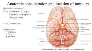
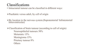
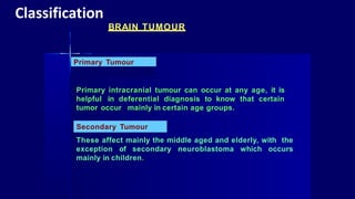
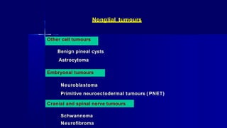
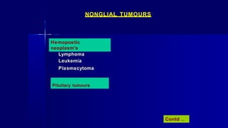
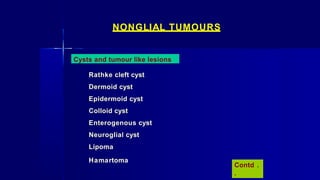
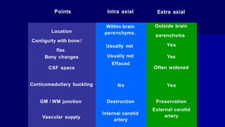
Brain Tumours
Dr. Fakir Mohan Sahu
SR MCh Neurosurgery
Dept of Neurosurgery
AIIMS Bhubaneswar
Learning Objectives
• Epidemiology and incidence
• Anatomical consideration and location
• Classification of brain tumours
• Clinical features
• Diagnostic imaging
• Individuals tumours
• Treatment modalities
• Recent Advances
Introduction
• A wide variety of tumors affect the CNS (Brain + Spinal cord)
• Primary benign and malignant tumors arise from the various elements of the CNS,
including neurons, glia, and meninges.
• Tumors metastasize to the CNS from many primary sources.
• Presentation varies widely depending on relevant neuroanatomy.
• Prognosis depends on histology and anatomy
• Team approaches to CNS tumors, as patients may require a combination:
Surgery,
Radiation Therapy
Chemotherapy and Research Protocol Enrolment.
Epidemiology of Intracranial tumour
• In general, primary brain tumors is higher in whites than in blacks
• Males are more likely to be diagnosed with brain tumors than females-( 1.5:1 )
• Meningiomas and pituitary adenomas are slightly more common in women than in men.
• Mortality is higher in males than in females.
• Brain tumours are responsible for 2% of all cancer deaths.
• The incidence varies with age.
In children CNS tumours comprise 20 % of all childhood malignancies.
Peak at 2 years followed by a decline for the rest of the first decade.
• The incidence then slowly increases, peaking at 20 per 100 000 in late adulthood.
Anatomic consideration and location of tumours
The brain consists of
• The Cerebrum - 2 Large
Cerebral Hemisphere
4 major lobes
• The Cerebellum
• Brainstem
• Midbrain
• Pons
• Medulla
Location
• In adults supratentorial tumours
out number posterior fossa
tumour by a ratio of 7 to 3
• But in children this ratio is
reversed and posterior Fossa
tumours are the most common.
Classifications
• Intracranial tumour can be classified in different ways:
Paediatric versus adult, by cell of origin
By location in the nervous system.(Supratentorial/ Infratentorial/
Intraventricular)
Classification of brain tumour (according to cell of origin)
Neuroepithelial tumours 50%
Metastatic 15%
Meningioma 15%
Pituitary tumour 8%
Others
Primary Tumour
Primary intracranial tumour can occur at any age, it is
helpful in deferential diagnosis to know that certain
tumor occur mainly in certain age groups.
BRAIN TUMOUR
Secondary Tumour
These affect mainly the middle aged and elderly, with the
exception of secondary neuroblastoma which occurs
mainly in children.
Classification
CLASSIFICATION OF INTRA CRANIAL TUMOURS:
There are several ways of classifying brain tumours:
primary versus secondary
intraxial ( arising from the brain parenchyma) versus extra
axial ( arising from tissue covering the brain such as dura)
and
various regional
classification
> Supratentorial
> Infratentorial
> Intraventricular
> Pineal region
> Sellar region tumours. Contd ..
Astrocytomas
Oligodendroglioma
Ependymal tumours
CLASSIFICATION ACCORDING TO HISTOLOGY
Primary brain tumours are subdivided into two basic groups:
Tumours of neuroglial origin (Glioma)
Non - glial tumours that are specified by a combination
of putative cell origin and specific location.
Glial tumors (gliomas)
Contd ..
NONGLIAL TUMOURS
Neuronal and mixed neuronal- glial tumours
Ganglioglioma
Gangliocytoma
Meningeal and mesenchymal tumours
Meningioma
Hemangiopericytoma
Hemangioblastoma
Fibrous histiocytoma Contd .
.
NONGLIAL TUMOURS
Pineal region tumours
Pineocytoma
Pineoblastoma
Pineal cell tumours
Choriocarcinoma
Teratoma
Germinoma
Germ cell tumours
Contd ..
Nonglial tumours
Other cell tumours
Benign pineal cysts
Astrocytoma
Embryonal tumours
Neuroblastoma
Primitive neuroectodermal tumours ( PNET)
Cranial and spinal nerve tumours
Schwannoma
Neurofibroma
NONGLIAL TUMOURS
Hemopoetic
neoplasm's
Lymphoma
Leukemia
Plasmacytoma
Pituitary tumours
Contd ..
NONGLIAL TUMOURS
Cysts and tumour like lesions
Rathke cleft cyst
Dermoid cyst
Epidermoid cyst
Colloid cyst
Enterogenous cyst
Neuroglial cyst
Lipoma
Hamartoma
Contd .
.
NONGLIAL TUMOURS Contd.
Local extensions from regional
tumours
Craniopharyngioma
Paraganglioma
Chordoma
According to location
Intra axial
Extra axial
Points Intra axial Extra axial
Within brain
parenchyma.
Outside brain
parenchyma
Yes
Location
Contiguity with bone /
flax
Bony changes
CSF space
Usually not
Usually not
Effaced
Yes
Often widened
Corticomedullary buckling No Yes
GM / WM junction Destruction
Vascular supply
Internal carotid
artery
Preservation
External carotid
artery
Supratentorial
• Meningeoma
• Dermoid
• Epidermoid
• Pitutary adenoma
• Pineal region tumour
• Craniopharyngeoma
• Chordoma
Infratentorial
• Brainstem glioma
• Cerebellar astrocytoma
• Medulloblastoma
• Ependymoma
• Meningioma
• Hemangioblastoma
• Dermoid
• Acoustic neuroma
• Meningioma
• Dermoid
• Chordoma
• Glomus jugular tumour
OTHER
CLASSIFICATION
Intraventricular tumour
Ependymoma
Choroid pluxus tumor
Colloid cysts
Meningioma
CLASSIFICATION – ACCORDING AGE
GROUP
5-15
YEARS CLASSIFICATION
0-5 Brain Stem Glioma, Optic Nerve Glioma
Medulloblastoma, Cerebellar Astrocytoma,
Craniopharyngma, Choroid Plexus Papiloma ,
Pinealoma.
15-30
30-60
60+
Ependymoma
Glioma, Meningioma, Acoustic neuroma,
Pitutary Tumour, Hemangioblastoma
Meningeoma, Acoustic Neuroma,
Glioblastoma
WHO CLASSIFICATION
CLASSIFICATION
Pilocytic astrocytoma
SGCA(subependymal giant cell astrocytoma)
Low grade astrocytoma
Anaplastic astrocytoma
GRADE
Grade 1
Grade 2
Grade 3
Grade 4 Glioblastoma multiforme
FEATURES OF LOW GRADE
GLIOMA
Age – Younger age group 20- 40 yrs
Incidence – 10- 20% of astrocytoma
Location –
Histology-
cerebral hemisphere
frontal and parietel lobe
temporal lobe
low grade malignancy
Presenting Symptom - Seizure
IMAGING STUDY LOW GRADE GLIOMA
CT
NECT : Iso/hypo
CECT : Little or no enhancement
IMAGING STUDY LOW GRADE GLIOMA
MRI
T1 : Iso/Hypointense
T2 : Homogenously
hyper
ANAPLASTIC ASTROCYTOMA
It usually occurs in the middle aged patients
Incidence- 20- 25%
Locations: cerebral hemispheres
frontal and
temporal lobe
Histology- malignant
Presenting Symptom- Seizure, focal neurological deficit
CT
IMAGING STUDY ANAPLASTIC ASTROCYTOMA
NECT : Iso/hypo In homogenous mixed density
CECT : Enhance strongly, inhomogenously
IMAGING STUDY ANAPLASTIC ASTROCYTOMA
MRI
T1: Hypo to iso intense
T2 : Heterogenously Hyperintense
As typically enhance strongly
but non uniformly following
contrast administration.
Irregular rim enhancement is
common
GLIOBLASTOMA MULTIFORME
Half of all astrocytoma are GBM. It is most common
supratentorial neoplasm in adult. The most common primary
brain tumour, it is also the most malignant astrocytoma
Incidence –
Age-
Locations-
Presenting symptom-
deficit,
40-50%.
> 50yrs.
cerebral hemisphere, Frontal and
temporal lobe, White mater
Seizure, Focal neurological
stroke like syndroms
IMAGING STUDY
Enhances strongly, inhomogenously.
Ring enhancing lesion – due to increased
cellularity and neovascularity.
Area of central necrosis shows hypodensity
Imaging study shows ‘multiforme’ appearance
CT
NECT : In homogenously mixed density
MRI
T1: T1 weighted image shows mixed signal mass with
necrosis or cyst formation and thick irregular wall.
Marked but in homogenous contrast enhancement is
present in majority of glioblastoma multiforme. These
tumours are hihgly vascular, haemorrhage of different
ages are often present.
T-1 T-1 C T-1 C
MRI
T2: T2- weighted image shows very heterogenous mass with
mixed signal intensity. Central necrosis is the hall mark.
Haemorrhage , necrosis, oedema are present in GBM
Angiograph
y
Alarge mass with striking tumours blush, Contrast stasis
and pooling in Bizarre vascular channel is typical
IMAGING STUDY Contd.
Spread
Through white mater
Sub ependymal seedling
Through CSF
Rarely through haemtogenous route
Extra cerebral metastasis- Lung, Liver, Bone
MRI
Sharply defined macrocystic mass
Mural nodule easily appreciate with
contrast enhancement
PILOCYTIC ASTROCYTOMA IMAGING STUDY
OLIGODENDROGLIOMA
Arise from a specific type of glial cell- Oligodendrocyte. These
are typically unencapsulated but well circumscribed focal
white matter tumours that may extent into the cortex and
leptomeninges. Foci of cystic degeneration common Hge,
necrosis uncommon
Incidence -
Age distribution-
Peak age
Location
-
-
5- 10% of gliomas
4th to 5th decade
35- 45 yrs.
85% supratentorial
Cerebral hemisphere- mostly
frontal lobe
IMAGING STUDY-
OLIGODENDROGLIOMA
CT
NECT- Prominent mass of calcification. Partially calcified
mixed density hemispheric mass that extends peripherally to
the cortex.
CECT- Mild to moderate contrast enhancement occurs
MRI
Heterogenous signal intensity due to calcifiacation
T1W iimage shows mixed hypo or iso intensity
T2W image shows hyper intense foci
Absent to slight enhancement is typical
T-1 T- 1 C T- 2
IMAGING STUDY-
OLIGODENDROGLIOMA
EPENDYMOMA
Ependymomas are tumours of the young and are third
most common intracranial tumour ofchildren
Age distribution-
Incidence-
1-5 yrs.
2-8% of gliomas
15% of pediatric brain tumour
Location- 60% infratentorial more common in children .
40% supratentorial more common inadult.
4th ventricle, C- P angle, in or near 3rd ventricle
IMAGING STUDY- EPENDYMOMA
CT
NECT-
CECT-
Mixed density, isodense or slightly hyperdense
Fine calcification seen in approximately 50% of the
patients.
May have cystic areas
More than 80% contrast enhancement occurs
MRI
T1- Heterogenous signal intensity markedly hypointense area
due to calcification
In homogenous enhancement with gadolinium
T2- Iso to hyper intensity
Histological feature shows uniform ependymal cells in pattern of
rosettes, canal or perivascular pseudorosettes
CHOROID PLEXUS PAPILLOMA
Tumours of choroid plexus are rare, accounting for 0.4-
0.6% of all intracranial tumour
Age distribution- > 85% occurs in children
Location In children MC site lateral ventricle
In adult most of the choroid plexus
papilloma occurs in 4th ventricle
IMAGING STUDY CHOROID PLEXUS
PAPILLOMA
CT
NECT
CECT
Iso or hyperdense, 3/4th hyperdense
heterogenous contrast enhancement
MRI
T1 weighted image shows- Predominently isointense
Intensely contrast enhancement
occurs
T2 weighted image shows- Iso to hyperintense,
occasionally signal void from the vascular pedicle
IMAGING STUDY CHOROID PLEXUS
PAPILLOMA
PINEAL TUMOUR , Cont
the incidence of
The pineal region tumours are rare tumour
which is 1% of all intra cranial tumour.
Gemcell tumour
Pineal cell tumour
Germinoma
Teratoma
Embryonal carcinoma
Choriocarcinoma
Pinealoblastoma
Pinealocytoma
Types of pineal region tumour are as follows
SELLAR/SUPRASELLAR MASSES
The sellar region is an anatomically complex area
composed of the bony sellaturcica, pituitary gland, and
adjacent structures
Pituitary adenoma
Older classification
Chromophobic
Acido philic
Basophilic
Mixed
New classification
Pituitary microadenoma ( size
<10mm)
Pituitary macroadenoma (size
IMAGING STUDY
X-ray
expansion of sellar cavity
thining of bony cortex
ballooning of sella
CT
Large, homogenously isodense, rounded midline mass
MRI
T 1 weighted image shows hypointense
T 2 weighted image showshyperintensity
Extension is better visualized after contrast
Both CT and MRI show strong contrast enhancement with
some what inhomogenously
MENINGEAL AND MESENCHYMAL TUMOUR
Meningiomas
Malignant mesenchymal tumour
Hemangiopericytoma
Hemangioblastoma
Meningiomas
Meningiomas are most common nonglial primary brain tumour
Most common extra axial tumour ( 13- 18%)
Age – adult tumour 40- 60yrs
Sex- more in female
Cytogenetics- chromosome 22 is important for pathogenesis
of meningioma
Location meningioma
Cerebral convexity 32- 45 %
Parasagital 26%
Sphenoid ridge
Juxtra sellar
20 %
10 %
olfactory groove 10 %
Posterior fossa 10 %
Tentorium
Pineal region
Others optic nerve sheath, intravetrricular
IMAGE STUDY
X-ray
Hyperostosis
Erosion
Enlarged vascular
Calcification
Pneumosinus dilatans
IMAGE STUDY
CT
70% to 75% hyperdense
20% to 25% calcified
90% enhance strongly , uniformly
10% to 15% cystic areas
60% peritumoral edema
Hemorrhage
rare
Area of haemorrhage, necrosis, cyst formation are common
which appear as hypodense within the tumour
IMAGE STUDY
Angiography
Dual vascular supply common
Sunburst of enlarged dural feeders in tumour
Extension
Into the suprasellar cystern forming the figure of eight( 8 )
Elevate and compress the optic chiasma and 3rd ventricle
Laterally into the cavernous sinus
May encase the ICA or narrow the vessels
Common CPA
masses
- Vestubular schwannoma ( acoustic neuroma)
- Meningioma
- Epidermoid
- Other schwannoma
- Less com.
- Arachnoid cyst
- Metastases
- Vascular
- Lipoma
- Dermoid.
CEREBELLOPONTINE ANGLE ( CPA) CISTERN MASSES
MEDULLOBLASTOMA
Most common malignant pediatric brain tumour.
Incidence: 15- 20% of intracranial tumour in children.
Male: Female 2:1.
Age: most in 1st decade. 75% in 4- 8 years.
Site: 75% arises in the cerebellar vermis mostly in midline,
in the apex of 4th ventricle.
25% arises in lateral cerebellum.
Highly radiosensitive and moderately chemo sensitive.
Metastasis occur early in the CSF.
Prognosis is very poor
CRANIOPHARYNGIOMA
Craniopharyngiomas arise from the squamous epithelial rests
along the involuted hypophyseal Rathke’s duct.
Incidence: 3- 5% of primary brain tumour.
50% of pediatric brain tumour.
Age: > 50% in children, peak between 8-12 years.
Location: 70 % combined suprasellar and intrasellar
Imaging study
CT scan of brain-
90% partially cystic,
90% calcification present,
90% nodular or rim enhancement occur
MRI of Brain
Variable signal, most common is hypointense in T 1 weighted
image and hyperintense in T2 weighted image
INTRACRANIAL METASTASES
Representing 1/ 4th to 1/ 3rd of all brain tumour.
Common:
Skull
Leptomeninges
Parenchymal (most common).
Less common:
Dural
Pial
Sub pial
Parenchymal metastases:
Location – any where but most common in cortico medullary
junction ( grey mater- white mater interface).
INTRACRANIAL METASTASES
Pathology
Welldefined circumscribed nodule of variable size
May be solid partially cystic, filled with mucinous material,
necrotic material, haemorrhagic fluid .
Imaging Study
CT:
NECT- mostly isodense lesion / hyperdense lesion-
Example- Thyroid carcinoma, Lung carcinoma, choriocarcinoma,
malignant melanoma, sarcoma
INTRACRANIAL METASTASES
Cystic metastasis
Mucin producing tumour adenocarcinoma arising from stomach,
small and large intestine, pancreas, ovary, breast cancer
Cystic and calcified metastasis
Rare- breast carcinoma, lung cancer.
CECT: Most enhance strongly, both solid and ring shaped
pattern are noted
MRI
T 1 weighted image- shows variable features most non
haemorrhagic tumour slightly hyper intense.
Some non haemorrhagic tumour – hyper intense
General
Consideration
Surgicalmanagement:Craniotomy+ Excision
Approaches
Craniotomy:- flap of bone is cutand
reflected.
- If necessary, combined with either a
stereotactic frame
- or preferably an image guided
system(frameless, steriotaxy) to give
accurate lesion localisation
Operative procedure:
Thesubsequent procedure
Biopsy Partial tumour removal Internal decompression Complete removal
Depends on the nature of the tumour &site.
Primary malignant tumour :complete removal of tumour is not possible due to its
infiltrative nature. So operation is restricted to -biopsy
-Tumourdecompression
Complete removal is done for
-Meningioma
-Craniopharyngioma
Radiotherapy
Aim :toprovide highest possible dose
tothe specificregion, while minimal
irradiation toadjacent normal brain.
Indication: In malignant tumour:
- malignant astrocytoma
- metastasis
- medulloblastoma
- germinoma
In benign tumours
- pituitary adenoma
- craniopharyngioma
 Malignant astrocytoma:
 -nitrosoureas are most effectivedrug
 commonly used treatmentforrelapsepatient
 Low gradetumour or benign tumour:
 chemotherapy has limitedvalue
 Medulloblastoma:
 respond totreatmentbut value of treatmentof patient survival – is unclear.
 Primary germ cell tumour & primary cerebral lymphoma:
chemotherapy has a role.
Chemotherapy
Prognostics Variables
For each tumour entity, combinations of parameters
WHO grade
Clinical findings- age/ neurologic performance Status
Tumour location
Radiological features - contrast enhancement
Extent of surgical resection
Proliferation indices
Genetic alterations
Recent advances
• Neuro-navigation
• Stereotactic surgery
• Endovascular embolization
• Endoscopic Surgery
Thank
You

Brain tumours

  • 1.
    Brain Tumours Dr. FakirMohan Sahu SR MCh Neurosurgery Dept of Neurosurgery AIIMS Bhubaneswar
  • 2.
    Learning Objectives • Epidemiologyand incidence • Anatomical consideration and location • Classification of brain tumours • Clinical features • Diagnostic imaging • Individuals tumours • Treatment modalities • Recent Advances
  • 3.
    Introduction • A widevariety of tumors affect the CNS (Brain + Spinal cord) • Primary benign and malignant tumors arise from the various elements of the CNS, including neurons, glia, and meninges. • Tumors metastasize to the CNS from many primary sources. • Presentation varies widely depending on relevant neuroanatomy. • Prognosis depends on histology and anatomy • Team approaches to CNS tumors, as patients may require a combination: Surgery, Radiation Therapy Chemotherapy and Research Protocol Enrolment.
  • 4.
    Epidemiology of Intracranialtumour • In general, primary brain tumors is higher in whites than in blacks • Males are more likely to be diagnosed with brain tumors than females-( 1.5:1 ) • Meningiomas and pituitary adenomas are slightly more common in women than in men. • Mortality is higher in males than in females. • Brain tumours are responsible for 2% of all cancer deaths. • The incidence varies with age. In children CNS tumours comprise 20 % of all childhood malignancies. Peak at 2 years followed by a decline for the rest of the first decade. • The incidence then slowly increases, peaking at 20 per 100 000 in late adulthood.
  • 5.
    Anatomic consideration andlocation of tumours The brain consists of • The Cerebrum - 2 Large Cerebral Hemisphere 4 major lobes • The Cerebellum • Brainstem • Midbrain • Pons • Medulla
  • 6.
    Location • In adultssupratentorial tumours out number posterior fossa tumour by a ratio of 7 to 3 • But in children this ratio is reversed and posterior Fossa tumours are the most common.
  • 7.
    Classifications • Intracranial tumourcan be classified in different ways: Paediatric versus adult, by cell of origin By location in the nervous system.(Supratentorial/ Infratentorial/ Intraventricular) Classification of brain tumour (according to cell of origin) Neuroepithelial tumours 50% Metastatic 15% Meningioma 15% Pituitary tumour 8% Others
  • 8.
    Primary Tumour Primary intracranialtumour can occur at any age, it is helpful in deferential diagnosis to know that certain tumor occur mainly in certain age groups. BRAIN TUMOUR Secondary Tumour These affect mainly the middle aged and elderly, with the exception of secondary neuroblastoma which occurs mainly in children. Classification
  • 9.
    CLASSIFICATION OF INTRACRANIAL TUMOURS: There are several ways of classifying brain tumours: primary versus secondary intraxial ( arising from the brain parenchyma) versus extra axial ( arising from tissue covering the brain such as dura) and various regional classification > Supratentorial > Infratentorial > Intraventricular > Pineal region > Sellar region tumours. Contd ..
  • 10.
    Astrocytomas Oligodendroglioma Ependymal tumours CLASSIFICATION ACCORDINGTO HISTOLOGY Primary brain tumours are subdivided into two basic groups: Tumours of neuroglial origin (Glioma) Non - glial tumours that are specified by a combination of putative cell origin and specific location. Glial tumors (gliomas) Contd ..
  • 11.
    NONGLIAL TUMOURS Neuronal andmixed neuronal- glial tumours Ganglioglioma Gangliocytoma Meningeal and mesenchymal tumours Meningioma Hemangiopericytoma Hemangioblastoma Fibrous histiocytoma Contd . .
  • 12.
    NONGLIAL TUMOURS Pineal regiontumours Pineocytoma Pineoblastoma Pineal cell tumours Choriocarcinoma Teratoma Germinoma Germ cell tumours Contd ..
  • 13.
    Nonglial tumours Other celltumours Benign pineal cysts Astrocytoma Embryonal tumours Neuroblastoma Primitive neuroectodermal tumours ( PNET) Cranial and spinal nerve tumours Schwannoma Neurofibroma
  • 14.
  • 15.
    NONGLIAL TUMOURS Cysts andtumour like lesions Rathke cleft cyst Dermoid cyst Epidermoid cyst Colloid cyst Enterogenous cyst Neuroglial cyst Lipoma Hamartoma Contd . .
  • 16.
    NONGLIAL TUMOURS Contd. Localextensions from regional tumours Craniopharyngioma Paraganglioma Chordoma According to location Intra axial Extra axial
  • 17.
    Points Intra axialExtra axial Within brain parenchyma. Outside brain parenchyma Yes Location Contiguity with bone / flax Bony changes CSF space Usually not Usually not Effaced Yes Often widened Corticomedullary buckling No Yes GM / WM junction Destruction Vascular supply Internal carotid artery Preservation External carotid artery
  • 18.
    Supratentorial • Meningeoma • Dermoid •Epidermoid • Pitutary adenoma • Pineal region tumour • Craniopharyngeoma • Chordoma
  • 19.
    Infratentorial • Brainstem glioma •Cerebellar astrocytoma • Medulloblastoma • Ependymoma • Meningioma • Hemangioblastoma • Dermoid • Acoustic neuroma • Meningioma • Dermoid • Chordoma • Glomus jugular tumour
  • 20.
  • 21.
    CLASSIFICATION – ACCORDINGAGE GROUP 5-15 YEARS CLASSIFICATION 0-5 Brain Stem Glioma, Optic Nerve Glioma Medulloblastoma, Cerebellar Astrocytoma, Craniopharyngma, Choroid Plexus Papiloma , Pinealoma. 15-30 30-60 60+ Ependymoma Glioma, Meningioma, Acoustic neuroma, Pitutary Tumour, Hemangioblastoma Meningeoma, Acoustic Neuroma, Glioblastoma
  • 22.
    WHO CLASSIFICATION CLASSIFICATION Pilocytic astrocytoma SGCA(subependymalgiant cell astrocytoma) Low grade astrocytoma Anaplastic astrocytoma GRADE Grade 1 Grade 2 Grade 3 Grade 4 Glioblastoma multiforme
  • 23.
    FEATURES OF LOWGRADE GLIOMA Age – Younger age group 20- 40 yrs Incidence – 10- 20% of astrocytoma Location – Histology- cerebral hemisphere frontal and parietel lobe temporal lobe low grade malignancy Presenting Symptom - Seizure
  • 24.
    IMAGING STUDY LOWGRADE GLIOMA CT NECT : Iso/hypo CECT : Little or no enhancement
  • 25.
    IMAGING STUDY LOWGRADE GLIOMA MRI T1 : Iso/Hypointense T2 : Homogenously hyper
  • 26.
    ANAPLASTIC ASTROCYTOMA It usuallyoccurs in the middle aged patients Incidence- 20- 25% Locations: cerebral hemispheres frontal and temporal lobe Histology- malignant Presenting Symptom- Seizure, focal neurological deficit
  • 27.
    CT IMAGING STUDY ANAPLASTICASTROCYTOMA NECT : Iso/hypo In homogenous mixed density CECT : Enhance strongly, inhomogenously
  • 28.
    IMAGING STUDY ANAPLASTICASTROCYTOMA MRI T1: Hypo to iso intense T2 : Heterogenously Hyperintense As typically enhance strongly but non uniformly following contrast administration. Irregular rim enhancement is common
  • 29.
    GLIOBLASTOMA MULTIFORME Half ofall astrocytoma are GBM. It is most common supratentorial neoplasm in adult. The most common primary brain tumour, it is also the most malignant astrocytoma Incidence – Age- Locations- Presenting symptom- deficit, 40-50%. > 50yrs. cerebral hemisphere, Frontal and temporal lobe, White mater Seizure, Focal neurological stroke like syndroms
  • 30.
    IMAGING STUDY Enhances strongly,inhomogenously. Ring enhancing lesion – due to increased cellularity and neovascularity. Area of central necrosis shows hypodensity Imaging study shows ‘multiforme’ appearance CT NECT : In homogenously mixed density
  • 31.
    MRI T1: T1 weightedimage shows mixed signal mass with necrosis or cyst formation and thick irregular wall. Marked but in homogenous contrast enhancement is present in majority of glioblastoma multiforme. These tumours are hihgly vascular, haemorrhage of different ages are often present. T-1 T-1 C T-1 C
  • 32.
    MRI T2: T2- weightedimage shows very heterogenous mass with mixed signal intensity. Central necrosis is the hall mark. Haemorrhage , necrosis, oedema are present in GBM Angiograph y Alarge mass with striking tumours blush, Contrast stasis and pooling in Bizarre vascular channel is typical
  • 33.
    IMAGING STUDY Contd. Spread Throughwhite mater Sub ependymal seedling Through CSF Rarely through haemtogenous route Extra cerebral metastasis- Lung, Liver, Bone
  • 34.
    MRI Sharply defined macrocysticmass Mural nodule easily appreciate with contrast enhancement PILOCYTIC ASTROCYTOMA IMAGING STUDY
  • 35.
    OLIGODENDROGLIOMA Arise from aspecific type of glial cell- Oligodendrocyte. These are typically unencapsulated but well circumscribed focal white matter tumours that may extent into the cortex and leptomeninges. Foci of cystic degeneration common Hge, necrosis uncommon Incidence - Age distribution- Peak age Location - - 5- 10% of gliomas 4th to 5th decade 35- 45 yrs. 85% supratentorial Cerebral hemisphere- mostly frontal lobe
  • 36.
    IMAGING STUDY- OLIGODENDROGLIOMA CT NECT- Prominentmass of calcification. Partially calcified mixed density hemispheric mass that extends peripherally to the cortex. CECT- Mild to moderate contrast enhancement occurs
  • 37.
    MRI Heterogenous signal intensitydue to calcifiacation T1W iimage shows mixed hypo or iso intensity T2W image shows hyper intense foci Absent to slight enhancement is typical T-1 T- 1 C T- 2 IMAGING STUDY- OLIGODENDROGLIOMA
  • 38.
    EPENDYMOMA Ependymomas are tumoursof the young and are third most common intracranial tumour ofchildren Age distribution- Incidence- 1-5 yrs. 2-8% of gliomas 15% of pediatric brain tumour Location- 60% infratentorial more common in children . 40% supratentorial more common inadult. 4th ventricle, C- P angle, in or near 3rd ventricle
  • 39.
    IMAGING STUDY- EPENDYMOMA CT NECT- CECT- Mixeddensity, isodense or slightly hyperdense Fine calcification seen in approximately 50% of the patients. May have cystic areas More than 80% contrast enhancement occurs
  • 40.
    MRI T1- Heterogenous signalintensity markedly hypointense area due to calcification In homogenous enhancement with gadolinium T2- Iso to hyper intensity Histological feature shows uniform ependymal cells in pattern of rosettes, canal or perivascular pseudorosettes
  • 41.
    CHOROID PLEXUS PAPILLOMA Tumoursof choroid plexus are rare, accounting for 0.4- 0.6% of all intracranial tumour Age distribution- > 85% occurs in children Location In children MC site lateral ventricle In adult most of the choroid plexus papilloma occurs in 4th ventricle
  • 42.
    IMAGING STUDY CHOROIDPLEXUS PAPILLOMA CT NECT CECT Iso or hyperdense, 3/4th hyperdense heterogenous contrast enhancement
  • 43.
    MRI T1 weighted imageshows- Predominently isointense Intensely contrast enhancement occurs T2 weighted image shows- Iso to hyperintense, occasionally signal void from the vascular pedicle IMAGING STUDY CHOROID PLEXUS PAPILLOMA
  • 44.
    PINEAL TUMOUR ,Cont the incidence of The pineal region tumours are rare tumour which is 1% of all intra cranial tumour. Gemcell tumour Pineal cell tumour Germinoma Teratoma Embryonal carcinoma Choriocarcinoma Pinealoblastoma Pinealocytoma Types of pineal region tumour are as follows
  • 45.
    SELLAR/SUPRASELLAR MASSES The sellarregion is an anatomically complex area composed of the bony sellaturcica, pituitary gland, and adjacent structures Pituitary adenoma Older classification Chromophobic Acido philic Basophilic Mixed New classification Pituitary microadenoma ( size <10mm) Pituitary macroadenoma (size
  • 46.
    IMAGING STUDY X-ray expansion ofsellar cavity thining of bony cortex ballooning of sella CT Large, homogenously isodense, rounded midline mass
  • 47.
    MRI T 1 weightedimage shows hypointense T 2 weighted image showshyperintensity Extension is better visualized after contrast Both CT and MRI show strong contrast enhancement with some what inhomogenously
  • 48.
    MENINGEAL AND MESENCHYMALTUMOUR Meningiomas Malignant mesenchymal tumour Hemangiopericytoma Hemangioblastoma Meningiomas Meningiomas are most common nonglial primary brain tumour Most common extra axial tumour ( 13- 18%) Age – adult tumour 40- 60yrs Sex- more in female Cytogenetics- chromosome 22 is important for pathogenesis of meningioma
  • 49.
    Location meningioma Cerebral convexity32- 45 % Parasagital 26% Sphenoid ridge Juxtra sellar 20 % 10 % olfactory groove 10 % Posterior fossa 10 % Tentorium Pineal region Others optic nerve sheath, intravetrricular
  • 50.
  • 51.
    IMAGE STUDY CT 70% to75% hyperdense 20% to 25% calcified 90% enhance strongly , uniformly 10% to 15% cystic areas 60% peritumoral edema Hemorrhage rare Area of haemorrhage, necrosis, cyst formation are common which appear as hypodense within the tumour
  • 52.
    IMAGE STUDY Angiography Dual vascularsupply common Sunburst of enlarged dural feeders in tumour Extension Into the suprasellar cystern forming the figure of eight( 8 ) Elevate and compress the optic chiasma and 3rd ventricle Laterally into the cavernous sinus May encase the ICA or narrow the vessels
  • 53.
    Common CPA masses - Vestubularschwannoma ( acoustic neuroma) - Meningioma - Epidermoid - Other schwannoma - Less com. - Arachnoid cyst - Metastases - Vascular - Lipoma - Dermoid. CEREBELLOPONTINE ANGLE ( CPA) CISTERN MASSES
  • 54.
    MEDULLOBLASTOMA Most common malignantpediatric brain tumour. Incidence: 15- 20% of intracranial tumour in children. Male: Female 2:1. Age: most in 1st decade. 75% in 4- 8 years. Site: 75% arises in the cerebellar vermis mostly in midline, in the apex of 4th ventricle. 25% arises in lateral cerebellum. Highly radiosensitive and moderately chemo sensitive. Metastasis occur early in the CSF. Prognosis is very poor
  • 55.
    CRANIOPHARYNGIOMA Craniopharyngiomas arise fromthe squamous epithelial rests along the involuted hypophyseal Rathke’s duct. Incidence: 3- 5% of primary brain tumour. 50% of pediatric brain tumour. Age: > 50% in children, peak between 8-12 years. Location: 70 % combined suprasellar and intrasellar Imaging study CT scan of brain- 90% partially cystic, 90% calcification present, 90% nodular or rim enhancement occur MRI of Brain Variable signal, most common is hypointense in T 1 weighted image and hyperintense in T2 weighted image
  • 56.
    INTRACRANIAL METASTASES Representing 1/4th to 1/ 3rd of all brain tumour. Common: Skull Leptomeninges Parenchymal (most common). Less common: Dural Pial Sub pial Parenchymal metastases: Location – any where but most common in cortico medullary junction ( grey mater- white mater interface).
  • 57.
    INTRACRANIAL METASTASES Pathology Welldefined circumscribednodule of variable size May be solid partially cystic, filled with mucinous material, necrotic material, haemorrhagic fluid . Imaging Study CT: NECT- mostly isodense lesion / hyperdense lesion- Example- Thyroid carcinoma, Lung carcinoma, choriocarcinoma, malignant melanoma, sarcoma
  • 58.
    INTRACRANIAL METASTASES Cystic metastasis Mucinproducing tumour adenocarcinoma arising from stomach, small and large intestine, pancreas, ovary, breast cancer Cystic and calcified metastasis Rare- breast carcinoma, lung cancer. CECT: Most enhance strongly, both solid and ring shaped pattern are noted MRI T 1 weighted image- shows variable features most non haemorrhagic tumour slightly hyper intense. Some non haemorrhagic tumour – hyper intense
  • 59.
  • 60.
    Surgicalmanagement:Craniotomy+ Excision Approaches Craniotomy:- flapof bone is cutand reflected. - If necessary, combined with either a stereotactic frame - or preferably an image guided system(frameless, steriotaxy) to give accurate lesion localisation
  • 61.
    Operative procedure: Thesubsequent procedure BiopsyPartial tumour removal Internal decompression Complete removal Depends on the nature of the tumour &site. Primary malignant tumour :complete removal of tumour is not possible due to its infiltrative nature. So operation is restricted to -biopsy -Tumourdecompression Complete removal is done for -Meningioma -Craniopharyngioma
  • 62.
    Radiotherapy Aim :toprovide highestpossible dose tothe specificregion, while minimal irradiation toadjacent normal brain. Indication: In malignant tumour: - malignant astrocytoma - metastasis - medulloblastoma - germinoma In benign tumours - pituitary adenoma - craniopharyngioma
  • 63.
     Malignant astrocytoma: -nitrosoureas are most effectivedrug  commonly used treatmentforrelapsepatient  Low gradetumour or benign tumour:  chemotherapy has limitedvalue  Medulloblastoma:  respond totreatmentbut value of treatmentof patient survival – is unclear.  Primary germ cell tumour & primary cerebral lymphoma: chemotherapy has a role. Chemotherapy
  • 64.
    Prognostics Variables For eachtumour entity, combinations of parameters WHO grade Clinical findings- age/ neurologic performance Status Tumour location Radiological features - contrast enhancement Extent of surgical resection Proliferation indices Genetic alterations
  • 65.
    Recent advances • Neuro-navigation •Stereotactic surgery • Endovascular embolization • Endoscopic Surgery
  • 66.