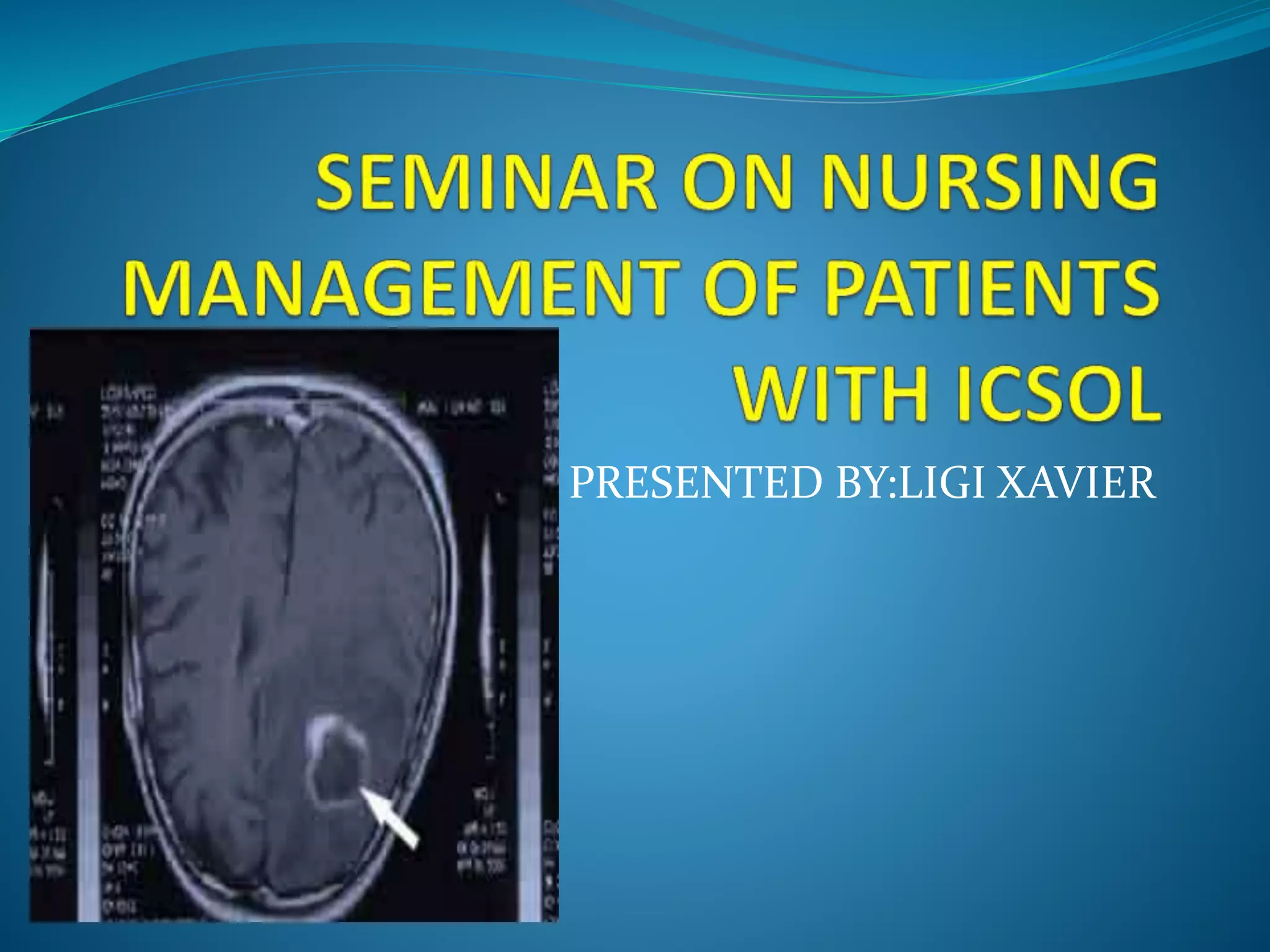
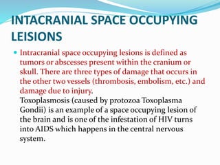
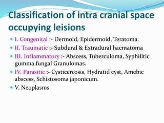
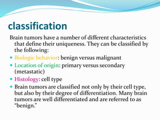
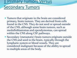
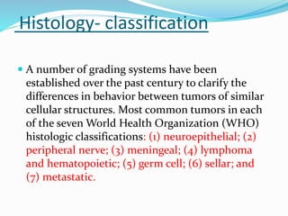
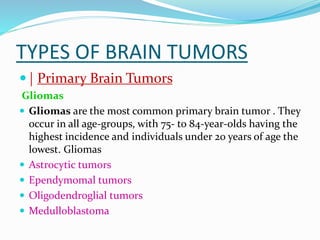
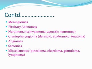
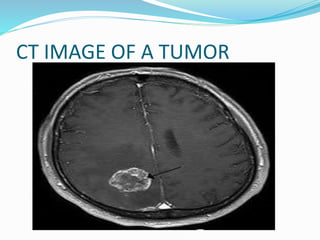
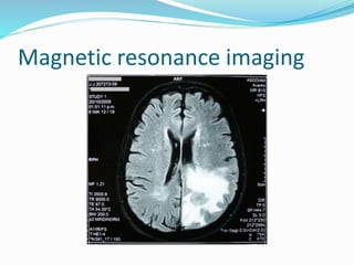
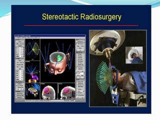
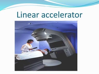
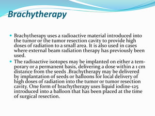
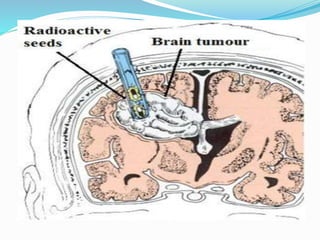
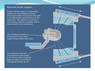
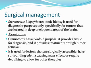
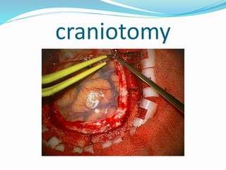
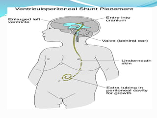
PRESENTED BY:LIGI XAVIER
INTACRANIAL SPACE OCCUPYING
LEISIONS
 Intracranial space occupying lesions is defined as
tumors or abscesses present within the cranium or
skull. There are three types of damage that occurs in
the other two vessels (thrombosis, embolism, etc.) and
damage due to injury.
Toxoplasmosis (caused by protozoa Toxoplasma
Gondii) is an example of a space occupying lesion of
the brain and is one of the infestation of HIV turns
into AIDS which happens in the central nervous
system.
Classification of intra cranial space
occupying leisions
 I. Congenital :- Dermoid, Epidermoid, Teratoma.
 II. Traumatic :- Subdural & Extradural haematoma
 III. Inflammatory :- Abscess, Tuberculoma, Syphilitic
gumma,fungal Granulomas.
 IV. Parasitic :- Cysticercosis, Hydratid cyst, Amebic
abscess, Schistosoma japonicum.
 V. Neoplasms
classification
Brain tumors have a number of different characteristics
that define their uniqueness. They can be classified by
the following:
 Biologic behavior: benign versus malignant
 Location of origin: primary versus secondary
(metastatic)
 Histology: cell type
 Brain tumors are classified not only by their cell type,
but also by their degree of differentiation. Many brain
tumors are well differentiated and are referred to as
“benign.”
| Primary Tumors Versus
Secondary Tumors
 Tumors that originate in the brain are considered
primary, brain tumors. They are derived from cells
found in the CNS. They do not seed or spread outside
of the CNS, although some brain tumors, such as
medulloblastomas and ependymomas, can spread
within the CNS along CSF pathways.
 Secondary (metastatic) brain tumors originate outside
the CNS and seed to the brain, typically through the
lymphatic system or blood vessels. They are
considered malignant because of the ability to spread
to multiple areas of the body.
Histology- classification
 A number of grading systems have been
established over the past century to clarify the
differences in behavior between tumors of similar
cellular structures. Most common tumors in each
of the seven World Health Organization (WHO)
histologic classifications: (1) neuroepithelial; (2)
peripheral nerve; (3) meningeal; (4) lymphoma
and hematopoietic; (5) germ cell; (6) sellar; and
(7) metastatic.
TYPES OF BRAIN TUMORS
 | Primary Brain Tumors
Gliomas
 Gliomas are the most common primary brain tumor . They
occur in all age-groups, with 75- to 84-year-olds having the
highest incidence and individuals under 20 years of age the
lowest. Gliomas
 Astrocytic tumors
 Ependymomal tumors
 Oligodendroglial tumors
 Medulloblastoma
Contd…………………..
 Meningiomas
 Pituitary Adenomas
 Neruinoma (schwannoma, acoustic neuronma)
 Craniopharyngioma (dermoid, epidermoid, teratoma)
 Angiomas
 Sarcomas
 Miscellaneous (pinealoma, chordoma, granuloma,
lymphoma)
Types of Benign Brain Tumors
 Chordomas
 Craniopharyngiomas
 Gangliocytomas
 Glomus jugulare
 Meningiomas
 Pineocytomas
 Pituitary adenomas
 Schwannomas
Types of Malignant Brain Tumors
 Gliomas
 Astrocytomas
 Ependymomas
 Oligodendrogliomas
 Glioblastoma multiforme
 Medullolastoma
Other tumors
 Hemangioblastoma:are slow-growing tumors,
commonly located in the cerebellum. They originate
from blood vessels, can be large in size and often are
accompanied by a cyst. These tumors are most
common in people ages 40 to 60 and more prevalent in
men than women.
 Rhabdoid tumors are rare, highly aggressive tumors
that tend to spread throughout the central nervous
system. They often appear in multiple sites in the
body, especially in the kidneys.
Pathophysiology
 . Many tumors cause blood-brain barrier (BBB) disruption,
with consequent cerebral edema and increased intracranial
pressure (ICP).
 As tumors grow, they displace or invade normal brain tissue.
Rapidly growing tumors can quickly cause significant
neurologic deficits.
 Although the incidence of symptomatic intracranial
hemorrhage caused by a tumor (or hemorrhage within a tumor)
is low, these situations can be acute and devastating.
 Tumor growth can also lead to obstruction of the flow of
cerebrospinal fluid (CSF), leading to hydrocephalus. ICP
increases as the tumor enlarges and can lead to herniation and
rapid neurologic deterioration.
Clinical manifestations
 Generalised signs
Many of these features may be due to raised intracranial pressure.
 Headache
 Vomiting may occur. This may be without accompanying nausea.
 Nausea may be a feature
 A change in mental status or behavioural change is a cause for
concern
 There may be weakness, ataxia or disturbance of gait
 Even deficits of speech or vision may be poorly localising signs
 There may be generalized convulsions
False localising signs and
lateralising signs
 diplopia and cranial nerve lesions.
 Horner's syndrome is not a good localising lesion as
the path of the sympathetic nerves is also long but as
there is no chiasma (fibres don't cross the midline) it is
a good lateralising sign
 If a headache is unilateral, this is often a good
indicator of the side of the lesion.
 Causes of cerebellar signs include acoustic
neuroma, Friedreich's ataxia, stroke,
haemangioma, tumours, multiple sclerosis,
chronic alcohol excess and abscess.
Localising signs
 Temporal lobe
 Temporal lobe lesions often present with rather vague
psychological problems.
 There may be depersonalisation, emotional changes, and
disturbances of behaviour.
 There can be hallucinations of smell, taste, sound and sight.
There may be Déja vu in which there is a feeling of familiarity as
if the present has happened before.
 Dysphasia may be noted.
 Visual field defects involve the contralateral upper quadrant.
 There may be convulsions.
 Other psychological problems include forgetfulness, fugue
 Frontal lobe
 Frontal lobe tumours can cause anosmia. This is
especially significant if it is unilateral.
 There may be a change in personality with the person
becoming indecent, indiscreet or dishonest.
 Dysphasia can occur if Broca's area is involved.
 Hemiparesis or fits may affect the contralateral side.
Parietal lobe-LESIONS
 Parietal lobe lesions can produce a very interesting
neurological picture.
 There may be hemisensory loss.
 Decreased two-point discrimination.
 Astereognosis.
 Extinction can be demonstrated.
 The patient may systematically ignore one side of his body,
called sensory inattention..
 Dysphasia may occur.
 Gerstmann's syndrome can be congenital or acquired. The 4
components are:
 Agraphia or dysgraphia
 Acalculia or dyscalculia
 Finger agnosia
 Left-right disorientation
Occipital lobe
 Visual loss
 Hemianopia
 Cerebellopontine angle
 The commonest pathology here is an acoustic neuroma.
Common features include:
 Ipsilateral deafness
 Tinnitus
 Nystagmus
 Reduced corneal reflex
 Facial and trigeminal nerve palsies
 Ipsilateral cerebellar signs
Midbrain
 The following features suggest a midbrain lesion:
 Unequal pupils
 Inability to direct the eyes up or down
 Amnesia for recent events with confabulation
 Somnolence
Lateral and Third Ventricles (patients with small tumors in this region
will likely be asymptomatic):
 Obstructive CSF disorders
 Headache
 Nausea and vomiting
 Rapidly increased ICP
Pituitary tumours
 Visual deficits
 Extra-occular muscle paralysis
 Headache
 Endocrine dysfunction
 Cushing’s Syndrome
 Giantism
 Acromegaly
 Hypopituitarism
Diagnostic measures
 ASSESSMENT
 Papilledema (swelling of the optic discs)
 Headaches (experienced by over 50% of patients
with brain tumors)
 Nausea and/or vomiting
 These three symptoms are referred to as the triad
of symptoms
 Changes in Mental Status.
 Focal neurological signs
 Neurodiagnostic and Laboratory Studies
CT
MRI
 Magnetic resonance angiography(MRA)
 Functional MRI(f MRI)
 Magnetic resonance spectrography(MRS)
PET(positron emission tomography)
 CT: used as a screening study to determine
whether a lesion is present (often at presentation
to an emergency department); also used to
evaluate bone, blood, and calcification
 MRI: localizes the lesion and assesses the amount
of edema and mass effect on surrounding
structures; preferred over CT because it evaluates
tumors in three planes: axial, coronal, and sagittal;
in addition, it provides greater anatomic detail
than CT,
 Magnetic resonance angiography (MRA):
provides a noninvasive method of assessing
vascular anatomy and identifying vessels that
supply blood to the tumor
 Functional MRI (fMRI): a type of imaging
that detects physiologic changes during the
performance of certain tasks and aids in the
preoperative assessment of language, motor
and sensory function, and tumor location
 Positron emission tomography (PET): measures
brain metabolism and cerebral blood flow and is used
to differentiate low-grade from high-grade tumors or
radiation necrosis from active malignant lesions; PET/
CT is now able to image tumor metabolism
 Magnetic resonance spectroscopy (MRS): uses
magnetic resonance to assess the metabolic activity of
abnormal areas on MRI and likewise is used to
differentiate treatment necrosis from active, tumor, as
well as high-grade from low-grade tumors
CT IMAGE OF A TUMOR
Magnetic resonance imaging
TREATMENT
Medical therapy
 Chemotherpy
 Corticosteroids
 Antiepileptic drugs
 Antacids
CONTD………..
• Investigational Therapies
 convection-enhanced delivery (CED)
 Viral-based gene therapy
 Tumor vaccination
Radiation therapy
 Radiation Therapy. Because many brain tumors
cannot be resected completely, it is essential to provide
therapy for residual neoplastic cells. The goal of
radiation is to kill tumor cells by damaging their
deoxyribonucleic acid (I)XA) while protecting the
surrounding normal tissue -For brain metastases,
particularly multiple metastases, shorter courses of
radiotherapy are generally used. Whole brain RT,
typically 10 treatments totaling 3000 cGy, is used .
Types
 Conventional Radiation Treatment
 fractionated (divided dose) radiotherapy
Intensity-Modulated Radiation Therapy
 Stereotactic Radiosurgery
 Linear accelerator: arc treatments using circular
collimators
 Cyber knife: linear accelerator with robotic arm
 Tomotherapy: mounted linear accelerator rotates
around the patient
Linear accelerator
Brachytherapy
 Brachytherapy uses a radioactive material introduced into
the tumor or the tumor resection cavity to provide high
doses of radiation to a small area. It is also used in cases
where external beam radiation therapy has previously been
used.
 The radioactive isotopes may be implanted on either a tem-
porary or a permanent basis, delivering a dose within a 1 cm
distance from the seeds .Brachytherapy may be delivered
by implantation of seeds or balloons for local delivery of
high doses of radiation into the tumor or tumor resection
cavity. One form of brachytherapy uses liquid iodine-125
introduced into a balloon that has been placed at the time
of surgical resection.
Radiosensitizers
 Substances are being evaluated for their ability to enhance
the effects of RT. It is thought that the addition of certain
agents may increase destruction of tumor cells while
preserving the integrity of normal cells. Multiple
substances have been used in other solid tumors to
potentiate the effects of radiation.
 Several recent studies are based on the hypothesis that
tumors are hypoxic and that hypoxia decreases the
effectiveness of RT. Introducing a substance that improves
the uploading of oxygen to the hypoxic tumor tissue will
enhance the effectiveness of RT. One such sensitizer,
efaproxiral (RSR13), has been administered in
conjunction with RT in phase I and phase 2
Boron Neutron Capture Therapy
 The basic premise of boron neutron capture therapy
(BNCT) is to use atomic energy and nuclear fission to
treat human subjects with various types of tumors.
Reactors have been used to produce particles that
concentrate in and kill tumor .commonly given
intravenously, followed by radiation (neutron beams)
delivered from a nuclear reactor. The boronated com-
pounds capture the neutrons to yield high-energy
radiation particles that are deposited in and treat a
very localized area, while sparing normal tissue.—
most commonly glioblastoma and melanoma
Gamma knife:
 spherical dose distribution is better described as
radiosurgery, which is a non invasive neurosurgical
procedure that uses powerful doses of radiation to
treat diseased brain tissue (both cancerous and
benign). This procedure is used for AVMs and deep
and difficult to access tumors. It focuses on being able
to use large doses of radiation while minimizing
damage to surrounding tissue This state of the art
technology, now lets physicians treat brain tumors
that in the past were thought to be inoperable.
Surgical management
 Stereotactic Biopsy:Stereotactic biopsy is used for
diagnostic-purposes only, specifically for tumors that
are located in deep or eloquent areas of the brain.
 Craniotomy
 Craniotomy has a twofold purpose: it provides tissue
for diagnosis, and it provides treatment through tumor
removal.
 It is used for lesions that are surgically accessible, have
surrounding edema causing mass effect, or require
debulking to allow for other therapies
Stereotactic biopsy
craniotomy
Implantable Devices for Delivery
of Local Therapy
 There have been recent advances in the treatment of
primary malignant brain tumors and metastatic
lesions with the use of local therapies introduced at
the time of tumor removal. Biodegradable
chemotherapy wafers, radiation- filled balloons, and
catheters able to deliver chemotherapeutic agents,
immunotherapy
Ventriculoperitoneal shunting
 The neurosurgeon may decide to use a shunt to divert
the spinal fluid away from the brain and, therefore,
reduce the pressure. The body cavity in which the CSF
is diverted usually is the peritoneal cavity (the area
surrounding the abdominal organs). The shunt usually
is permanent.
Post operative complications
 anesthetic complications.
 Respiratory complications
 Cardiovascular complications. Gastrointestinal
complications—gastric stress ulceration and
hemorrhage
 Endocrine complications
Diabetes insipidus
Infectious complications
 1.Meningitis
 2. brain ascess
Hematological complications
 Deep vein thrombosis (DVT)
 Hemorrhage
Cerebral complications
 Increased intracranial pressure
 Cerebral infarct: Cerebral infarction
Contd……………..
 . Pneumocephalus
 Tension pneumocephalus
 Hydrocephalus
 Seizure
 CSF leak
 Cranial nerve deficits

Icsol

  • 1.
  • 2.
    INTACRANIAL SPACE OCCUPYING LEISIONS Intracranial space occupying lesions is defined as tumors or abscesses present within the cranium or skull. There are three types of damage that occurs in the other two vessels (thrombosis, embolism, etc.) and damage due to injury. Toxoplasmosis (caused by protozoa Toxoplasma Gondii) is an example of a space occupying lesion of the brain and is one of the infestation of HIV turns into AIDS which happens in the central nervous system.
  • 3.
    Classification of intracranial space occupying leisions  I. Congenital :- Dermoid, Epidermoid, Teratoma.  II. Traumatic :- Subdural & Extradural haematoma  III. Inflammatory :- Abscess, Tuberculoma, Syphilitic gumma,fungal Granulomas.  IV. Parasitic :- Cysticercosis, Hydratid cyst, Amebic abscess, Schistosoma japonicum.  V. Neoplasms
  • 4.
    classification Brain tumors havea number of different characteristics that define their uniqueness. They can be classified by the following:  Biologic behavior: benign versus malignant  Location of origin: primary versus secondary (metastatic)  Histology: cell type  Brain tumors are classified not only by their cell type, but also by their degree of differentiation. Many brain tumors are well differentiated and are referred to as “benign.”
  • 5.
    | Primary TumorsVersus Secondary Tumors  Tumors that originate in the brain are considered primary, brain tumors. They are derived from cells found in the CNS. They do not seed or spread outside of the CNS, although some brain tumors, such as medulloblastomas and ependymomas, can spread within the CNS along CSF pathways.  Secondary (metastatic) brain tumors originate outside the CNS and seed to the brain, typically through the lymphatic system or blood vessels. They are considered malignant because of the ability to spread to multiple areas of the body.
  • 6.
    Histology- classification  Anumber of grading systems have been established over the past century to clarify the differences in behavior between tumors of similar cellular structures. Most common tumors in each of the seven World Health Organization (WHO) histologic classifications: (1) neuroepithelial; (2) peripheral nerve; (3) meningeal; (4) lymphoma and hematopoietic; (5) germ cell; (6) sellar; and (7) metastatic.
  • 7.
    TYPES OF BRAINTUMORS  | Primary Brain Tumors Gliomas  Gliomas are the most common primary brain tumor . They occur in all age-groups, with 75- to 84-year-olds having the highest incidence and individuals under 20 years of age the lowest. Gliomas  Astrocytic tumors  Ependymomal tumors  Oligodendroglial tumors  Medulloblastoma
  • 8.
    Contd…………………..  Meningiomas  PituitaryAdenomas  Neruinoma (schwannoma, acoustic neuronma)  Craniopharyngioma (dermoid, epidermoid, teratoma)  Angiomas  Sarcomas  Miscellaneous (pinealoma, chordoma, granuloma, lymphoma)
  • 9.
    Types of BenignBrain Tumors  Chordomas  Craniopharyngiomas  Gangliocytomas  Glomus jugulare  Meningiomas  Pineocytomas  Pituitary adenomas  Schwannomas
  • 10.
    Types of MalignantBrain Tumors  Gliomas  Astrocytomas  Ependymomas  Oligodendrogliomas  Glioblastoma multiforme  Medullolastoma
  • 11.
    Other tumors  Hemangioblastoma:areslow-growing tumors, commonly located in the cerebellum. They originate from blood vessels, can be large in size and often are accompanied by a cyst. These tumors are most common in people ages 40 to 60 and more prevalent in men than women.  Rhabdoid tumors are rare, highly aggressive tumors that tend to spread throughout the central nervous system. They often appear in multiple sites in the body, especially in the kidneys.
  • 12.
    Pathophysiology  . Manytumors cause blood-brain barrier (BBB) disruption, with consequent cerebral edema and increased intracranial pressure (ICP).  As tumors grow, they displace or invade normal brain tissue. Rapidly growing tumors can quickly cause significant neurologic deficits.  Although the incidence of symptomatic intracranial hemorrhage caused by a tumor (or hemorrhage within a tumor) is low, these situations can be acute and devastating.  Tumor growth can also lead to obstruction of the flow of cerebrospinal fluid (CSF), leading to hydrocephalus. ICP increases as the tumor enlarges and can lead to herniation and rapid neurologic deterioration.
  • 13.
    Clinical manifestations  Generalisedsigns Many of these features may be due to raised intracranial pressure.  Headache  Vomiting may occur. This may be without accompanying nausea.  Nausea may be a feature  A change in mental status or behavioural change is a cause for concern  There may be weakness, ataxia or disturbance of gait  Even deficits of speech or vision may be poorly localising signs  There may be generalized convulsions
  • 14.
    False localising signsand lateralising signs  diplopia and cranial nerve lesions.  Horner's syndrome is not a good localising lesion as the path of the sympathetic nerves is also long but as there is no chiasma (fibres don't cross the midline) it is a good lateralising sign  If a headache is unilateral, this is often a good indicator of the side of the lesion.  Causes of cerebellar signs include acoustic neuroma, Friedreich's ataxia, stroke, haemangioma, tumours, multiple sclerosis, chronic alcohol excess and abscess.
  • 15.
    Localising signs  Temporallobe  Temporal lobe lesions often present with rather vague psychological problems.  There may be depersonalisation, emotional changes, and disturbances of behaviour.  There can be hallucinations of smell, taste, sound and sight. There may be Déja vu in which there is a feeling of familiarity as if the present has happened before.  Dysphasia may be noted.  Visual field defects involve the contralateral upper quadrant.  There may be convulsions.  Other psychological problems include forgetfulness, fugue
  • 16.
     Frontal lobe Frontal lobe tumours can cause anosmia. This is especially significant if it is unilateral.  There may be a change in personality with the person becoming indecent, indiscreet or dishonest.  Dysphasia can occur if Broca's area is involved.  Hemiparesis or fits may affect the contralateral side.
  • 17.
    Parietal lobe-LESIONS  Parietallobe lesions can produce a very interesting neurological picture.  There may be hemisensory loss.  Decreased two-point discrimination.  Astereognosis.  Extinction can be demonstrated.  The patient may systematically ignore one side of his body, called sensory inattention..  Dysphasia may occur.  Gerstmann's syndrome can be congenital or acquired. The 4 components are:  Agraphia or dysgraphia  Acalculia or dyscalculia  Finger agnosia  Left-right disorientation
  • 18.
    Occipital lobe  Visualloss  Hemianopia  Cerebellopontine angle  The commonest pathology here is an acoustic neuroma. Common features include:  Ipsilateral deafness  Tinnitus  Nystagmus  Reduced corneal reflex  Facial and trigeminal nerve palsies  Ipsilateral cerebellar signs
  • 19.
    Midbrain  The followingfeatures suggest a midbrain lesion:  Unequal pupils  Inability to direct the eyes up or down  Amnesia for recent events with confabulation  Somnolence Lateral and Third Ventricles (patients with small tumors in this region will likely be asymptomatic):  Obstructive CSF disorders  Headache  Nausea and vomiting  Rapidly increased ICP
  • 20.
    Pituitary tumours  Visualdeficits  Extra-occular muscle paralysis  Headache  Endocrine dysfunction  Cushing’s Syndrome  Giantism  Acromegaly  Hypopituitarism
  • 21.
    Diagnostic measures  ASSESSMENT Papilledema (swelling of the optic discs)  Headaches (experienced by over 50% of patients with brain tumors)  Nausea and/or vomiting  These three symptoms are referred to as the triad of symptoms
  • 22.
     Changes inMental Status.  Focal neurological signs  Neurodiagnostic and Laboratory Studies CT MRI  Magnetic resonance angiography(MRA)  Functional MRI(f MRI)  Magnetic resonance spectrography(MRS) PET(positron emission tomography)
  • 23.
     CT: usedas a screening study to determine whether a lesion is present (often at presentation to an emergency department); also used to evaluate bone, blood, and calcification  MRI: localizes the lesion and assesses the amount of edema and mass effect on surrounding structures; preferred over CT because it evaluates tumors in three planes: axial, coronal, and sagittal; in addition, it provides greater anatomic detail than CT,
  • 24.
     Magnetic resonanceangiography (MRA): provides a noninvasive method of assessing vascular anatomy and identifying vessels that supply blood to the tumor  Functional MRI (fMRI): a type of imaging that detects physiologic changes during the performance of certain tasks and aids in the preoperative assessment of language, motor and sensory function, and tumor location
  • 25.
     Positron emissiontomography (PET): measures brain metabolism and cerebral blood flow and is used to differentiate low-grade from high-grade tumors or radiation necrosis from active malignant lesions; PET/ CT is now able to image tumor metabolism  Magnetic resonance spectroscopy (MRS): uses magnetic resonance to assess the metabolic activity of abnormal areas on MRI and likewise is used to differentiate treatment necrosis from active, tumor, as well as high-grade from low-grade tumors
  • 26.
    CT IMAGE OFA TUMOR
  • 27.
  • 28.
    TREATMENT Medical therapy  Chemotherpy Corticosteroids  Antiepileptic drugs  Antacids
  • 29.
    CONTD……….. • Investigational Therapies convection-enhanced delivery (CED)  Viral-based gene therapy  Tumor vaccination
  • 30.
    Radiation therapy  RadiationTherapy. Because many brain tumors cannot be resected completely, it is essential to provide therapy for residual neoplastic cells. The goal of radiation is to kill tumor cells by damaging their deoxyribonucleic acid (I)XA) while protecting the surrounding normal tissue -For brain metastases, particularly multiple metastases, shorter courses of radiotherapy are generally used. Whole brain RT, typically 10 treatments totaling 3000 cGy, is used .
  • 31.
    Types  Conventional RadiationTreatment  fractionated (divided dose) radiotherapy Intensity-Modulated Radiation Therapy  Stereotactic Radiosurgery  Linear accelerator: arc treatments using circular collimators  Cyber knife: linear accelerator with robotic arm  Tomotherapy: mounted linear accelerator rotates around the patient
  • 33.
  • 34.
    Brachytherapy  Brachytherapy usesa radioactive material introduced into the tumor or the tumor resection cavity to provide high doses of radiation to a small area. It is also used in cases where external beam radiation therapy has previously been used.  The radioactive isotopes may be implanted on either a tem- porary or a permanent basis, delivering a dose within a 1 cm distance from the seeds .Brachytherapy may be delivered by implantation of seeds or balloons for local delivery of high doses of radiation into the tumor or tumor resection cavity. One form of brachytherapy uses liquid iodine-125 introduced into a balloon that has been placed at the time of surgical resection.
  • 36.
    Radiosensitizers  Substances arebeing evaluated for their ability to enhance the effects of RT. It is thought that the addition of certain agents may increase destruction of tumor cells while preserving the integrity of normal cells. Multiple substances have been used in other solid tumors to potentiate the effects of radiation.  Several recent studies are based on the hypothesis that tumors are hypoxic and that hypoxia decreases the effectiveness of RT. Introducing a substance that improves the uploading of oxygen to the hypoxic tumor tissue will enhance the effectiveness of RT. One such sensitizer, efaproxiral (RSR13), has been administered in conjunction with RT in phase I and phase 2
  • 37.
    Boron Neutron CaptureTherapy  The basic premise of boron neutron capture therapy (BNCT) is to use atomic energy and nuclear fission to treat human subjects with various types of tumors. Reactors have been used to produce particles that concentrate in and kill tumor .commonly given intravenously, followed by radiation (neutron beams) delivered from a nuclear reactor. The boronated com- pounds capture the neutrons to yield high-energy radiation particles that are deposited in and treat a very localized area, while sparing normal tissue.— most commonly glioblastoma and melanoma
  • 39.
    Gamma knife:  sphericaldose distribution is better described as radiosurgery, which is a non invasive neurosurgical procedure that uses powerful doses of radiation to treat diseased brain tissue (both cancerous and benign). This procedure is used for AVMs and deep and difficult to access tumors. It focuses on being able to use large doses of radiation while minimizing damage to surrounding tissue This state of the art technology, now lets physicians treat brain tumors that in the past were thought to be inoperable.
  • 41.
    Surgical management  StereotacticBiopsy:Stereotactic biopsy is used for diagnostic-purposes only, specifically for tumors that are located in deep or eloquent areas of the brain.  Craniotomy  Craniotomy has a twofold purpose: it provides tissue for diagnosis, and it provides treatment through tumor removal.  It is used for lesions that are surgically accessible, have surrounding edema causing mass effect, or require debulking to allow for other therapies
  • 42.
  • 43.
  • 44.
    Implantable Devices forDelivery of Local Therapy  There have been recent advances in the treatment of primary malignant brain tumors and metastatic lesions with the use of local therapies introduced at the time of tumor removal. Biodegradable chemotherapy wafers, radiation- filled balloons, and catheters able to deliver chemotherapeutic agents, immunotherapy
  • 45.
    Ventriculoperitoneal shunting  Theneurosurgeon may decide to use a shunt to divert the spinal fluid away from the brain and, therefore, reduce the pressure. The body cavity in which the CSF is diverted usually is the peritoneal cavity (the area surrounding the abdominal organs). The shunt usually is permanent.
  • 47.
    Post operative complications anesthetic complications.  Respiratory complications  Cardiovascular complications. Gastrointestinal complications—gastric stress ulceration and hemorrhage  Endocrine complications Diabetes insipidus
  • 48.
    Infectious complications  1.Meningitis 2. brain ascess Hematological complications  Deep vein thrombosis (DVT)  Hemorrhage Cerebral complications  Increased intracranial pressure  Cerebral infarct: Cerebral infarction
  • 49.
    Contd……………..  . Pneumocephalus Tension pneumocephalus  Hydrocephalus  Seizure  CSF leak  Cranial nerve deficits