Downloaded 205 times


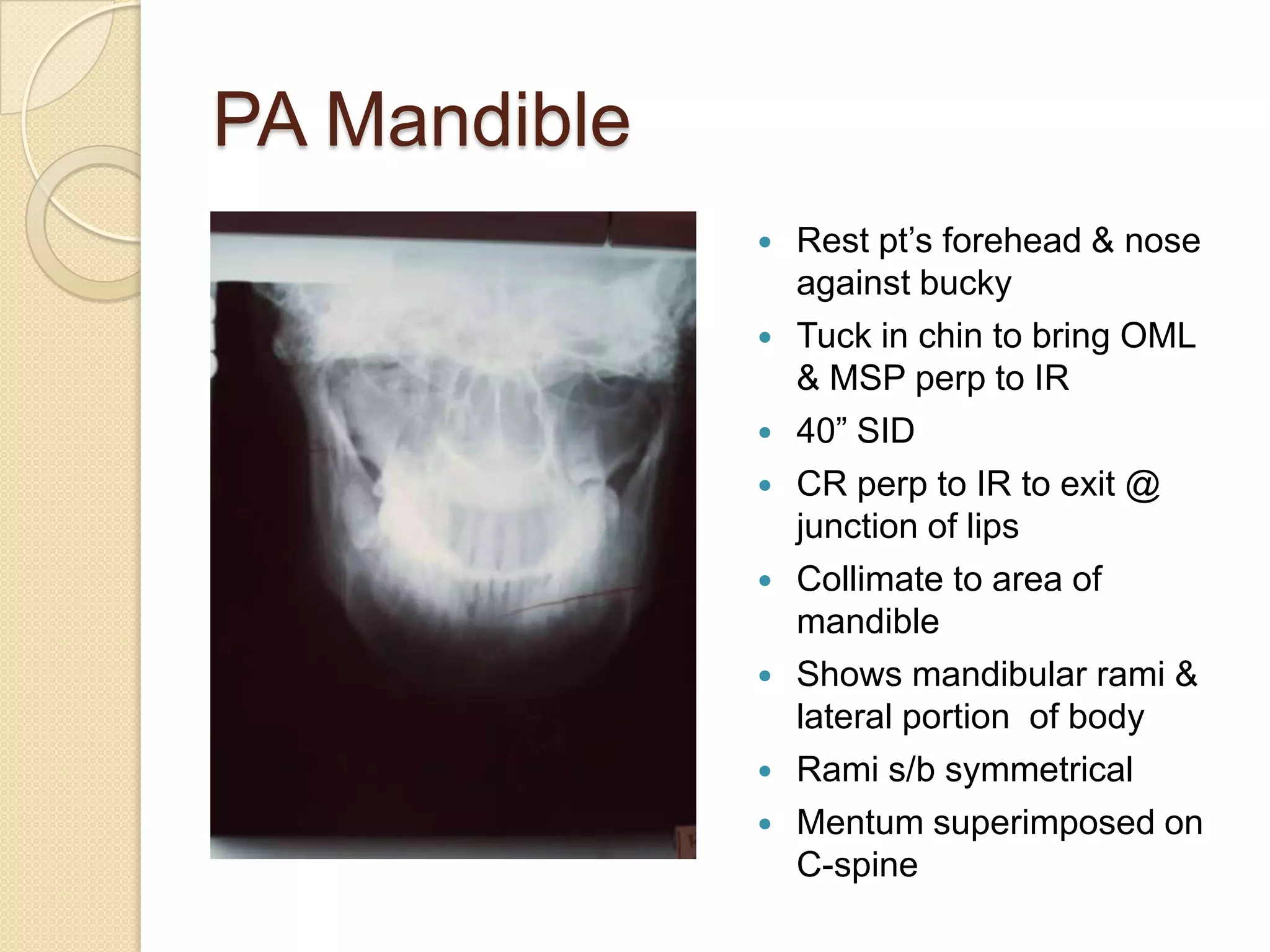


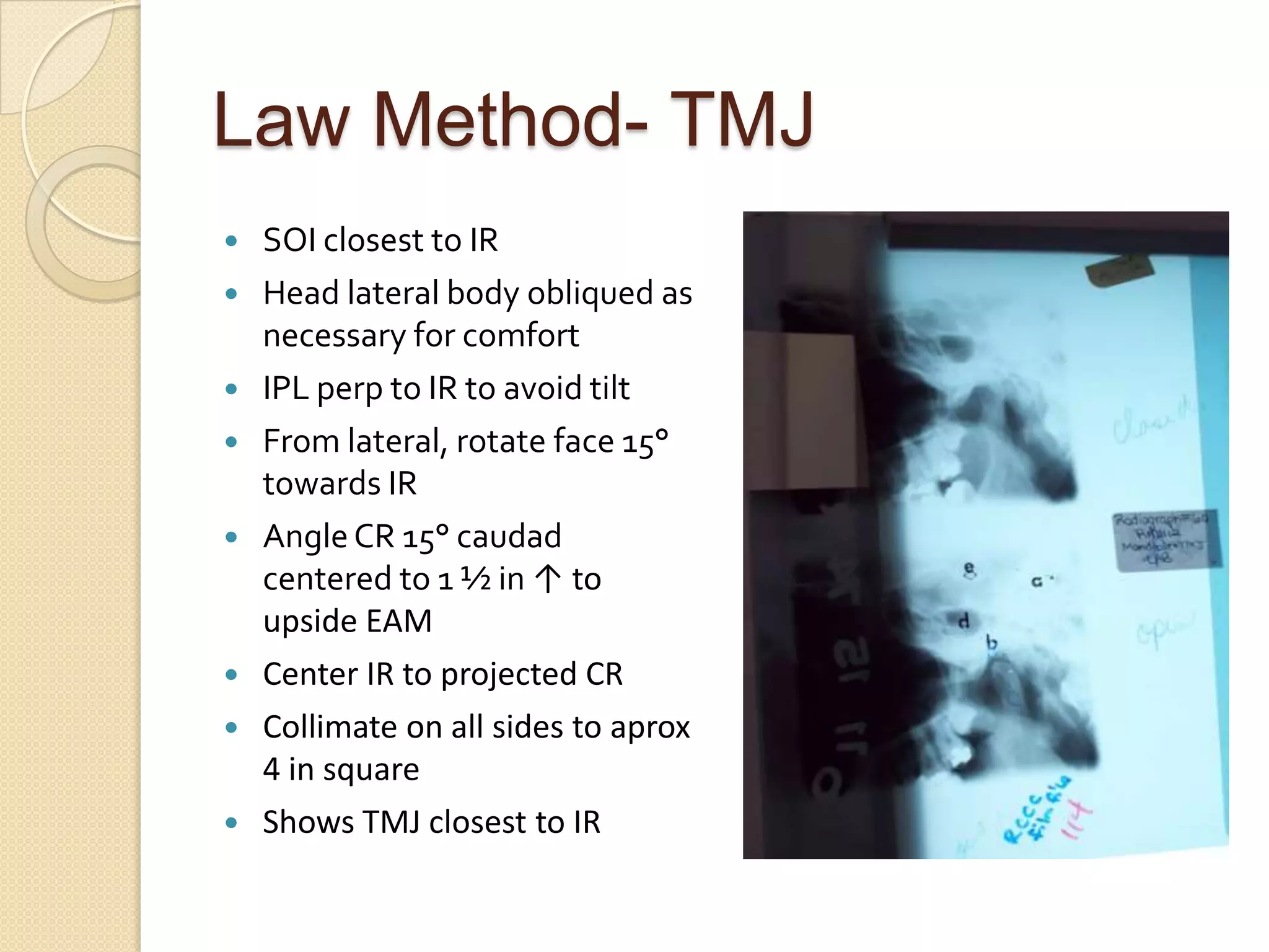
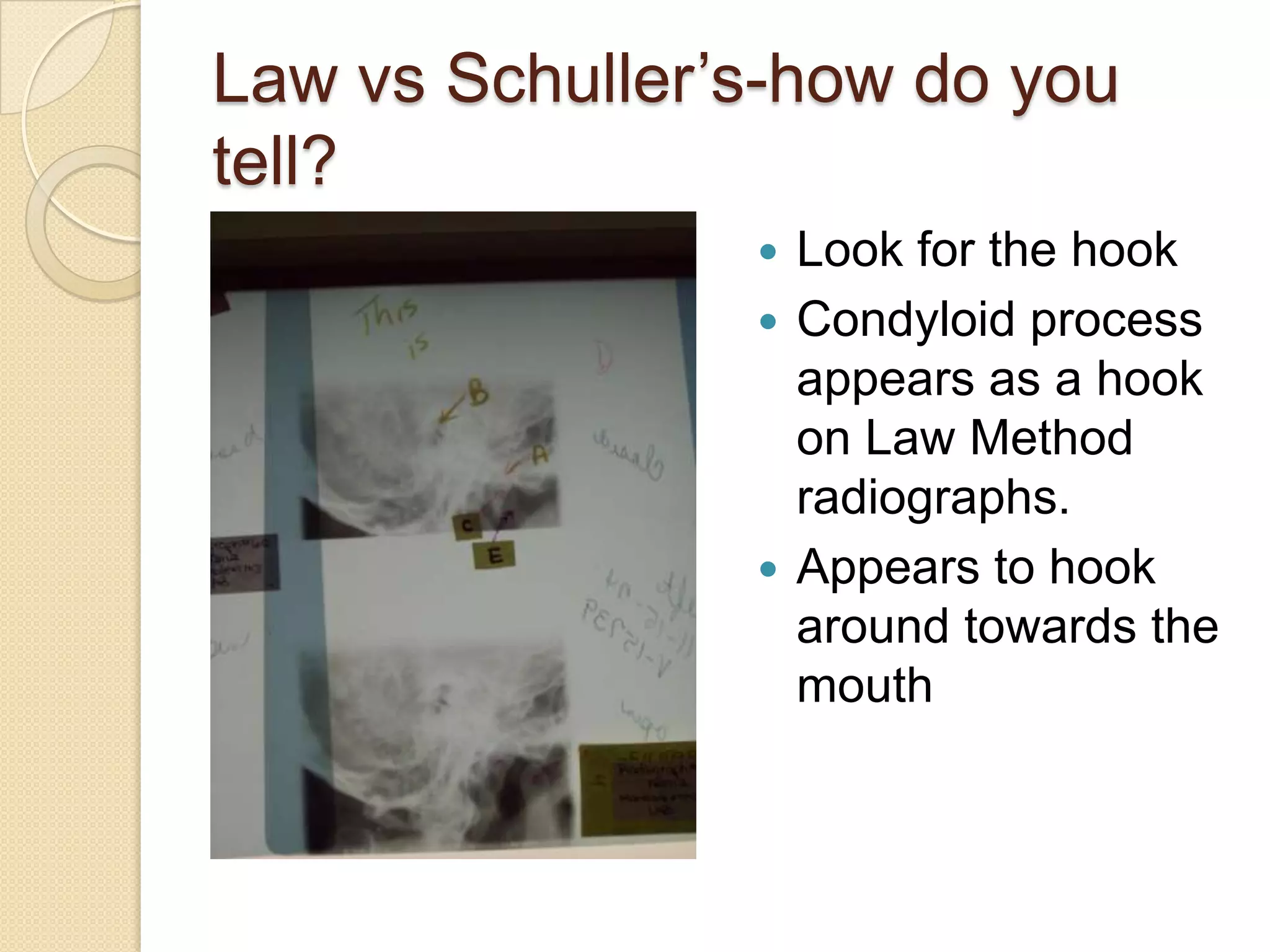
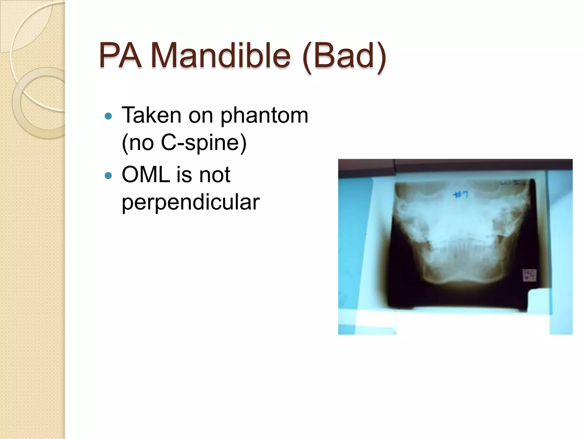

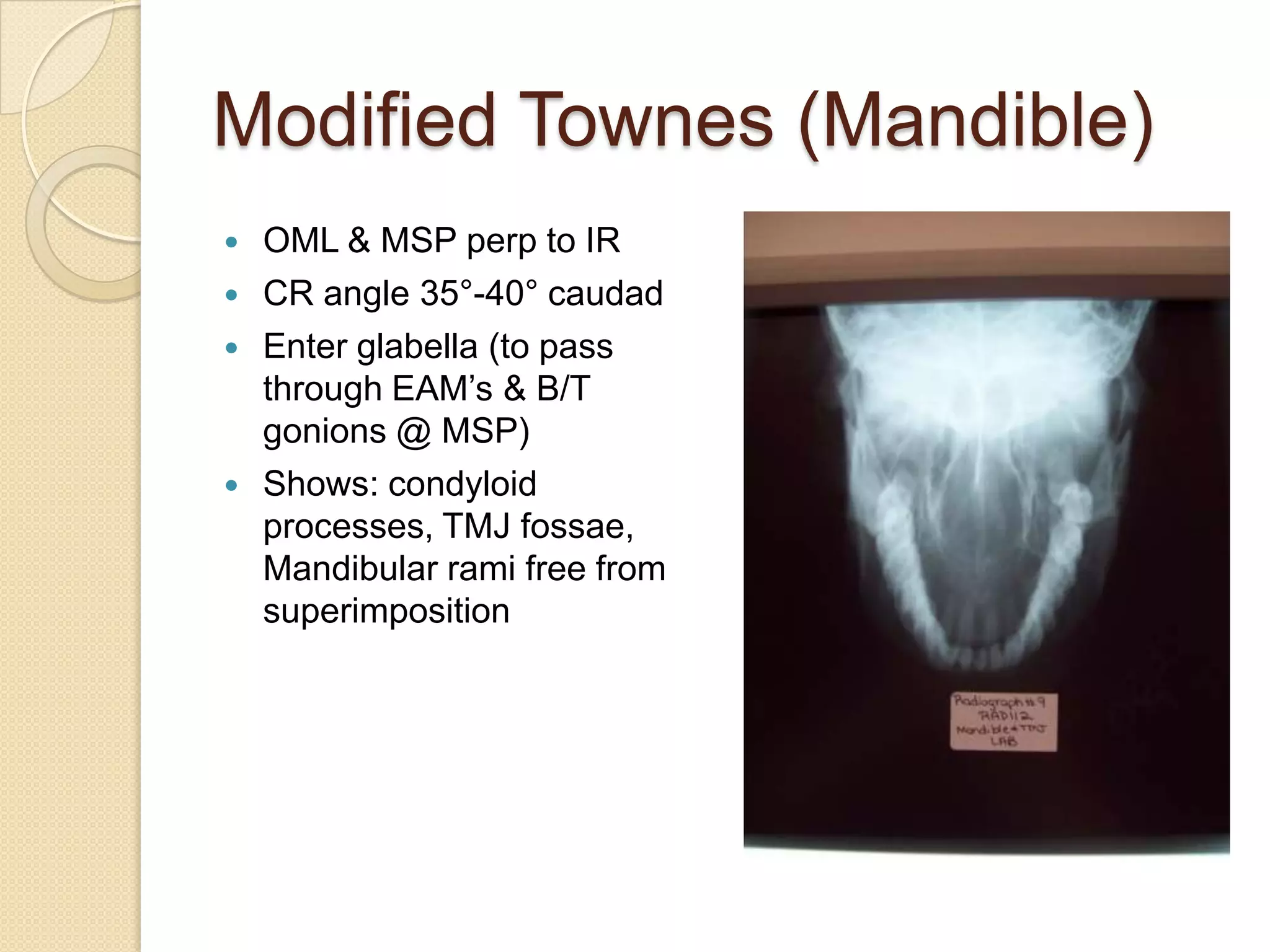


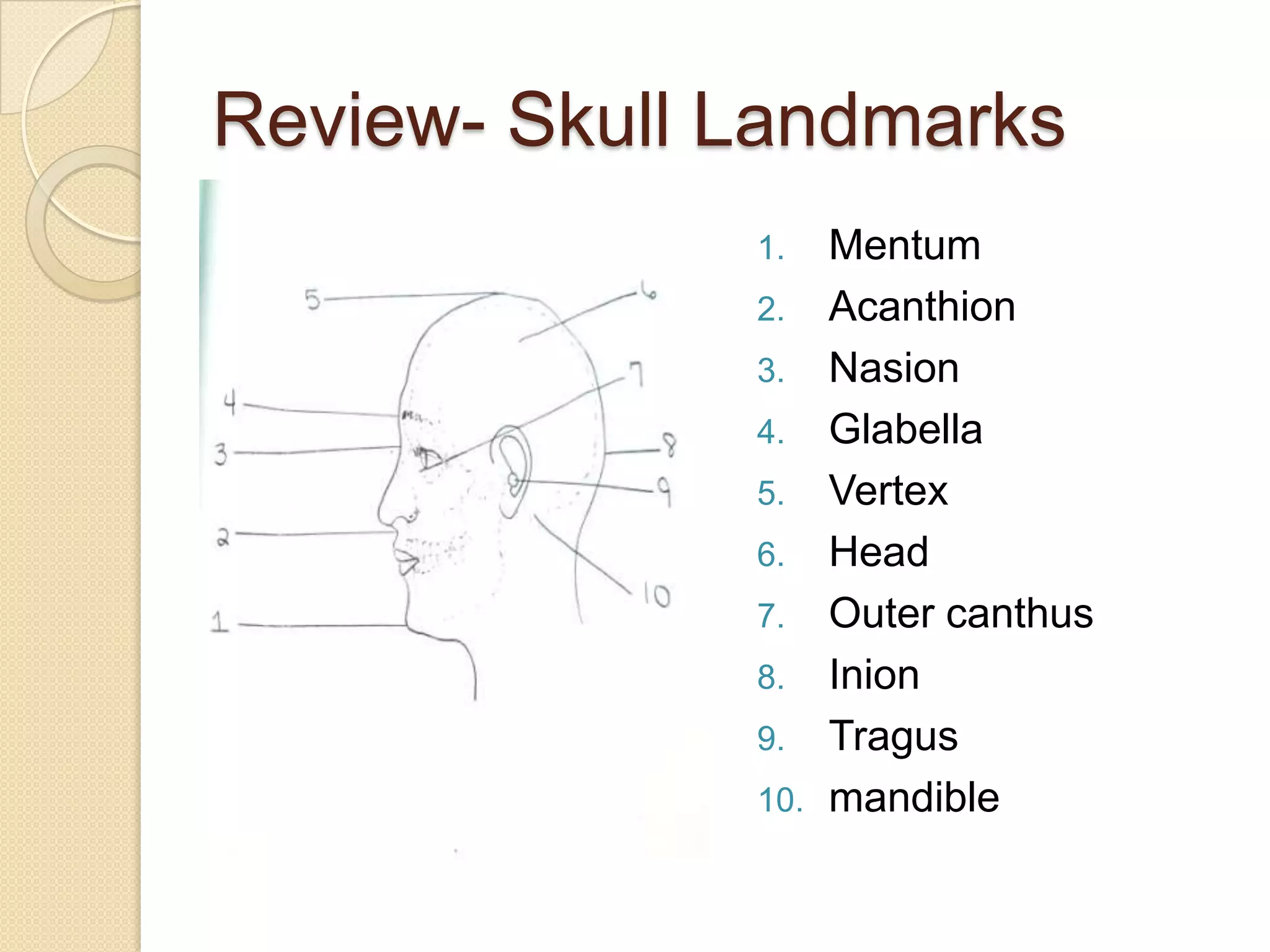
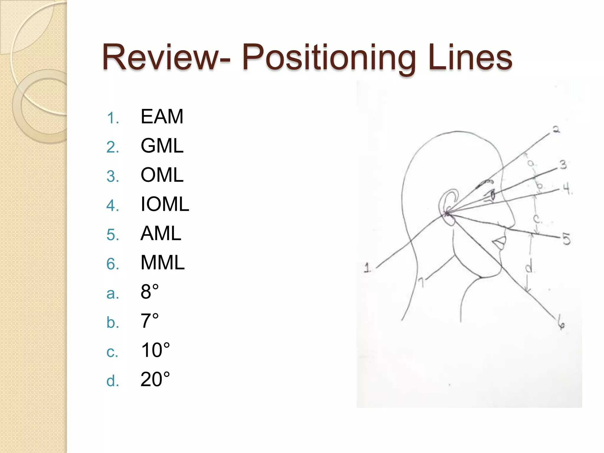

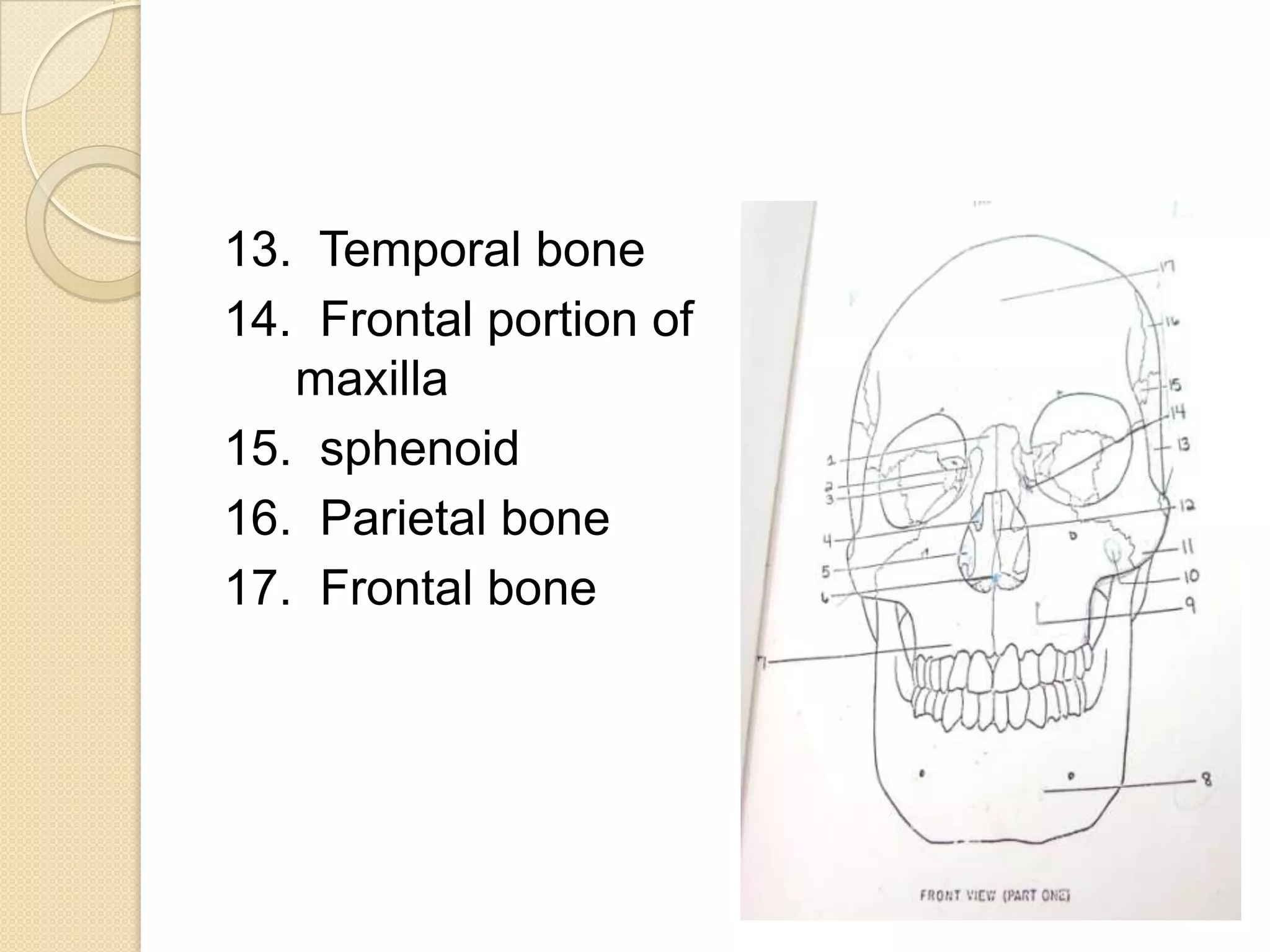


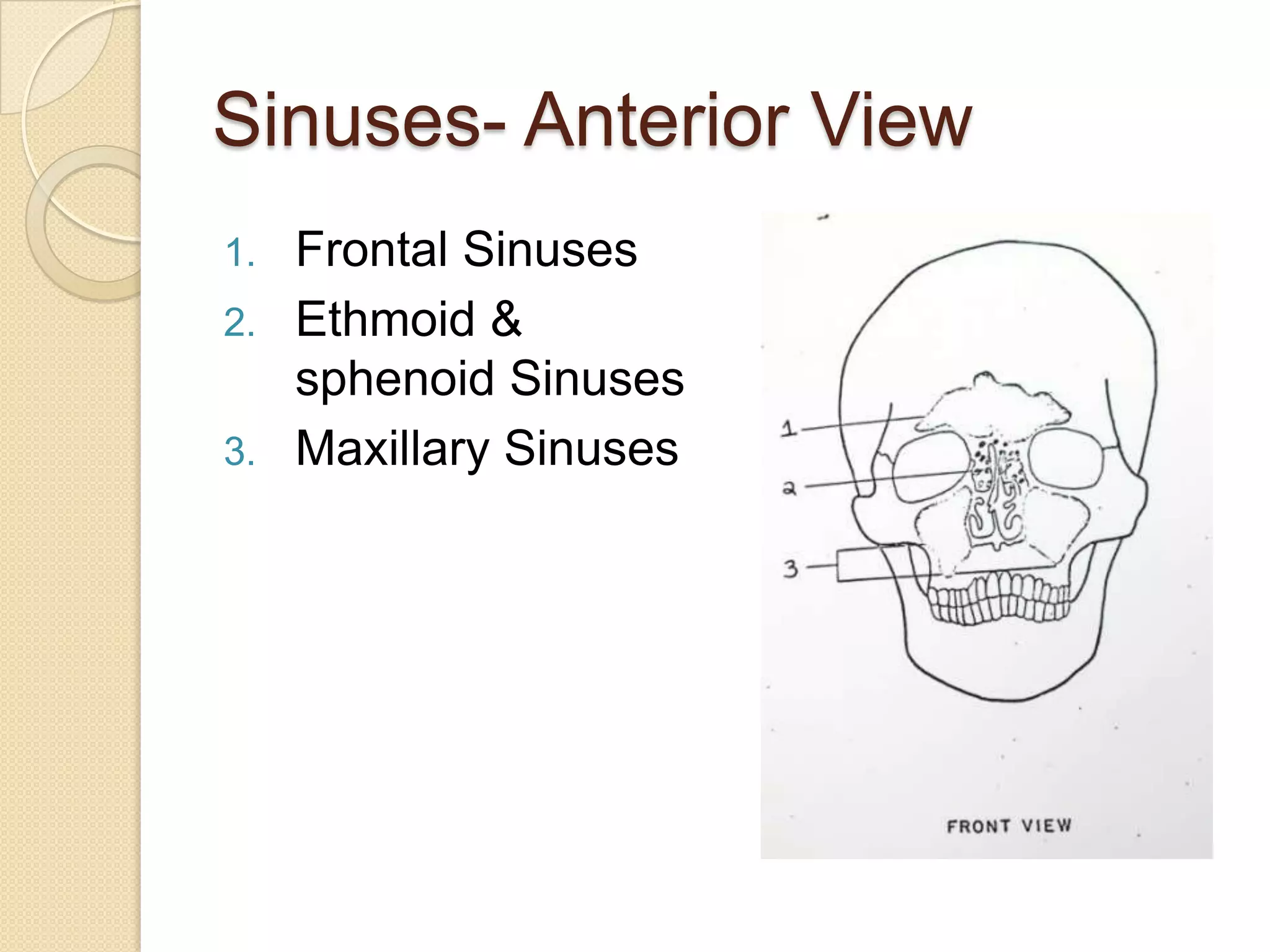


This document provides diagrams and descriptions of radiographic views of the mandible and temporomandibular joint (TMJ). It includes positioning, anatomical landmarks, and indications for various projections including posteroanterior, lateral, oblique, and panoramic views of the mandible and TMJ. Common radiographic techniques are outlined such as the Towne, Law, and Schuller methods for imaging the TMJ. Anatomical structures of the skull, sinuses, and mandible are also labeled on diagrams.


