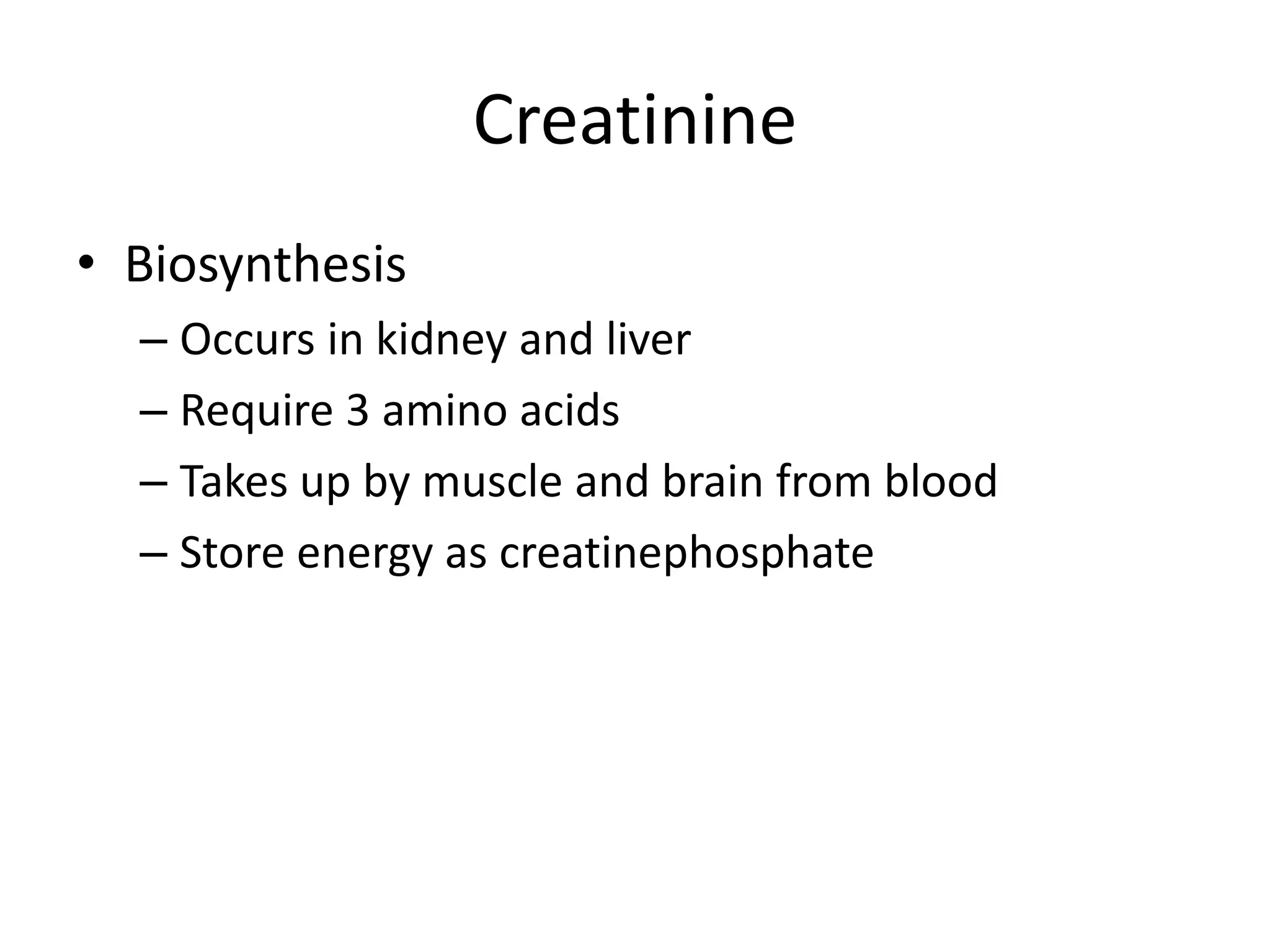
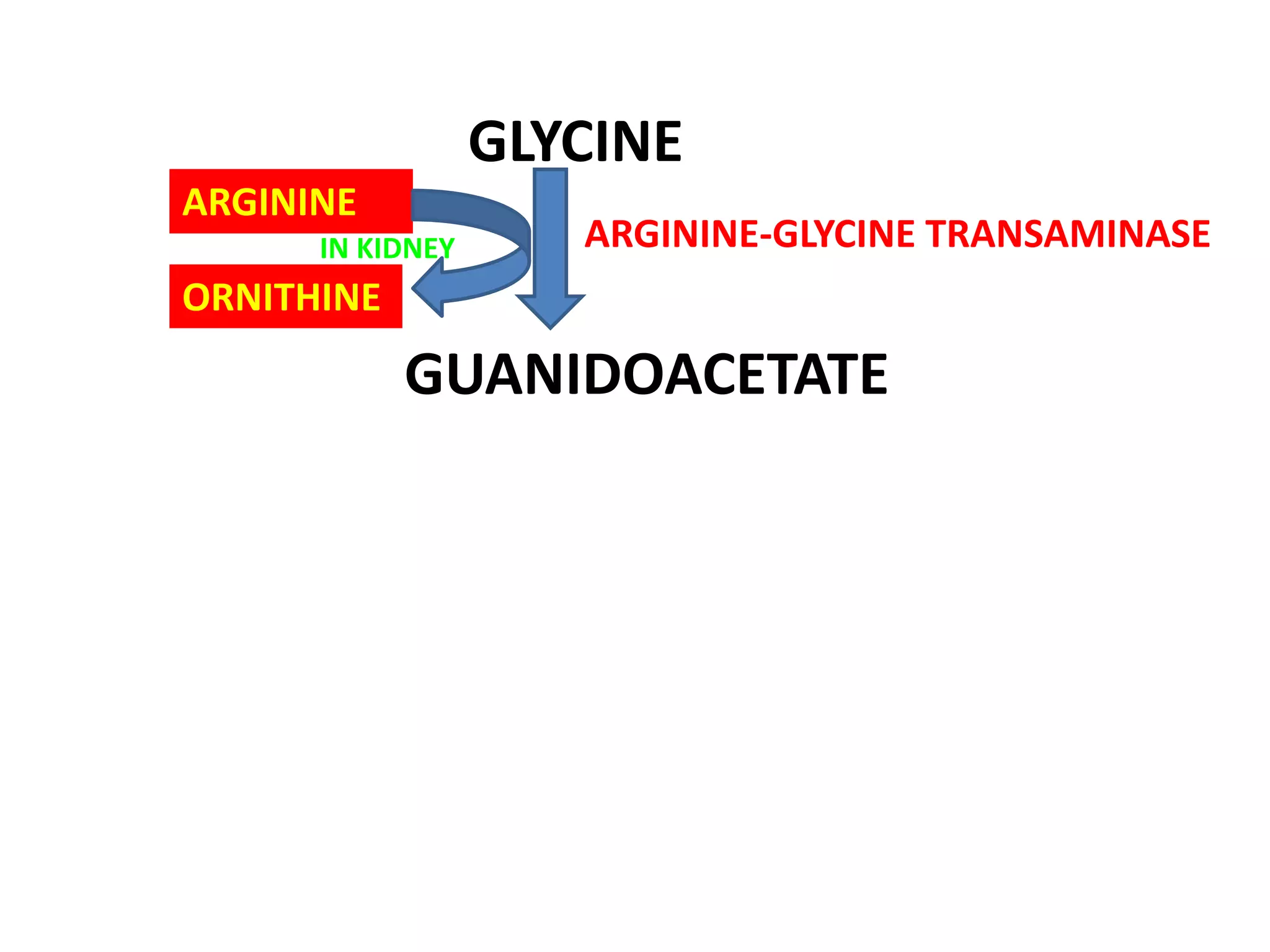
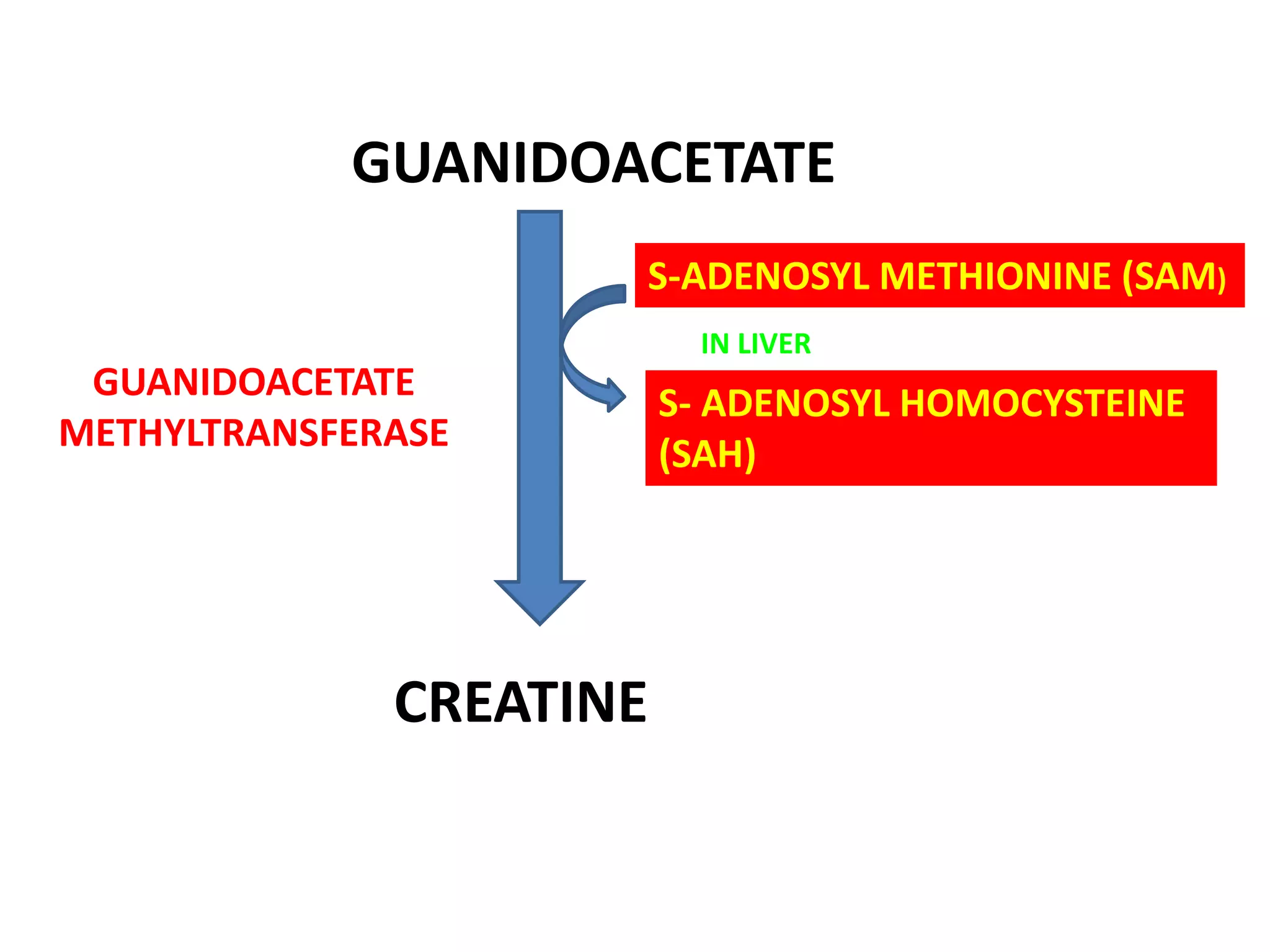
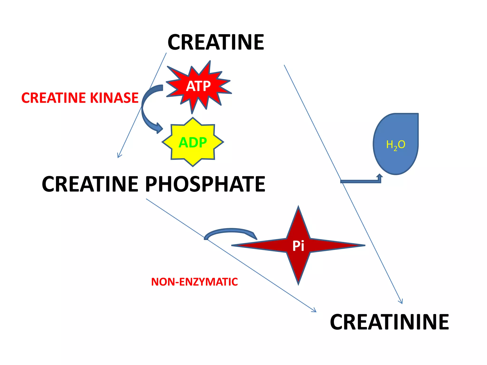
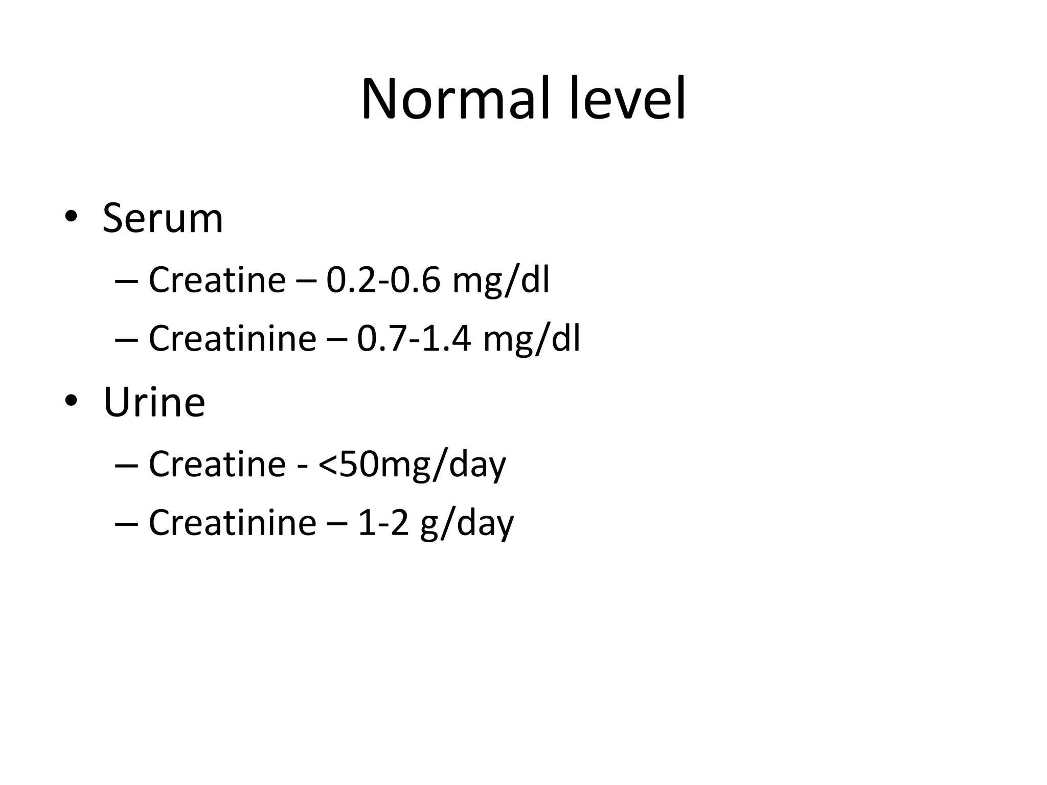
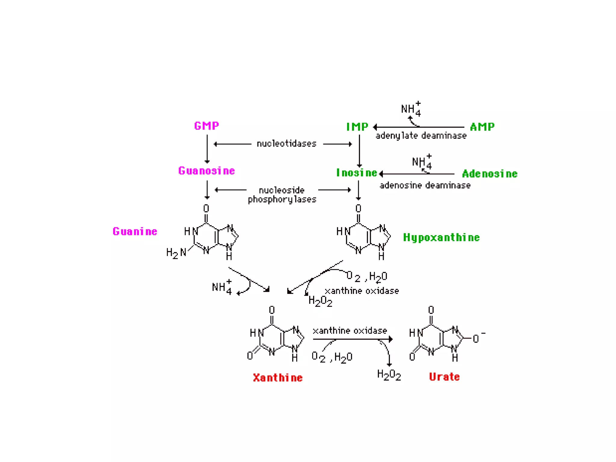
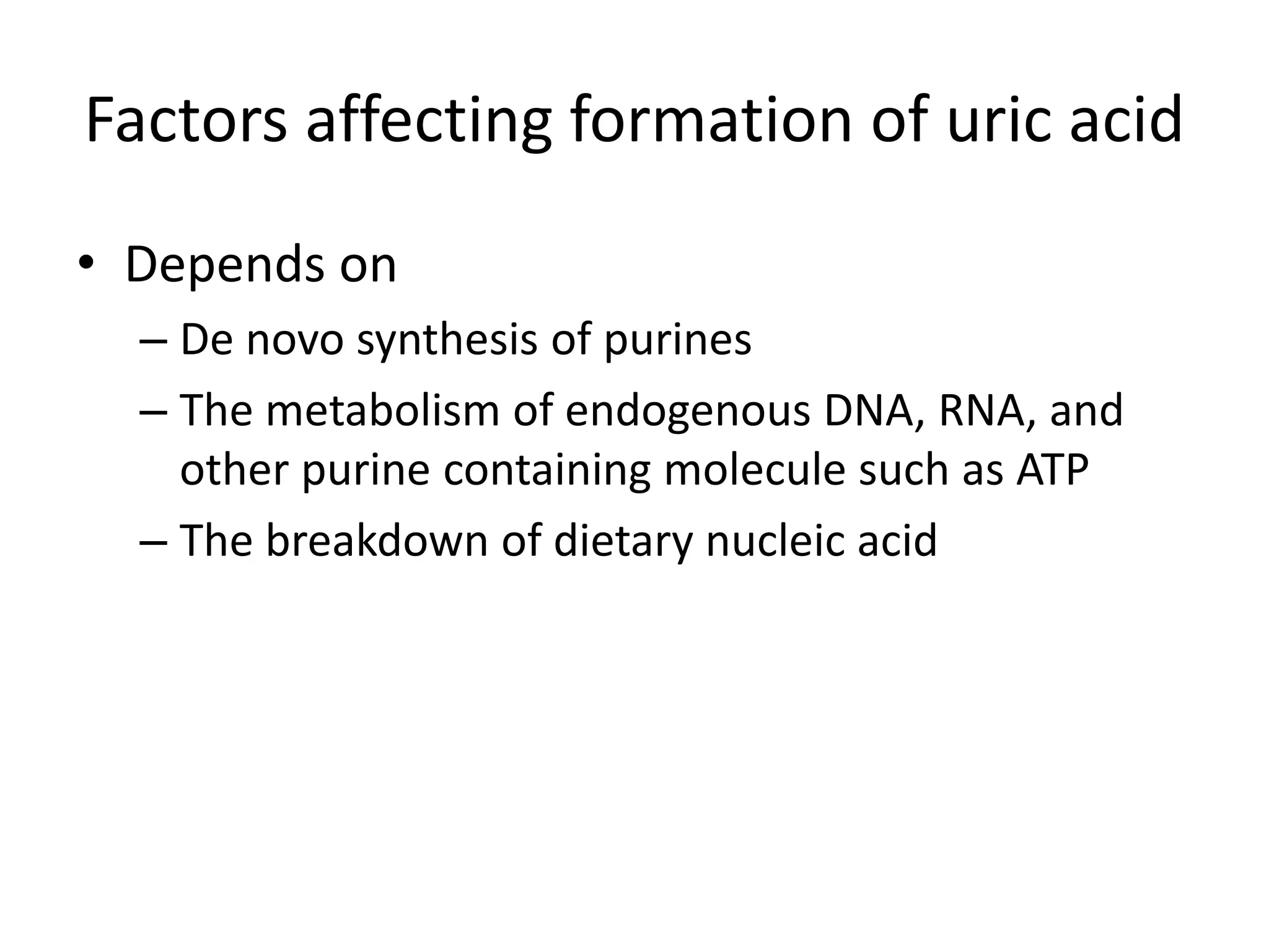
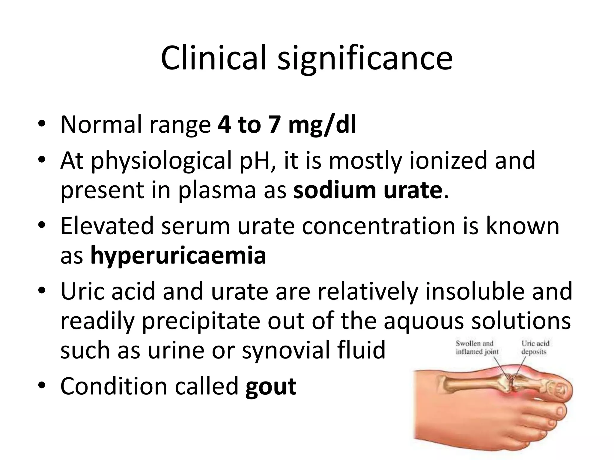
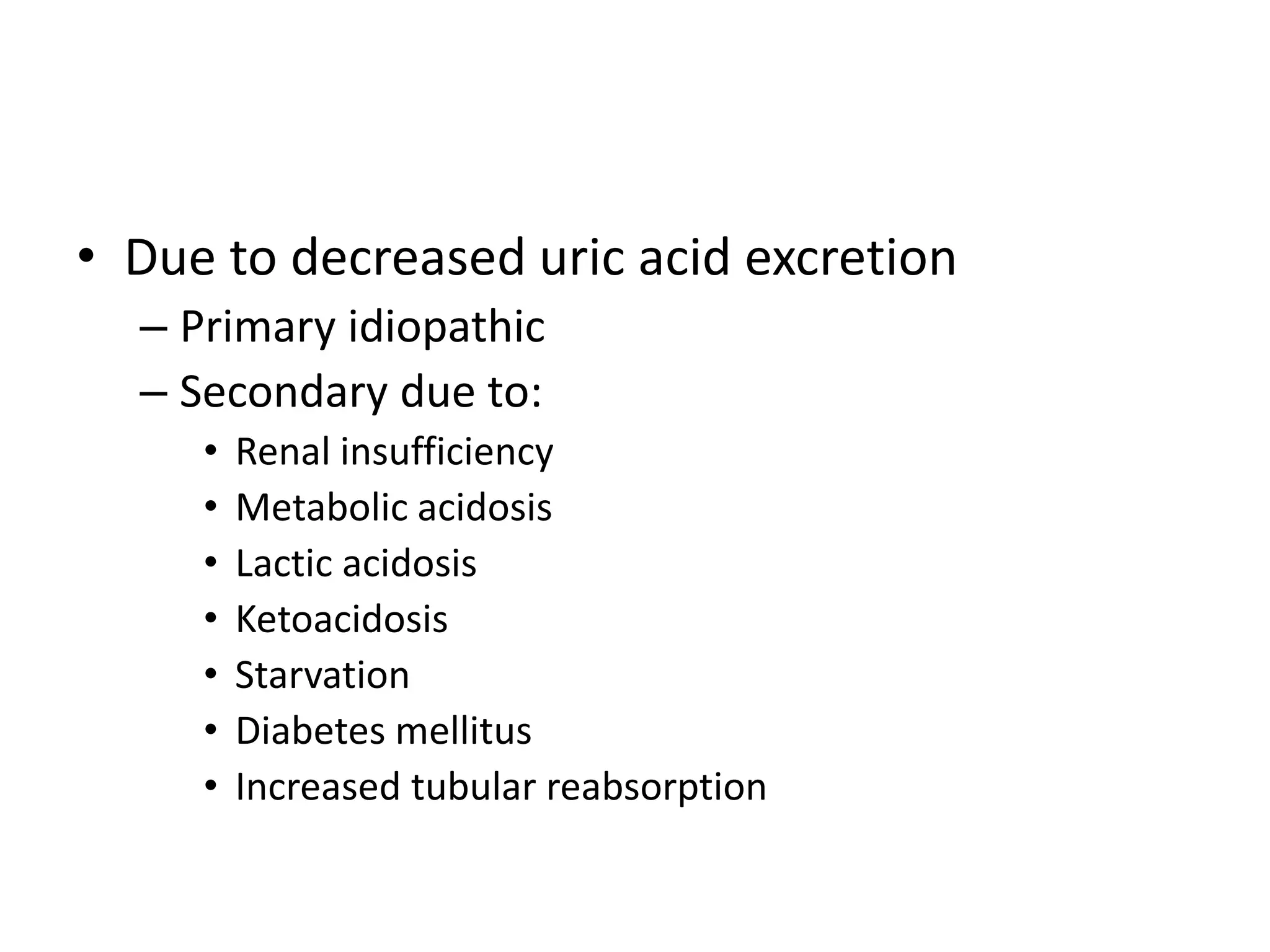
CREATININE METABOLISM
Creatinine
• Biosynthesis
– Occurs in kidney and liver
– Require 3 amino acids
– Takes up by muscle and brain from blood
– Store energy as creatinephosphate
GLYCINE
ARGININE
GUANIDOACETATE
ORNITHINE
ARGININE-GLYCINE TRANSAMINASE
IN KIDNEY
GUANIDOACETATE
CREATINE
S-ADENOSYL METHIONINE (SAM)
S- ADENOSYL HOMOCYSTEINE
(SAH)
GUANIDOACETATE
METHYLTRANSFERASE
IN LIVER
CREATINE
CREATINE PHOSPHATE
ATP
ADP
CREATININE
H2O
Pi
CREATINE KINASE
NON-ENZYMATIC
Normal level
• Serum
– Creatine – 0.2-0.6 mg/dl
– Creatinine – 0.7-1.4 mg/dl
• Urine
– Creatine - <50mg/day
– Creatinine – 1-2 g/day
Uric Acid Biosynthesis and Its Disorder
Factors affecting formation of uric acid
• Depends on
– De novo synthesis of purines
– The metabolism of endogenous DNA, RNA, and
other purine containing molecule such as ATP
– The breakdown of dietary nucleic acid
Significance of uric acid
• Potent antioxidant
– Very effective scavenger of free radicals.
Excretion
• Via the kidney : major
• Via the gut : smaller amount
Clinical significance
• Normal range 4 to 7 mg/dl
• At physiological pH, it is mostly ionized and
present in plasma as sodium urate.
• Elevated serum urate concentration is known
as hyperuricaemia
• Uric acid and urate are relatively insoluble and
readily precipitate out of the aquous solutions
such as urine or synovial fluid
• Condition called gout
Cause of hyperuricaemia
• Due to increased uric acid formation
– Primary (genetic)
• Due to enzyme defects in:
– PRPP synthase
– PRPP aminotransferase
– HGPRTase
– Secondary
• Due to increased:
– Dietary intake
– Nucleic acid turnover
– ATP breakdown
– Deficiency of glucoase -6-phosphatase
• Due to decreased uric acid excretion
– Primary idiopathic
– Secondary due to:
• Renal insufficiency
• Metabolic acidosis
• Lactic acidosis
• Ketoacidosis
• Starvation
• Diabetes mellitus
• Increased tubular reabsorption
Gout
• Elevated uric acid level associated with either
increased formation of uric acid or decreased
renal excretion
• Classification
– Primary gout
– Secondary gout
• Symptoms
– Patients with primary gout often show deposition of
urate as tophi in soft tissues that affects the joints and
leads to painful arthritis
– Kidneys are also affected lead to renal failure
Primary Gout
• Inherited due to an inborn error of
metabolism caused by defective enzymes of
purine biosynthesis
– Partial deficiency of HGPRTase
– Loss of feed back regulation
• PRPP synathase
• PRPP amidotransferase
Secondary Gout
• Elevated destruction of cells or defective elimination of uric
acid
– Over production of urate due to increased destruction or
turnover of cells
• Myeloproliferative disorders ( e.g. leukaemia, polycythaemia)
• Cytotoxic drug ( antimetabolites) therapy
• Psoriasis
– Defective elimination of uric acid due to
• Reduced glomerular filtration rate
• In diuretic therapy due to reduced distal tubular secretion of uric acid
– Hyper-catabolic states and starvation
• Lactic acidosis and ketoacidosis
– Inherited metabolic disorders
• Eg. Von gierke’s disease
Treatment
• Foods rich in nucleotides and nucleic acids
such as liver or coffee and tea, which contain
the purines caffeine and theobromin are
withheld from the diet
• Use of drug allopurinol
LESCH-NYHAN SYNDROME
• Inherited X-linked disorder
• Complete deficiency of HGPRTase
• Symptoms
– Hyperuricaemia
– Gout
– Urinary tract stones
– Mental retardation
– Spasticity
– Self-mutilation
• Treatment
– allopurinol
Hypouricaemia
• Decreased serum uric acid less than 2mg/dl
• Causes
– Xanthinuria
– Adenosine deaminase (ADA) deficiency
– Purine nucleoside phosphorylase deficiency
Melanin synthesis
Albinism
• greek: albino – white
• Inborn error, due to alck of synthesis of the
pigment melanin
• Autosomal recessive disorder with frequency
of 1 in 20000
• Causes
– Deficiency or lack of the enzyme tyrosinase
– Decrease in melanosomes of melanocytes
– Impairment in melanin polymerization
– Lack of protein matrix in melanosomes
– Limitation of substrate (tyrosine) availability
– Presence of inhibitors of tyrosinase
• Clinical manifestation
– Skin cancer
– Photophobia
– White hair

CREATININE METABOLISM, uric acid biosynthesis and melanin synthesis.pptx

  • 1.
  • 2.
    Creatinine • Biosynthesis – Occursin kidney and liver – Require 3 amino acids – Takes up by muscle and brain from blood – Store energy as creatinephosphate
  • 3.
  • 4.
    GUANIDOACETATE CREATINE S-ADENOSYL METHIONINE (SAM) S-ADENOSYL HOMOCYSTEINE (SAH) GUANIDOACETATE METHYLTRANSFERASE IN LIVER
  • 5.
  • 6.
    Normal level • Serum –Creatine – 0.2-0.6 mg/dl – Creatinine – 0.7-1.4 mg/dl • Urine – Creatine - <50mg/day – Creatinine – 1-2 g/day
  • 7.
    Uric Acid Biosynthesisand Its Disorder
  • 9.
    Factors affecting formationof uric acid • Depends on – De novo synthesis of purines – The metabolism of endogenous DNA, RNA, and other purine containing molecule such as ATP – The breakdown of dietary nucleic acid
  • 10.
    Significance of uricacid • Potent antioxidant – Very effective scavenger of free radicals.
  • 11.
    Excretion • Via thekidney : major • Via the gut : smaller amount
  • 12.
    Clinical significance • Normalrange 4 to 7 mg/dl • At physiological pH, it is mostly ionized and present in plasma as sodium urate. • Elevated serum urate concentration is known as hyperuricaemia • Uric acid and urate are relatively insoluble and readily precipitate out of the aquous solutions such as urine or synovial fluid • Condition called gout
  • 13.
    Cause of hyperuricaemia •Due to increased uric acid formation – Primary (genetic) • Due to enzyme defects in: – PRPP synthase – PRPP aminotransferase – HGPRTase – Secondary • Due to increased: – Dietary intake – Nucleic acid turnover – ATP breakdown – Deficiency of glucoase -6-phosphatase
  • 14.
    • Due todecreased uric acid excretion – Primary idiopathic – Secondary due to: • Renal insufficiency • Metabolic acidosis • Lactic acidosis • Ketoacidosis • Starvation • Diabetes mellitus • Increased tubular reabsorption
  • 15.
    Gout • Elevated uricacid level associated with either increased formation of uric acid or decreased renal excretion • Classification – Primary gout – Secondary gout • Symptoms – Patients with primary gout often show deposition of urate as tophi in soft tissues that affects the joints and leads to painful arthritis – Kidneys are also affected lead to renal failure
  • 16.
    Primary Gout • Inheriteddue to an inborn error of metabolism caused by defective enzymes of purine biosynthesis – Partial deficiency of HGPRTase – Loss of feed back regulation • PRPP synathase • PRPP amidotransferase
  • 17.
    Secondary Gout • Elevateddestruction of cells or defective elimination of uric acid – Over production of urate due to increased destruction or turnover of cells • Myeloproliferative disorders ( e.g. leukaemia, polycythaemia) • Cytotoxic drug ( antimetabolites) therapy • Psoriasis – Defective elimination of uric acid due to • Reduced glomerular filtration rate • In diuretic therapy due to reduced distal tubular secretion of uric acid – Hyper-catabolic states and starvation • Lactic acidosis and ketoacidosis – Inherited metabolic disorders • Eg. Von gierke’s disease
  • 18.
    Treatment • Foods richin nucleotides and nucleic acids such as liver or coffee and tea, which contain the purines caffeine and theobromin are withheld from the diet • Use of drug allopurinol
  • 19.
    LESCH-NYHAN SYNDROME • InheritedX-linked disorder • Complete deficiency of HGPRTase • Symptoms – Hyperuricaemia – Gout – Urinary tract stones – Mental retardation – Spasticity – Self-mutilation • Treatment – allopurinol
  • 20.
    Hypouricaemia • Decreased serumuric acid less than 2mg/dl • Causes – Xanthinuria – Adenosine deaminase (ADA) deficiency – Purine nucleoside phosphorylase deficiency
  • 21.
  • 23.
    Albinism • greek: albino– white • Inborn error, due to alck of synthesis of the pigment melanin • Autosomal recessive disorder with frequency of 1 in 20000
  • 24.
    • Causes – Deficiencyor lack of the enzyme tyrosinase – Decrease in melanosomes of melanocytes – Impairment in melanin polymerization – Lack of protein matrix in melanosomes – Limitation of substrate (tyrosine) availability – Presence of inhibitors of tyrosinase
  • 25.
    • Clinical manifestation –Skin cancer – Photophobia – White hair