
Recommended
More Related Content
What's hot
What's hot (20)
Similar to White blood cells - morphology, functions and variations
Similar to White blood cells - morphology, functions and variations (20)
More from Jilsha Cecil
Recently uploaded
Recently uploaded (20)
White blood cells - morphology, functions and variations
- 2. CONTENTS Introduction Normal values CLASSIFICATION Morphological features Functions Physiological and pathological variations
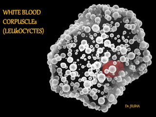
- 3. INTRODUCTION Why are they called White Blood Cells ? After centrifugation, WBCs are found in the buffy coat, a thin white layer of cells between the sedimented RBCs and the plasma.
- 4. INTRODUCTION… WBCs are nucleated cells that perform the defense functions of the body They destroy the invading micro- organisms and destroy abnormal cells like cancer cells.
- 5. NORMAL VALUES Total Leukocycte count – Normal WBC count : Adults : 4000 – 11,000 / mm3 of blood At Birth : 10,000 – 25,000 / mm3 of blood (count decreases after 2nd week and reaches the normal adult value at 5 – 10 yrs.)
- 6. CONTD.. CELL ABSOLUTECOUNT (/cu.mm) DIFFERENTIALCOUNT(%) Neutrophils 3000 - 6000 50 – 70 % Eosinophils 150 - 300 1 – 4% Basophils 10 - 100 < 1 % Lymphocytes 1500 - 2700 20 – 40 % Monocytes 300 - 600 2 – 8 %
- 8. VARIATIONS IN WBC COUNT Leukocytosis : refers to increase in WBC count above 11,000 / mm3 Physiological causes : Newborn Exercise Mental stress Pregnancy After food intake Pathological Causes Acute bacterial infections Acute hemorrhage Burns Tuberculosis
- 9. Leukopenia : refers to WBC count below 4000 / mm3 Causes : Typhoid fever Starvation Viral and protozoal infections Bone marrow depression FACT Leukemia is a cancerous condition of blood in which the TLC is more than 50,000 / cu.mm ASSOCIATED WITH THE PRESENCE OF IMMATURE WBCs in the peripheral smear
- 10. CLASSIFICATION
- 12. MORPHOLOGY Morphological features of various types of WBCs are studied under microscope with Leishman’s staining and hematoxylin – eosin staining. Size : 10 – 14 µm diameter Nucleus : Young neutrophil – horseshoe shaped. As the cells grow older nucleus becomes multi lobed (2-6 lobes). Mature neutrophil has purple colored nucleus.
- 13. MORPHOLOGY
- 14. MORPHOLOGY Cytoplasm : Granular, slight bluish in color. Granules Fine sand like particles Take both acidic and basic stains. Hence the name neutrophils (neutrophilic in nature) Contains variety of enzymes like glycosidases, sulphatases, phosphatases, nucleases, proteolytic enzymes etc. Hence can lyse any substance. Also liberate histamine and peroxidase enzymes which helps in killing the ingested bacteria
- 16. LIFESPANANDFATE Neutrophils like all other leukocyctes have four stages in their life : Marrow Pool Circulation pool : 8 -10 hours Marginated pool Tissue pool : either destroyed during phagocytosis or die due to senescence after 4 – 5 days. A huge number of neutrophils is also eliminated daily, mainly into the intestine and some into the respiratory secretions.
- 17. FUNCTIONS Neutrophils are actively phagocytic. They are considered as the first line defense against acute bacterial infections. They contain a fever producing substance called endogenous pyrogen which is an important mediator of febrile response to bacterial pyrogens.
- 18. NEUTROPHIL PHAGOCYTOSIS Phagocytosis is the process of ingestion and killing of microbes or a foreign substance by a phagocyte. Steps of phagocytosis : Chemotaxis Diapedesis Adherence Ingestion Killing
- 19. Chemotaxis It is the process by which the neutrophils are attracted towards bacteria at the site of inflammation. It is mediated by chemotactic agents called chemotaxins. They are usually the microbial products or chemicals released from damaging tissues. These include Leukotriene B4 , Complement proteins like C5a and C3. Neutrophils change their shape and become amoeboid. Leukopoiesis is stimulated and more neutrophils are produced.
- 20. Contd..
- 21. Diapedesis The process by which neutrophils pass through the capillary endothelial cells to reach the invader in the tissue is called diapedesis. The activated neutrophils first marginate (margination and pavementing) and adhere tightly to endothelial lining with the help of L – selectins. Then, by their amoeboid movement they squeeze through the space between the endothelial cells (emigration and diapedesis)
- 22. CONTD..
- 23. Opsonization and Adherence The process by which the bacteria are made tasty to the phagocyte is called Opsonization. In this process, antigen is coated by opsonins. Immunoglobulin G antibody and complement proteins are some examples. The attachment of membrane of phagocyte to the membrane of microbe is called adherence.
- 24. CONTD..
- 25. Ingestion (Endocytosis) The membrane of phagocyte extends projections from both the sides to encroach on to the microbe. These extensions are called pseudopodia. Pseudopodia finally surround the microbe and form phagocytic vesicle. The phagocytic vesicle fuse with the lysosome to form phagolysosome.
- 26. CONTD..
- 27. Killing or degradation The bactericidal mechanism can be broadly classified into : o Non – oxidative and o Oxidative mechanisms Non – oxidative mechanism Neutrophil granules contain a variety of anti – bacterial chemicals such as degradative enzymes, proteases, defensins, cationic proteins. Lysozyme – hydrolyzes the cell wall, Lactoferrin – sequestrates iron – non –oxidative components. Defensins – kill bacteria by disrupting their outer membrane and breaking single strand DNA structure.
- 28. Oxidative mechanisms: Following this, the cell membrane bound enzyme NADPH oxidase is activated. This causes a sharp uptake in O2. This is called respiratory burst. This leads to formation of superoxide radical (O2 -) and Hydrogen peroxide (H2O2) as follows : 𝑁𝐴𝐷𝑃𝐻 + 𝐻+ + 2𝑂2 → 𝑁𝐴𝐷𝑃+ + 2𝐻+ + 2𝑂2 2𝑂2 − + 2𝐻+ → 𝐻2 O2 O2 + 𝐻2 O2 → OH - + OH- + O2 Hydroxyl radical is a very potent bactericidal agent.
- 29. CONTD..
- 30. VARIATIONS Neutrophilia Exercise Cold Stress Acute pyogenic Bacterial infections Burns Hemorrhage Drugs like epinephrine, glucocorticoids
- 31. Neutropenia Typhoid and paratyphoid fever Malaria Aplastic anemia Viral infections like measles, influenza Drugs like chloramphenicol, pheytoin
- 32. EOSINOPHILS
- 33. MORPHOLOGY Size : 10 – 14 µm diameter Nucleus : Bilobed – spectacle shaped. Purple colored nucleus. Cytoplasm : Acidophilic, light pink in color. Granules : Coarse, stain bright red with eosin. Granules contain chemicals like: Major basic protein Eosinophil catonic protein Eosinophil derived peroxidase Eosinophil derived neurotoxin Cytokines
- 34. MORPHOLOGY
- 35. FUNCTIONS Eosinophils participate in two important defense mechanisms of the body Against helminthic / parasitic infections Against allergy They attack parasites that are too large to be engulfed by phagocytosis. The granules release peroxidase which are toxic to larvae of parasites. Eosinophils collect at the site of allergic reactions to release enzymes such as histaminase and thus limit the effects of inflammatory mediators.
- 36. VARIATIONS Eosinophilia : Allergic conditions like bronchial asthma, allergic rhinitis, urticarial Parasitic infections like filariasis, ascariasis Tropical pulmonary eosinophilia Drug reactions. Eosinopenia Glucocorticoid therapy Cushing’s syndrome
- 37. BASOPHILS
- 38. MORPHOLOGY Size : 10 – 14 µm diameter Nucleus : Bilobed – S shaped. Purple colored nucleus. Cytoplasm : Slight basophilic, blue in color. Granules : Coarse, stains purple or blue with methylene blue. Granules are plenty and overcrowd the nucleus. Granules contain histamine, heparin, serotonin and eosinophil chemotactic factor ( ECF –A ) of anaphylaxis.
- 39. CONTD..
- 40. FUNCTIONS Mild phagocytosis Liberates histamine and ECF – A during allergic reaction which leads to allergic manifestations. Mild urticaria to severe anaphylactic shock. Liberates heparin which acts as an anti – coagulant.
- 41. VARIATIONS Basophilia : Chickenpox Small pox Tuberculosis Influenza Ulcerative colitis Drug and food hypersensitivity Basopenia : Glucocorticoids Hyperthyroidism Cushing’s syndrome
- 42. SUMMARY
- 43. LYMPHOCYTES
- 44. morphology Lymphocytes are of two types : Large lymphocytes : 10 – 14 µm diameter Small lymphocytes : 7 – 10 µm diameter Nucleus : Single, big, Purple colored nucleus. Round, oval or indented. Central in position and occupies whole of the cell leaving marginal cytoplasm at one end of it or all around it. Nuclear chromatin is coarse and lumpy. Cytoplasm : Pale blue, scanty.
- 45. CONTD..
- 46. FUNCTIONS Fuctionally, lymphocytes are divided into three categories : B cells, T cells and Natural killer cells (NK cells). B cells : secrete antibodies. Mediates humoral or antibody – mediated immunity. T cells : mediate cell – mediated immunity. NK cells : mediate natural and nonspecific immunity.
- 47. VARIATIONS Lymphocytosis Children – relative lymphocytosis (60%) Chronic infections like TB Lymphatic leukemia Viral infections. Lymphocytopenia Hypoplastic bone marrow AIDS
- 48. MONOCYTES
- 49. MORPHOLOGY Size : 10 – 18 µm diameter. Largest of WBCs. Nucleus : Single, round or indented, eccentric, pale staining. Cytoplasm : pale blue in color, clear. Sometimes contain fine dust like granules – Azur granules.
- 50. CONTD..
- 51. FUNCTIONS Monocyte is an active phagocyte. It is the second line of defense against infections. It is an important Antigen Presenting Cell (APC) Monocytes secrete various chemokines that carry out different physiological functions. Life span : In circulation : 10 – 72 hrs. Then they enter tissues. Average lifespan is 3 months. In tissues they become tissue macrophages.
- 52. VARIATIONS Monocytosis : Acute monocytic leukemia Hodgkin’s disease Polycythemia vera Malaria Kala – azar Monocytopenia: Aplastic anemia Hairy cell leukemia Septicemia
- 53. SUMMARY
- 54. THANK YOU !